FIGURE 1. Geographical and geological location of Bogovina Cave. A) Terrain in the vicinity of Bogovina Cave with simplified geological map (after Veselinović et al., 1964). Elevation of the terrain is exaggerated several times. B) Position of Bogovina Cave in the central part of the Balkan Peninsula.
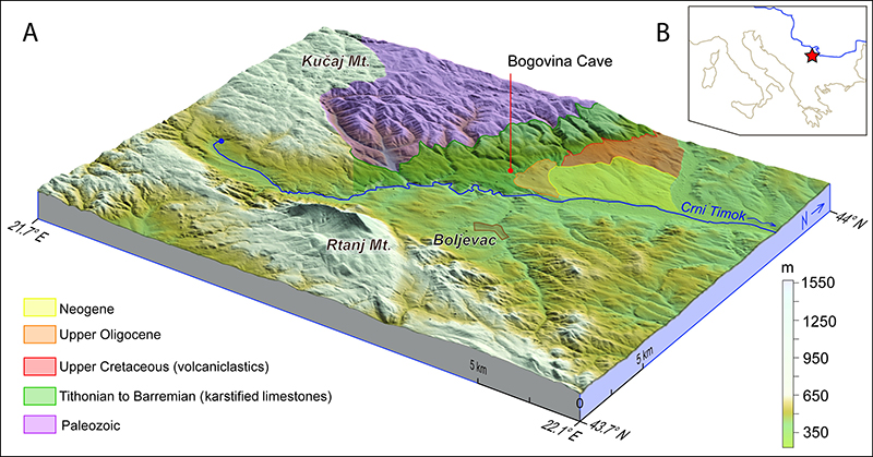
FIGURE 2. Dental terminology of rhinoceros cheek teeth (figure adapted from Fukuchi et al., 2009). A) Upper dentition: 1, protocone; 2, paracone; 3, metacone; 4, hypocone; 5, protoloph; 6, ectoloph; 7, metaloph; 8, parastyle; 9, metastyle; 10, paracone rib; 11, metacone rib; 12, crochet; 13, antecrochet; 14, crista; 15, lingual valley; 16, medisinus; 17, anterior fossetta; 18, postfossetta; 19, paracone fold; 20, protocone constriction; 21, mesial cingulum. B) Lower dentition: 22, paraconid; 23, protoconid; 24, metaconid; 25, hypoconid; 26, entoconid; 27, paralophid; 28, metalophid; 29, hypolophid; 30, mesial valley; 31, distal valley; 32, vestibular syncline.
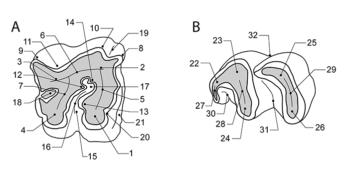
FIGURE 3. Upper premolars of Stephanorhinus hundsheimensis from Bogovina Cave: left P3/P4 NMKVRS.P13 in (A) occlusal, (B) vestibular, (C) lingual, (D) mesial and (E) distal views; right P3/P4 HMP-139b in (F) occlusal, (G) lingual, and (H) distal views.

FIGURE 4. Upper molars of Stephanorhinus hundsheimensis from Bogovina Cave: right M1/M2 HMP-131 in (A) occlusal, (B) vestibular, (C) lingual, (D) mesial and (E) distal views; right M1/M2 HMP-X in (F) occlusal, (G) vestibular, (H) lingual, (I) mesial and (J) distal views; left M1/M2 HMP-135 in (K) occlusal, (L) vestibular, (M) lingual, (N) mesial, and (O) distal views.
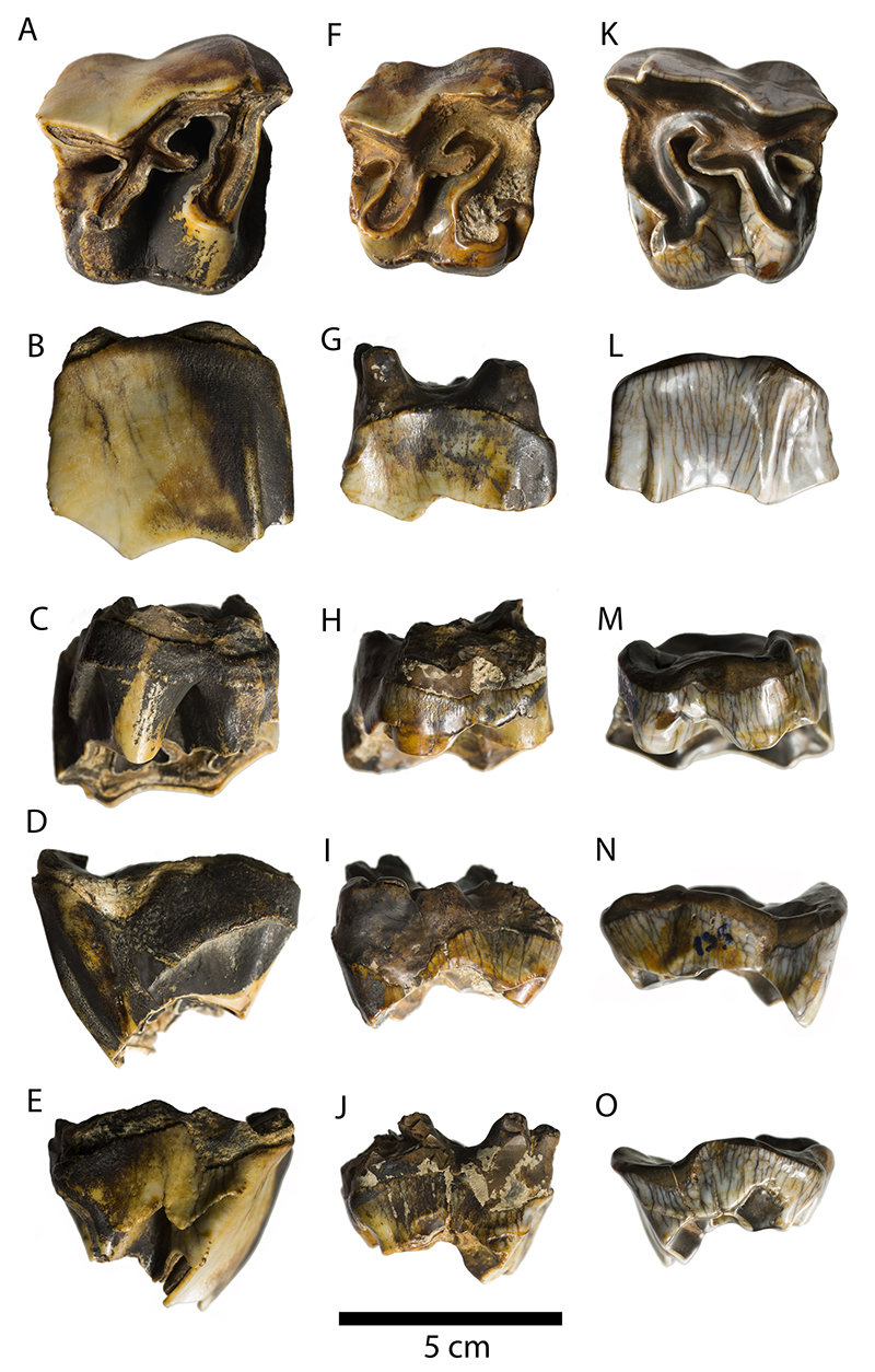
FIGURE 5. Fragmented upper teeth of Stephanorhinus hundsheimensis from Bogovina Cave: right M1/M2 HMP-136 in (A) occlusal and (B) lingual views; right M HMP-132 in (C) occlusal and (D) vestibular views; right M HMP-134 in (E) occlusal and (F) vestibular views; right DP4 HMP-130 in (G) occlusal and (H) vestibular views.
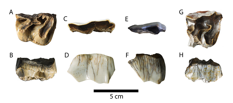
FIGURE 6. Lower teeth of Stephanorhinus hundsheimensis from Bogovina Cave: right m1/m2 HMP-129 in (A) occlusal, (B) vestibular, (C) lingual, (D) mesial and (E) distal views; right p3/p4 HMP-407 in (F) occlusal, (G) vestibular, (H) lingual, (I) mesial and (J) distal views; right p2 HMP-139a in (K) occlusal, (L) vestibular, (M) lingual, (N) mesial, and (O) distal views.
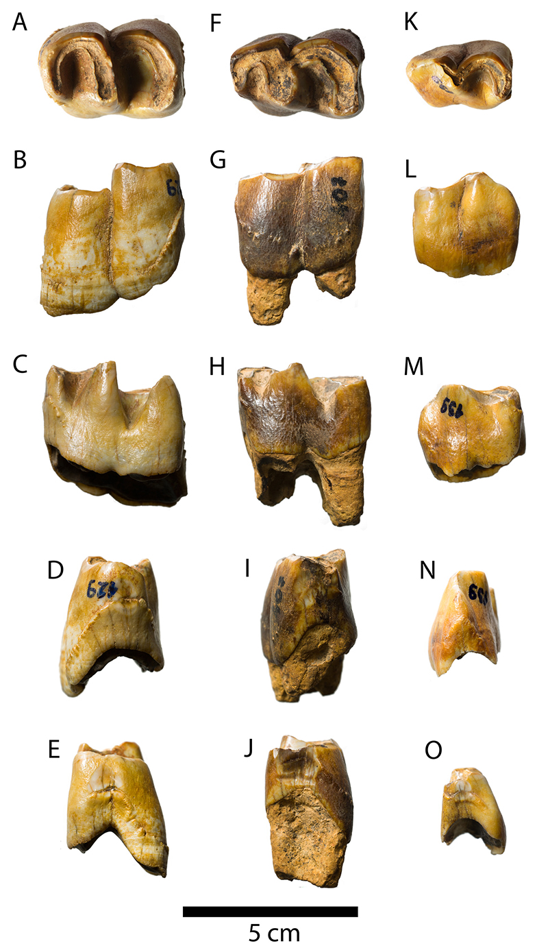
FIGURE 7. Equid and bovid dental specimens from Bogovina Cave: left lower molar of a caballoid horse NMKVRS.P12 in (A) occlusal view; indeterminate bovid right p4 NMKVRS.P14 in (B) occlusal view; indeterminate bovid left M1/M2 NMKVRS.P15 in (C) occlusal view.
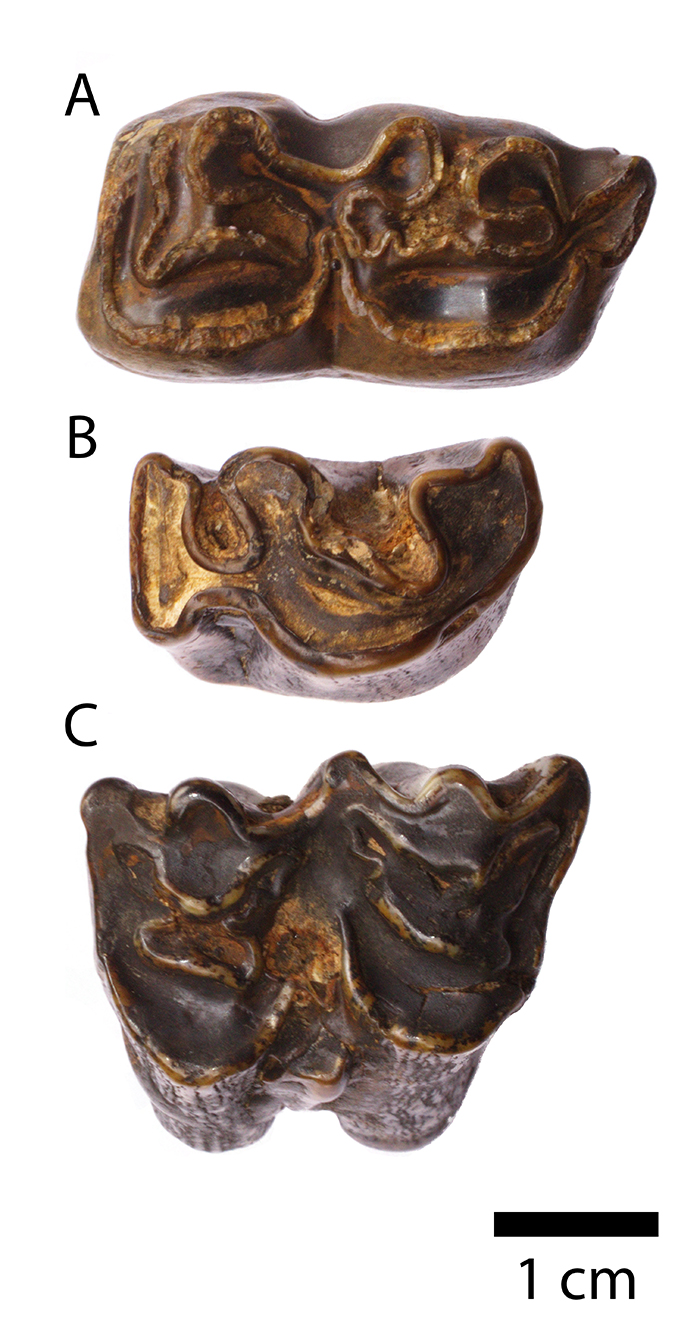
FIGURE 8. Chronological and biochronological ranges of the four Stephanorhinus species known from the territory of Europe. Marine isotope stages according to Lisiecki and Raymo (2005).
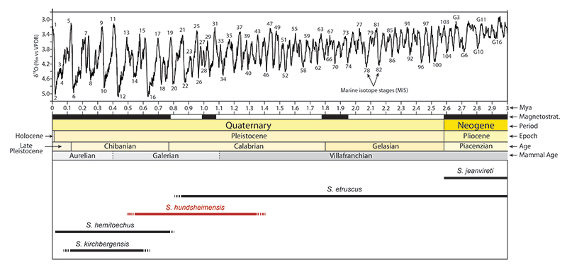
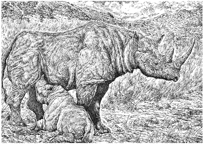
FIGURE 9. A reconstruction of Stephanorhinus hundsheimensis (Toula, 1902) with its calf in its hypothetical palaeoenvironment (after Gianfranco Mensi, 2015; this unpublished illustration is used here through the courtesy of the artist, all rights reserved).