FIGURE 1. Paleotemperatures (ice-free deep-ocean T°/ tropical sea surface T°) and Thermic events during the “doubthouse” conditions of Eocene period (from Cramwinckel et al., 2018 modified with events dating from Hollis et al., 2019). Stratigraphically and geographical locations of the main deposits with Elasmobranch associations along the southwestern Tethys. Abbreviations: DAK: Dakhla (Adnet et al., 2010), GEN: Genam (Zouhri et al., 2017, in press); AZ: Aznag (Tabuce et al., 2005) PM: Phosphate ores (see Noubhani and Cappetta, 1997), Morocco; GAF: Gafsa basin (see Arambourg, 1952); KEBAR: Kébar (this work and Adnet et al., 2019); MBK: Mabrouk (see Sweydan et al., 2019), Tunisia; EG: ElGedida (see Strougo et al., 2007); KM: KM11 (see Adnet et al., 2011) MT: Minqar Tabaghbagh (see Zalmout et al., 2012); BQ: Birquet Qarun QS: Quar et Sa; GE: Genahamm Fm.; MI: Midawara FM. from Wadi al Hitan, see Underwood et al., 2011), Egypt; QD: Qa Faydat al Dahikya, Jordania, see Mustafat and Zalmout, 2002).
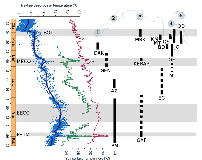
FIGURE 2. Locality of KEB-1, Djebel el Kébar, Kasserine region, Tunisia. A, Simplified topographic map of Tunisia locating the Djebel el Kébar in central Tunisia; B, simplified stratigraphical position of fossiliferous level having yielded the fossil-bearing KEB-1 locality; C, photograph showing the typical badlands of variegated clays from the early Tertiary sequence of Djebel el Kébar. See Merzeraud et al. (2016) for precise details about geological settings.
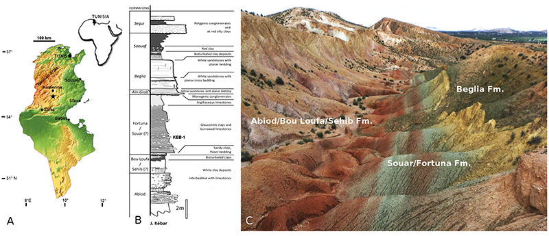
FIGURE 3. A-N: Carcharhinus kasserinensis nov. sp. A. Parasymphyseal upper tooth KEB 1-086, A1. labial view, A2. Lingual view; B. HOLOTYPE Anterior upper tooth KEB 1-087, B1. Labial view, B2. Lingual view, B3. Magnificence of mesial cutting edge; C. Antero-lateral upper tooth KEB 1-088, C1. Labial view, C2. Lingual view; D. Antero-lateral upper tooth KEB 1-089, D1. Labial view, D2. Lingual view; E. Antero-lateral upper tooth KEB 1-226, E1 Lingual view, E2. Labial view; F. Antero-lateral upper tooth KEB 1-227; G. Antero-lateral upper tooth KEB 1-090, G1. Labial view, G2. Lingual view; H. Lateral upper tooth KEB 1-091, labial view; I Posterior upper tooth KEB 1-092, lingual view; J. Symphyseal abnormal lower tooth KEB 1-093, J1. Labial view, J2. Lingual view; K. Anterior lower tooth KEB 1-094, labial view; L. Anterior lower tooth KEB 1-095, L1. Labial view, L2. Lingual view; M. Antero-lateral lower tooth KEB 1-096, M1. Labial view, M2. Lingual view; N. Antero-lateral lower tooth KEB 1-097, N1. Lingual view, N2. Labial view, N3. Profile.
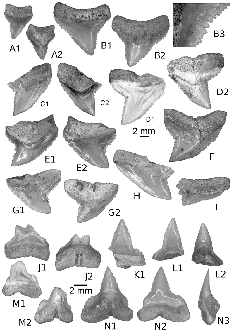
FIGURE 4. A-B: Carcharhinus kasserinensis nov. sp., A. Antero-lateral upper tooth of young specimen KEB 1-098, A1. Lingual view, A2. Labial view; B. Antero-lateral upper tooth of young specimen KEB 1-099, labial view. C-G: Carcharhinus frequens. C. Lateral upper tooth KEB 1-100, C1. Labial view, C2. Lingual view; D. Lateral upper tooth KEB 1-101, D1. Labial view, D2. Lingual view; E. Anterior lower tooth KEB 1-102, E1. Labial view, E2. Lingual view; F. Lateral lower tooth KEB 1-103, F1. Labial view, F2. Lingual view; G. Posterior lower tooth KEB 1-104, lingual view; H-K: Misrichtys sp. H. Anterior lower tooth KEB 1-105, lingual view; I. Lateral upper tooth KEB 1-106, labial view; J. Posterior lower tooth KEB 1-107, j1. Labial view, J2. Lingual view; K. Lateral upper tooth KEB 1-108, labial view; L-R: Rhizoprionodon sp. L. Anterior upper tooth KEB 1-126, labial view; M. lateral upper tooth KEB 1-127, labial view; N. Lateral upper tooth KEB 1-128, labial view; O. Anterior lower tooth KEB 1-129, labial view; P. Lateral lower tooth KEB 1-130, P1. Labial view, P2. Lingual view; Q. Posterior lower tooth KEB 1-131, labial view; R. Antero-lateral lower tooth KEB 1-132, R1. Labial view, R2. Lingual view.
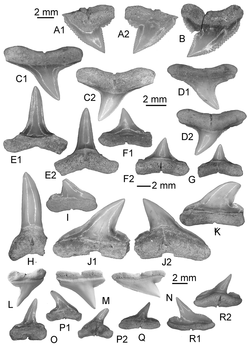
FIGURE 5. A-K: Sphyrna guinoti nov. sp. A. Anterior upper tooth KEB 1-109, A1. Labial view, A2. Lingual view; B. Lateral upper tooth KEB 1-110, B1. Labial view, B2. Lingual view; C. Anterior upper tooth KEB 1-111, C1. Lingual view, C2. Labial view; D. Lateral lower tooth KEB 1-112, D1. Labial view, D2. Lingual view; E. lateral upper tooth KEB 1-113, E1. Lingual view, E2. Labial view; F. Lateral upper tooth KEB 1-114, F1. Labial view, F2. Lingual view; G. Posterior upper tooth KEB 1-115, lingual view; H. Anterior lower tooth KEB 1-116, H1. Labial view, H2. Lingual view; I. Antero-lateral lower tooth KEB 1-117, I1. Labial view, I2. Lingual view; J. (HOLOTYPE) Antero-lateral lower tooth KEB 1-118, J1. Labial view, J2. Lingual view; K. Lateral lower tooth KEB 1-119, K1. Labial view, K2. Lingual view.
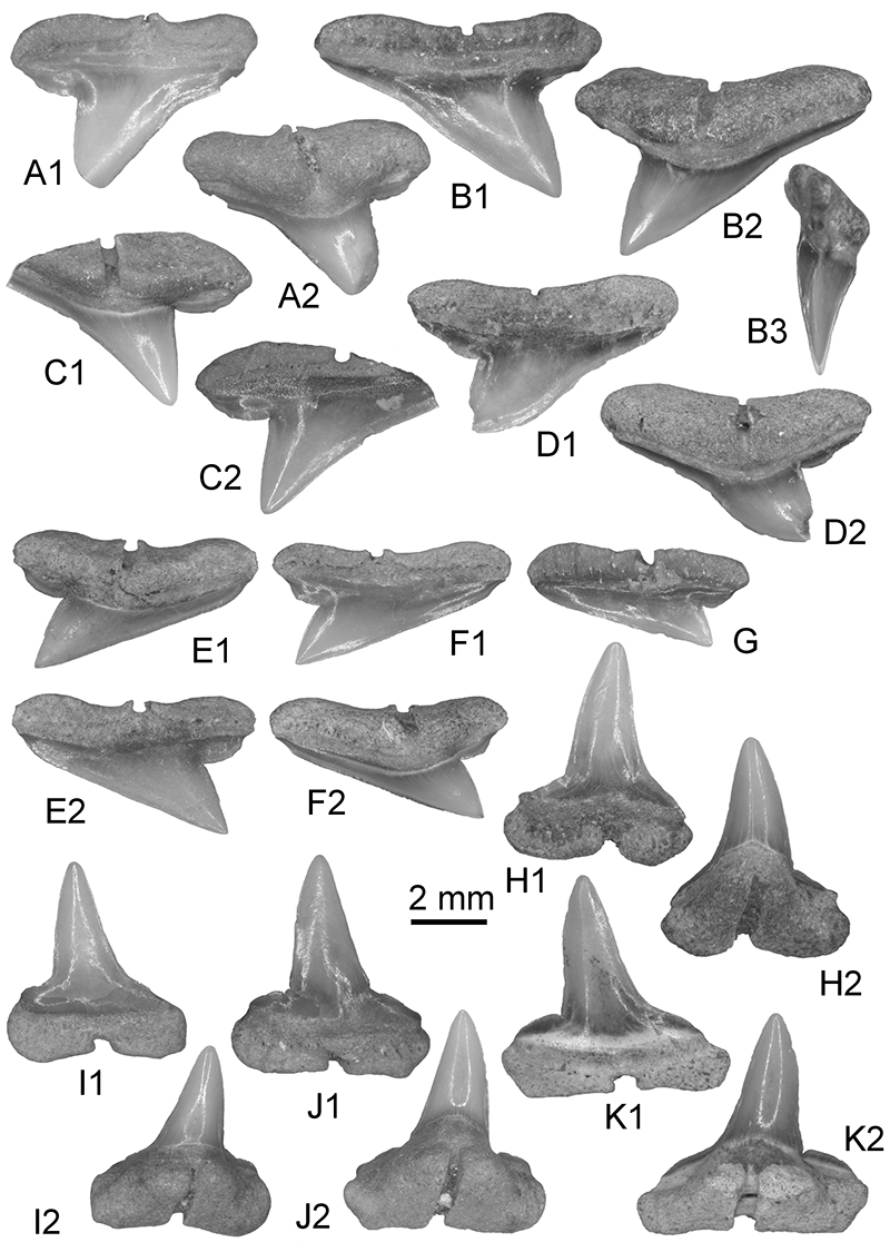
FIGURE 6. A-C: Sphyrna guinoti nov. sp. A. Anterior upper tooth of Young specimen KEB 1-120, A1. Lingual view, A2. Labial view; B. Lateral lower tooth of Young specimen KEB 1-121, B1. Labial view, B2. Lingual view; C. Lateral upper tooth KEB 1-122, C1. Labial view, C2. Lingual view; D-F: ? Sphyrna sp. 4. D. ?Antero-lateral upper tooth KEB 1-123, D1. Lingual view, D2. Labial view; E. ?Antero-lateral upper tooth KEB 1-124, E1. Labial view, E2. Lingual view; F. Lateral upper tooth KEB 1-125, lingual view.
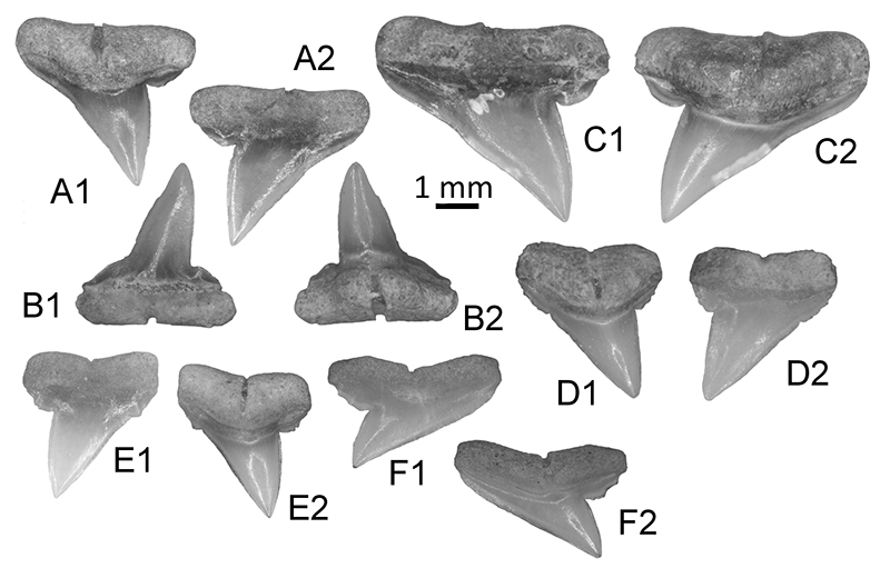
FIGURE 7. A-C: Hemipristis curvatus. A. Antero-lateral upper tooth KEB 1-133, A1. Lingual view, A2. Labial view; B. Lateral upper tooth of young specimen KEB 1-134, labial view; C. Anterior lower tooth of young specimen KEB 1-135, labial view; D-E: Moerigaleus sp. D. Lateral upper tooth KEB 1-136, D1. Lingual view, D2. Labial view; E. Lateral upper tooth KEB 1-137, E1. Lingual view, E2. Labial view; F-K: Leptocharias tunisiensis nov. sp. F. (HOLOTYPE) Antero-lateral lower tooth KEB 1-138, F1. Profile, F2. Lingual view, F3. Labial view; G. Antero-lateral upper tooth KEB 1-139, G1. Labial view, G2. Profile, G3. Lingual view; H. Anterro-lateral lower tooth KEB 1-140, labial view; I. Lateral lower tooth KEB 1-141, labial view; J. More lateral tooth KEB 1-142, J1. Labial view, J2. Lingual view, J3 occlusal view; K. Posterior tooth KEB 1-143, K1. Labial view, K2. Lingual view.
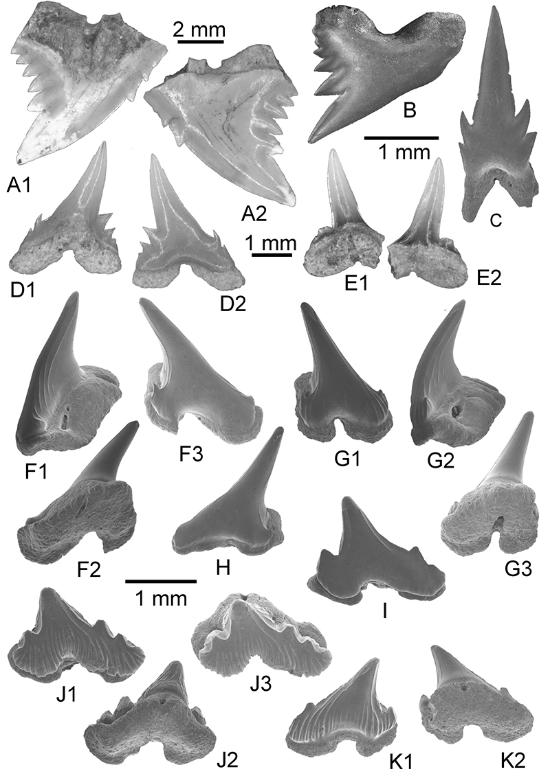
FIGURE 8. A-C: Nebrius sp. A. Anterior tooth KEB 1-144, A1. Labial view, A2. Lingual view, A3. profile; B. Antero-lateral tooth KEB 1-145, B1. Labial view, B2. Basal view; C. Lateral tooth KEB 1-146, C1. Labial view, C2. Lingual view; D-G: Stegostoma tethysiensis nov. sp. D. (HOLOTYPE) Anterior tooth KEB 1-147, D1. Labial view, D2. Lingual view, D3. Basal view; E. Antero-lateral tooth KEB 1-148, E1. Labial view, E2. Occlusal viex, E3. profile; F. Lateral tooth KEB 1-149, labial view; G. Posterior tooth KEB 1-150, labial view; H: Hemiscyllium sp. 8. Antero-lateral tooth KEB 1-151, labial view; I-N: Odontorythys pappenheimi I. ?Anterior tooth KEB 1-152, I1. Profile, I2. Labial view; J. ?Latero-posterior tooth KEB 1-153, lingual view; K. ?Latero-posterior tooth KEB 1-154, profile; L. ?Latero-posterior tooth KEB 1-155, profile; M. ?Latero-posterior tooth KEB 1-156, profile; N. ? lateral or posterior tooth KEB 1-157, N1. Profile, N2. Labial view, N3. Lingual view.
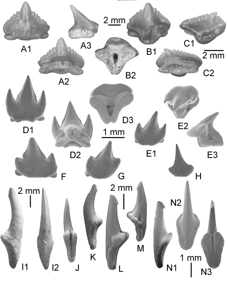
FIGURE 9. A-F: Propristis cf. schweinfurti, A. Anterior oral tooth KEB 1-166, A1. Lingual view, A2. Occlusal view, A3. profile; B. Antero-lateral oral tooth KEB 1-167, B1. Occlusal view, B2. Lingual view, B3. Labial view; C. Anterior oral tooth KEB 1-168, C1. Lingual view, C2. Basal view, C3. Profile; D. lateral tooth KEB 1-169, D1. Lingual view, D2. Occlusal view, D3. Basal view, D4. Magnificence of crown-root boundary of D3; E. ?male lateral tooth KEB 1-170, E1. Occlusal tooth, E2. Basal view; F. ?male anterior tooth KEB 1-171, F1. Lingual view, F2. Occlusal view; G-L: Pristis sp. G. porterior tooth KEB 1-158, G1. Occlusal view, G2. Basal view; H. anterior tooth KEB 1-159, occlusal view; I. lateral tooth KEB 1-160, I1 occlusal view, I2., lingual view. J lateral tooth KEB 1-161, occlusal view; K. anterior tooth KEB 1-162, occlusal view; L. lateral tooth of ?juvenile KEB 1-163, L1. Occlusal view, L2. Basal view; M. lateral tooth of juvenile KEB 1-164, M1. Occlusal view, M2. Profile.
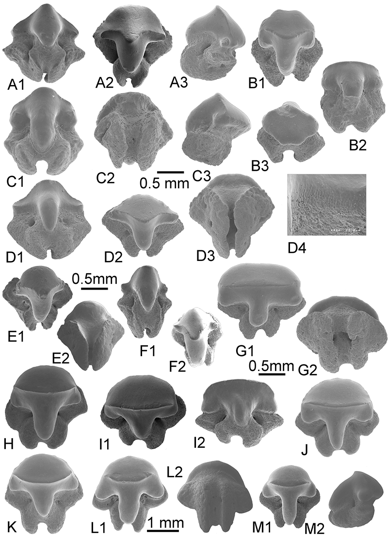
FIGURE 10. A-D: Propristis cf. schweinfurti. A. Rostral denticle KEB 1-172, A1. Profile, A2. dorsal view; B. Rostral denticle KEB 1-173, Profile; C. Rostral denticle KEB 1-174, C1. Profile, C2. Dorsal view, C3. basal view; D. Rostral denticle KEB 1-175, D1. profile. D2. dorsal view; E: Pristis sp. Rostral denticle, KEB 1-165, dorsal view; F-G. Rhynchobatus cf. vincenti. F. anterior tooth KEB 1-176, F1. Occlusal view, F2. Basal view; G. anterior tooth KEB 1-177, occlusal view; H-I. ?Torpedo sp. H. lateral tooth KEB 1-178, occlusal view, I. lateral tooth KEB 1-179, occlusal view.
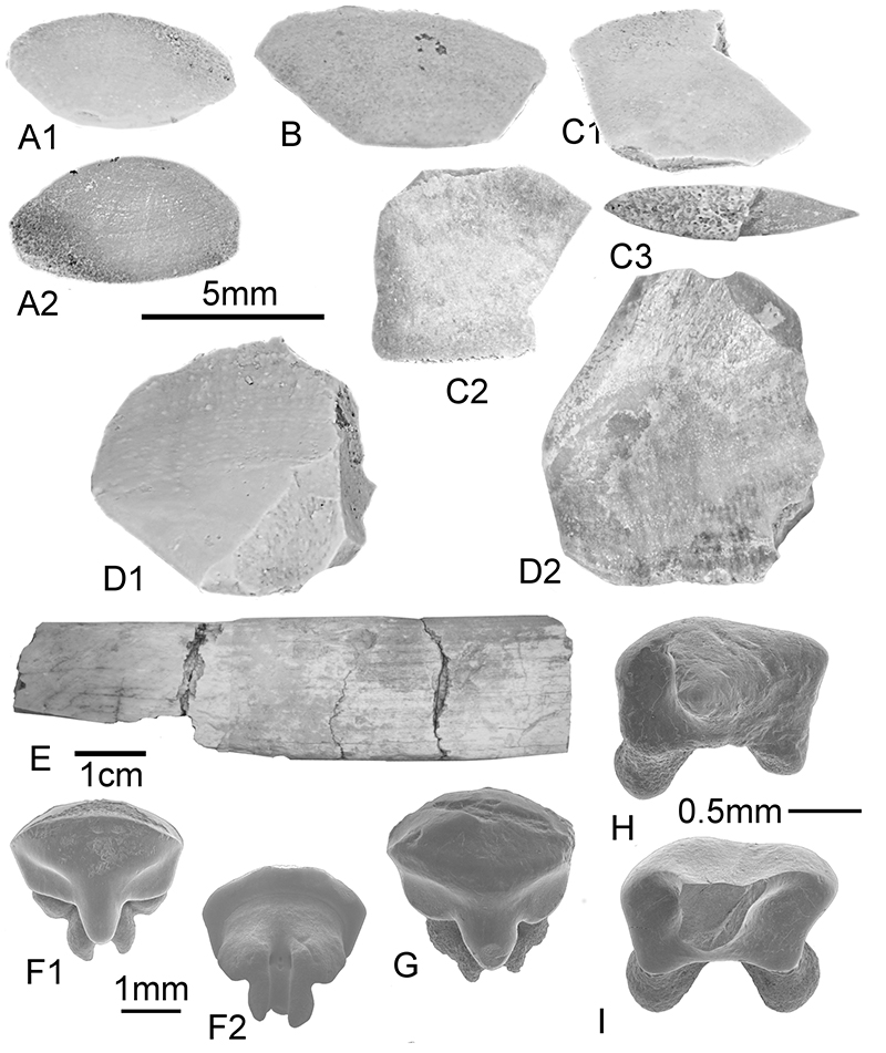
FIGURE 11. A-C. Ouledia lacuna nov. sp. A. ?Anterior tooth KEB 1-180, A1. Occlusal view, A2. Lingual view, A3. Magnificence of crown-root boundary in A2, A4. profile; B. ?lateral tooth KEB 1-181, B1. Occlusal view, B2. labial view, C. ?Anterior tooth KEB 1-182, C1. Labial view, C2. Basal view; D-H. Pachygymnura attiai nov. gen. D. Anterior tooth KEB 1-183, D1. Lingual view, D2. Occlusal view, D3. Labial view, D4. Basal view; E. Antero-lateral tooth KEB 1-184, E1. Lingual view, E2. Near labial view, F. lateral tooth KEB 1-185, F1. Occlusal view, F2. Profile, F3. Basal view; G. lateral tooth KEB 1-186, G1. Lingual view, G2. Occlusal view; H. Anterior tooth KEB 1-187, H1. Profile, H2. Occlusal view.
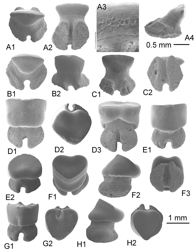
FIGURE 12. A-C. Mecotrygon asperodentulus nov. gen nov. sp. A. anterior tooth KEB 1-188, A1. occlusal view, A2. Profile, A3. Basal view; B. lateral tooth KEB 1-189, B1. occlusal view, B2. labial view, B3. Profile; C. lateral tooth KEB 1-190, HOLOTYPE, C1. Occlusal view, C2. Lingual view, C3. Basal view, C4. Profile; D-M. Himantura souarfortuna nov. sp. D. ?posterior tooth KEB 1-191, D1. Occlusal view, D2. Profile, D3. Basal view; E. antero-lateral tooth KEB 1-192, occlusal view; F. anterior tooth KEB 1-193, F1. Occlusal view, F2. Labial view; G. antero-lateral tooth KEB 1-194, G1. Occlusal view, G2. Profile; H. anterior tooth KEB 1-195, occlusal view; I. lateral tooth KEB 1-196, occlusal view; J. A. anterior tooth KEB 1-197, occlusal view; K. lateral tooth KEB 1-198, K1. Occlusal view, K2. Basal view; L. A. anterior tooth KEB 1-199, occlusal view; M. lateral tooth KEB 1-200, occlusal view. N-O. Dasyatoid indet. N. antero-lateral tooth KEB 1-201, N1. Occlusal view, N2. Basal view; O. A. antero-lateral tooth KEB 1-202, occlusal view; P-Q. Arechia sp. P. lateral tooth KEB 1-203, occlusal view; Q. A. anterior tooth KEB 1-204, Q1. Occlusal view, Q2. Lingual view.
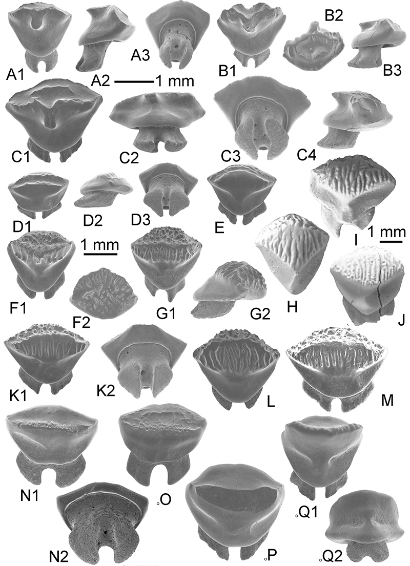
FIGURE 13. A-E. Coupatezia cristata nov. sp. A. antero-lateral tooth KEB 1-205, HOLOTYPE, A1.occlusal view, A2. Profile, A3. Labial view; B. Anterior tooth KEB 1-206, B1. Lingual view, B2. Basal view, C. lateral tooth KEB 1-207, C1. Occlusal view, C2. Magnificence of C1, C3. Lingual view; D. lateral tooth KEB 1-208, D1. Occlusal view, D2. Magnificence of D1, D3. Lingual view; E. lateral tooth KEB 1-209, E1. Occlusal view, E2., lingual view, E3. Basal view. F-G. Rhinoptera sp. F. medium tooth KEB 1-210, lingual view, G. medium tooth KEB 1-211, labial view; H-J. Myliobatis sp., H. lateral tooth KEB 1-212, lingual view, I. medio-lateral tooth KEB 1-213, lingual view, J lateral tooth KEB 1-214, lingual view; K. Aetobatus sp. KEB 1-215, ligual view, L-M. Garabatis sp., L. KEB 1-216, occlusal view, M. KEB 1-217, L1. occlusal view, L2. Magnificence of central part, L3. Magnificence of marginal part of crown.
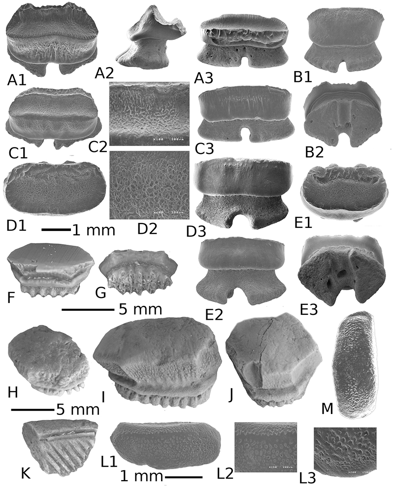
FIGURE 14. A-G. Amamriabatis heni nov gen. nov. sp. A. anterior tooth KEB 1-218, A1. Occlusal view, A2. Basal view, A3. Lingual view, B. antero-lateral tooth KEB 1-219 (Holotype), B1. Occlusal view, B2. Lingual view, B3. Basal view, C. juvenile tooth KEB 1-220, occlusal view, D. antero-lateral tooth KEB 1-221, D1. Occlusal view, D2. Basal view, D3. Labial view, E. lateral tooth KEB 1-222, E1. Occlusal view, E2. Lingual view, E3. Basal view, F. antero-lateral tooth KEB 1-223, F1. Occlusal view, F2. Basal view, F3. Labial view, G. lateral tooth KEB 1-224, G1. Occlusal view, G2. Basal view, G3. Lingual view; H. Archaeomanta sp. KEB 1-225, H1. Lateral view, H2. Labial view.
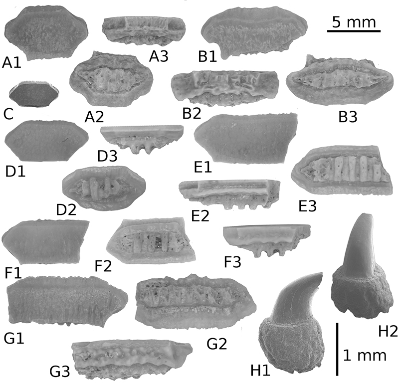
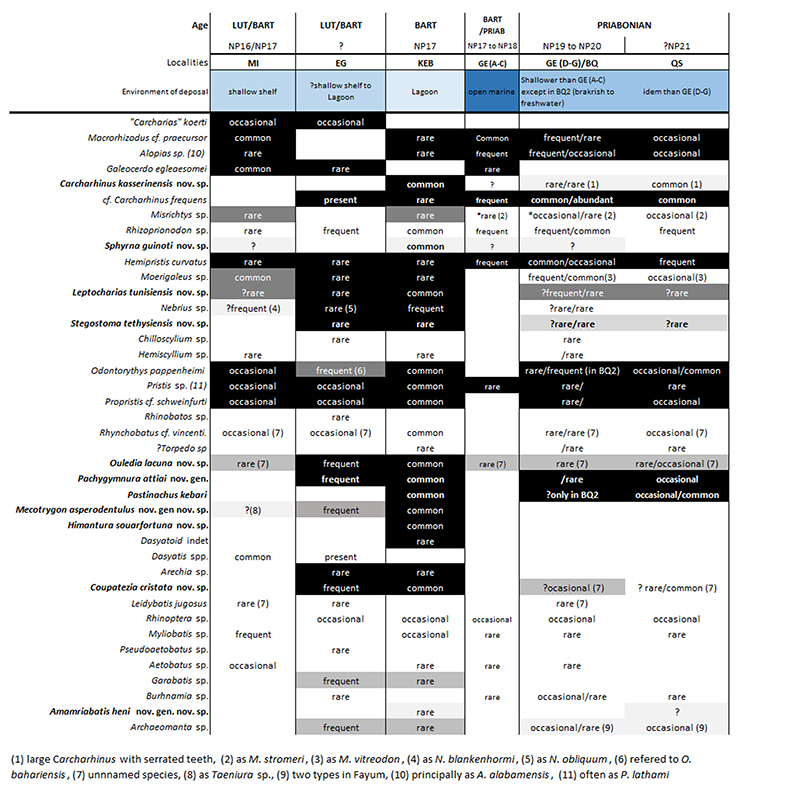
FIGURE 15. Distribution of Elasmobranch taxa within localities of KEB-1, Tunisia (KEB, this work) and El Gedida (EG), Baharia (Egypt, from Strougo et al., 2007 updated) with report of their occurences in the other late Middle - Late Eocene tropical assemblages from Egypt (MI, GE, BQ, QS). Frequencies of taxa are somewhat subjective due to the different sources and/or sampling methods applied. Black-Grey intensity indicating the fossil completeness between KEB/EG and the other localities due to uncertainty/resolution of taxonomic attributions (See systematic palaeontology for detail, black: certain to light: possible). (1) Medium-Large Carcharhinus sp. with serrated teeth, (2) as Misrichtys stromeri, (3) as Moerigaleus vitreodon, (4) as Nebrius blankenhormi, (5) as Nebrius sp., (6) refered to Odontorhytis bahariensis, (7) unnnamed species, (8) possibly as Taeniura sp., (9) two types in Fayum.