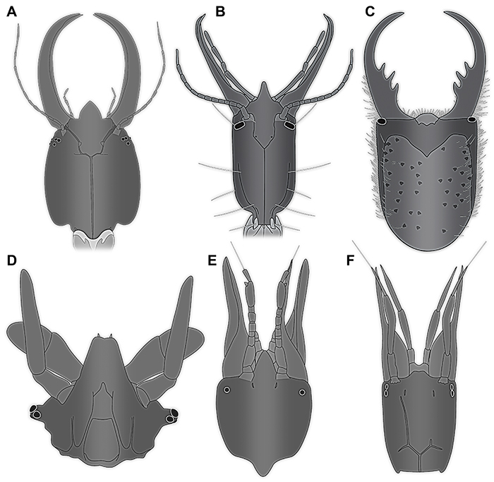
FIGURE 1. All known extant long-nosed antlion larvae, i.e., larvae of silky lacewings (Psychopsidae). Drawings partly simplified. A. Specimen 1, from Froggatt (1907). B-E. All from Tillyard (1918). B1. Specimen 2. B2. Specimen 2 in the same scale as the other specimens from Tillyard (1918). C. Specimen 3. D. Specimen 4. E. Specimen 5. F. Specimen 6, from Withycombe (1925). G. Specimen 7, from Macleod (1964). H. Specimen 8, from New (1989). I. Specimen 9, from New (1991). J. Specimen 10, from Aspöck and Aspöck (1999). K. Specimen 11, from Badano et al. (2017). L. Specimen 12, from Bakkes et al. (2017).
FIGURE 2. Fossils of long-nosed antlion larvae, i.e., larvae of silky lacewings (Psychopsidae). A. Specimen 13, Natural History Museum (Museum für Naturkunde) Berlin, MBI 5648. B. Colour-marked version of A. C. Specimen 14, MCZ (Museum of Comparative Zoology) Harvard, PALE 18004. D. Colour-marked version of C. Abbreviations: ad = abdomen; at = antenna; hc = head capsule; ms = mesothorax; mt = metathorax; pt = prothorax; sy = stylet; th = thorax.
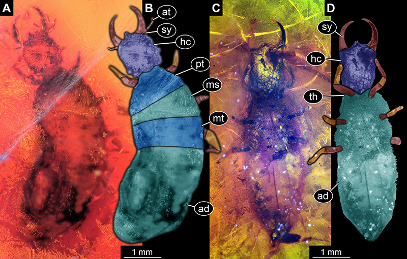
FIGURE 3. Fossils of long-nosed antlion larvae, i.e., larvae of silky lacewings (Psychopsidae), continued. Drawings partly simplified. A. Specimen 15, from Weitschat and Wichard (1998, 2002). B. Specimen 17, from Scheven (2004). C. Specimen 18, from Engel and Grimaldi (2008). D. Specimen 19, from Gröhn (2015). E. Specimen 20, from Zhang (2017). F. Specimen 21, from Badano et al. (2018). G. Specimen 22, from Makarkin (2018). H. Specimen 23, from Makarkin (2018).
FIGURE 4. Specimen 24 (Gröhn 2994); Baltic amber. A. Dorsal view. B. Dorsal view, colour marked. C. Ventral view. D. Close-up of labrum in dorsal view (arrow). E. Close-up of antenna; arrow points to spine-like seta. F. Close-up of tubercles on head capsule in dorsal view. Abbreviations: ad = abdomen; at = antenna; hc = head capsule; ms = mesothorax; mt = metathorax; pt = prothorax; sy = stylet.
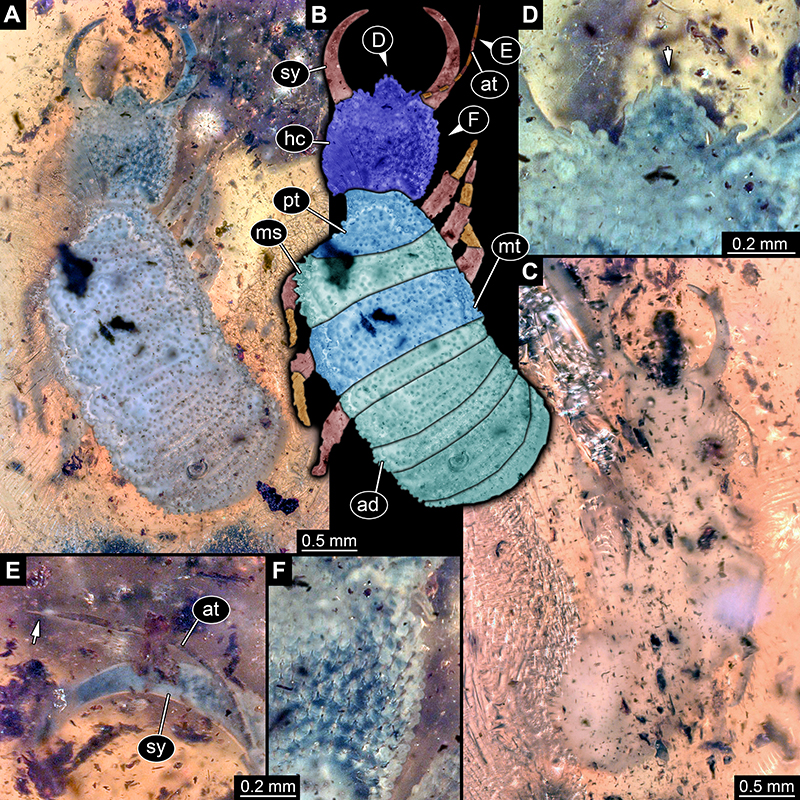
FIGURE 5. Specimen 25 (Gröhn 7639); Baltic amber. A. Dorsal view. B. Dorsal view, colour marked. C. Ventral view. D. Close-up of antenna; arrow points to spine-like seta. E. Close-up of labrum in dorsal view (arrow). F. Close-up of empodium (arrow) of first walking appendage. G. Close-up of empodium (arrow) of second walking appendage. Abbreviations: ad = abdomen; at = antenna; hc = head capsule; ms = mesothorax; mt = metathorax; pt = prothorax; sy = stylet.
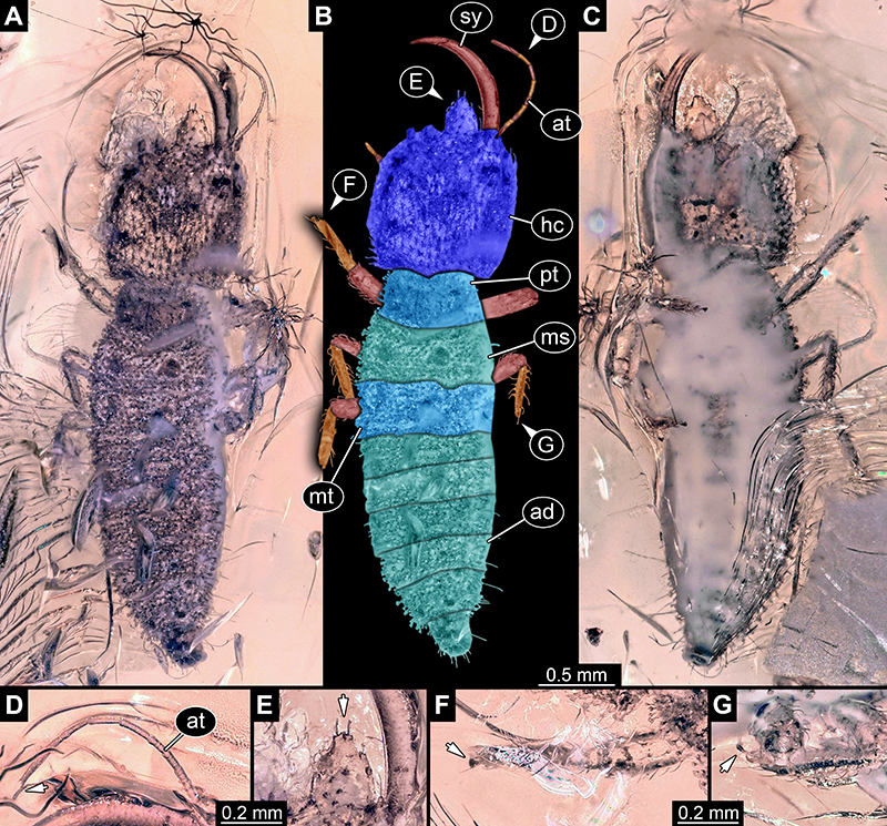
FIGURE 6. Specimens in Baltic amber, continued. A. Specimen 26 (Gröhn 7507), ventral view, strongly verlumt. B. Specimen 27; image from Jonas Damzen. C. Specimen 28. D. Specimen 29. E. Specimen 30. C-E. Images by Marius Veta.
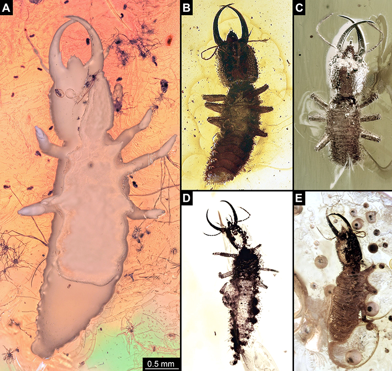
FIGURE 7. Specimen 16 (IGR.ARC-205.2); Charentese amber. A. Ventral view. B. Dorsal view. C. Dorsal view, colour marked. D. Close-up of labrum (arrow) in ventral view. E. Close-up of antenna in dorsal view; arrow points to spine-like seta. F. Close-up of empodium (arrow) of first walking appendage. Abbreviations: ad = abdomen; at = antenna; hc = head capsule; ms = mesothorax; mt = metathorax; pt = prothorax; sy = stylet.
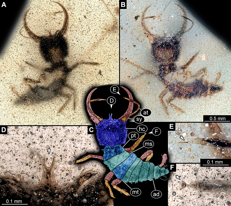
FIGURE 8. Specimen 16 (IGR.ARC-205.2), continued. A-B. Composite fluorescence images in dorsal view, note the surface details. A. Blue light (GFP). B. Green light (TRITC). C. Composite bright-field image.
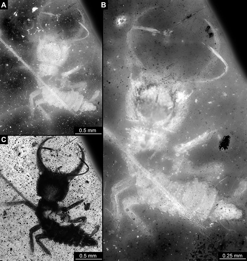
FIGURE 9. Specimen 31 (BUB 3356); Burmese amber. A. Dorsal view. B. Dorsal view, colour marked. C. Close-up of labrum (arrow) in ventral view. D. Ventral view. Abbreviations: ad = abdomen; at = antenna; hc = head capsule; lp = labial palp; ms = mesothorax; mt = metathorax; pt = prothorax; sy = stylet.
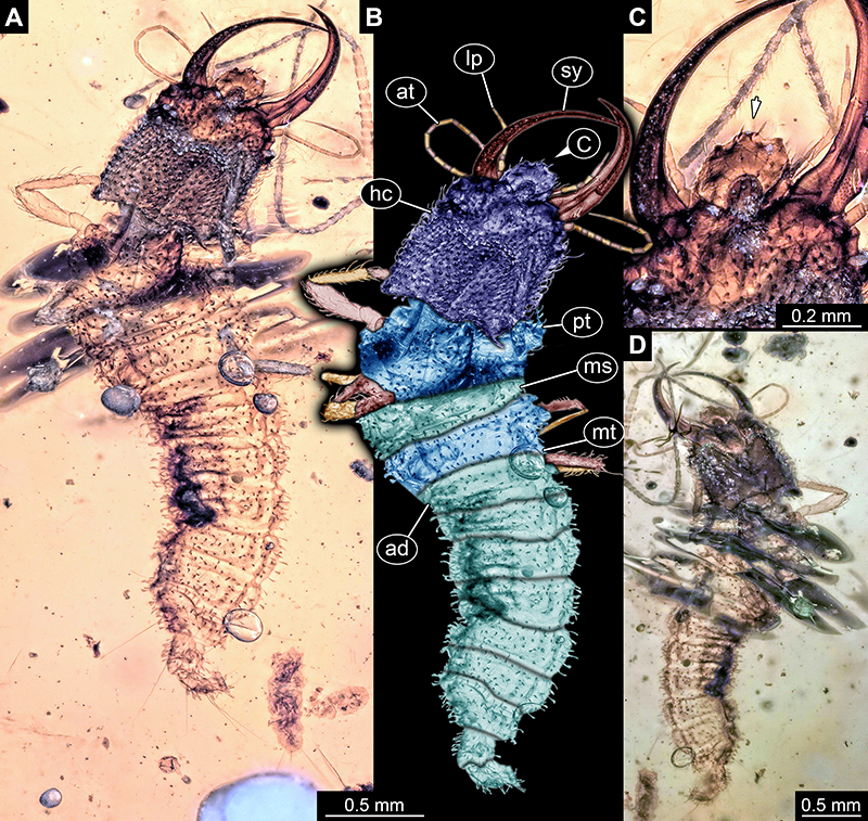
FIGURE 10. Specimen 32 (PED 0055); Burmese amber. A. Dorsal view. B. Dorsal view, colour marked. C. Ventral view. D. Close-up of labrum (arrow) in dorsal view. E. Close-up of empodium (arrow) of third walking appendage. F. Close-up of abdomen. Abbreviations: ad = abdomen; at = antenna; hc = head capsule; lp = labial palp; ms = mesothorax; mt = metathorax; pt = prothorax; sy = stylet.
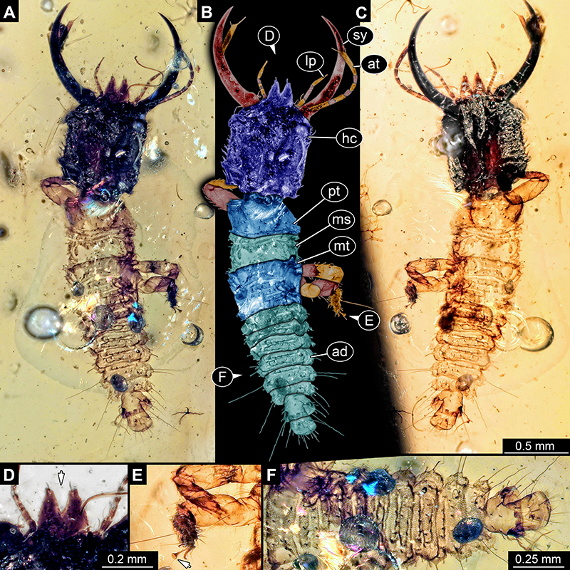
FIGURE 11. Specimen 33 (PED 0082); Burmese amber. A. Ventral view. B. Dorsal view. C. Dorsal view, colour marked. D. Close-up of antenna; arrow points to spine-like seta. E. Close-up of empodia (arrows) of first and second walking appendage. F. Close-up of labrum (arrow) in dorsal view. Abbreviations: ad = abdomen; at = antenna; hc = head capsule; ms = mesothorax; mt = metathorax; pt = prothorax; sy = stylet.
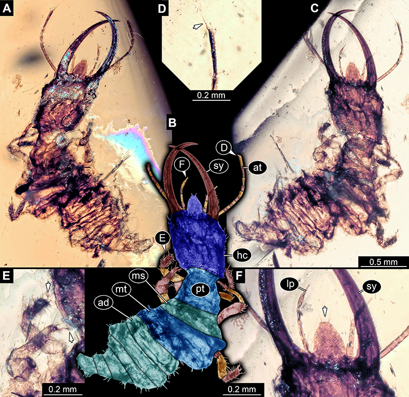
FIGURE 12. Specimen 34 (PED 0131); Burmese amber. A. Dorsal view. B. Dorsal view, colour marked. C. Close-up of antenna; arrow points to spine-like seta. D. Close-up of empodium (arrow) of second walking appendage. E. Close-up of empodium (arrow) of third walking appendage. Abbreviations: ad = abdomen; at = antenna; hc = head capsule; lp = labial palp; ms = mesothorax; mt = metathorax; pt = prothorax; sy = stylet.
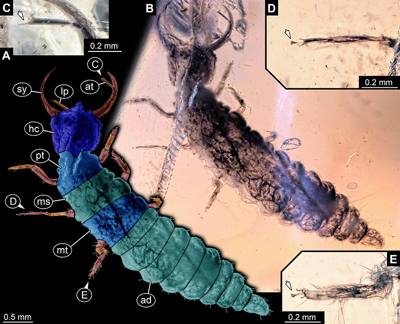
FIGURE 13. Specimen 35 (PED 0080); Burmese amber. A-C. Dorsal view. A. Cross-polarised light. B. Colour marked. C. Ring light. D. Close-up of labrum (arrow) in dorsal view. E. Close-up of empodium (arrow) of third walking appendage. Abbreviations: ad = abdomen; at = antenna; cl = claw; hc = head capsule; lp = labial palp; sy = stylet; th = thorax.
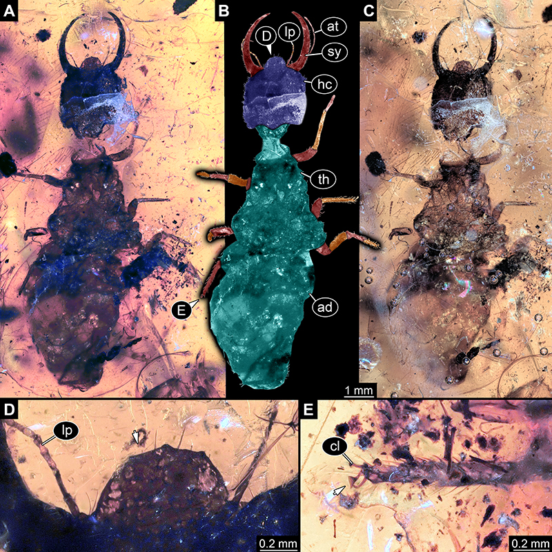
FIGURE 14. Specimen 36 (PED 0137); Burmese amber. A. Dorsal view. B. Dorsal view, colour marked. C. Ventral view. D. Close-up of labrum (arrow) in dorsal view. E. Close-up of empodium (arrow) of second walking appendage. Abbreviations: ad = abdomen; at = antenna; cl = claw; hc = head capsule; lp = labial palp; ms = mesothorax; mt = metathorax; pt = prothorax; sy = stylet.
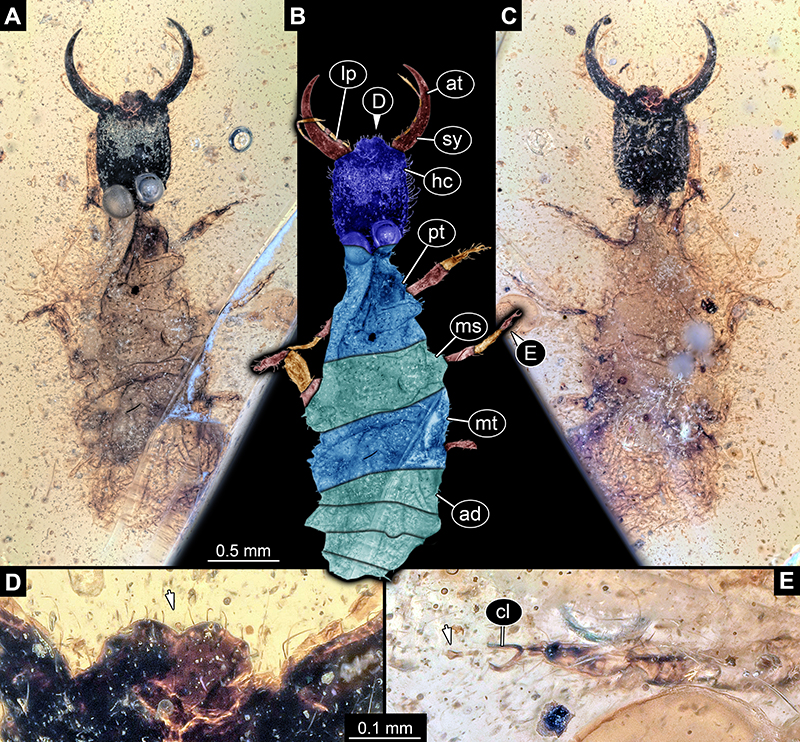
FIGURE 15. Specimen 37 (PED 0205); Burmese amber. A. Ventral view. B. Dorsal view, colour marked. C. Dorsal view. D. Close-up of antenna; arrow points to spine-like seta. E. Close-up of labrum (arrow) in dorsal view. F. Close-up of empodium (arrow) of second walking appendage. Abbreviations: ad = abdomen; at = antenna; hc = head capsule; ms = mesothorax; mt = metathorax; pt = prothorax; sy = stylet; ta = tarsus; ti = tibia.
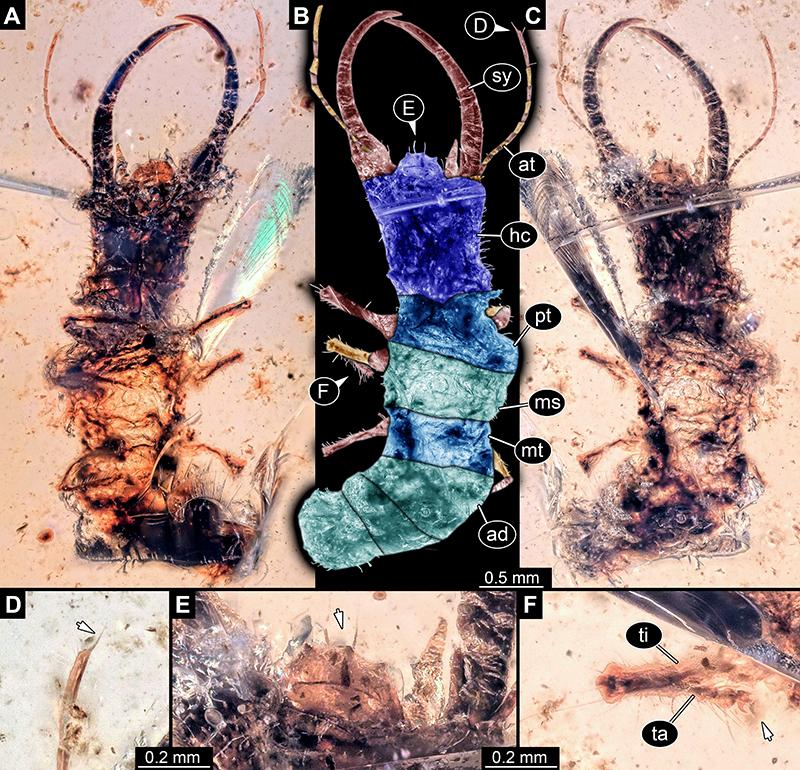
FIGURE 16. Specimen 38 (BUB 3389); Burmese amber. A. Ventral view. B. Dorsal view, colour marked. C. Dorsal view. D. Close-up of antenna in dorsal view; arrow points to spine-like seta. E. Close-up of labrum (arrow) in dorsal view. F. Close-up of tubercles (arrow) on head capsule in dorsal view. Abbreviations: ad = abdomen; at = antenna; hc = head capsule; lp = labial palp; sy = stylet; th = thorax.
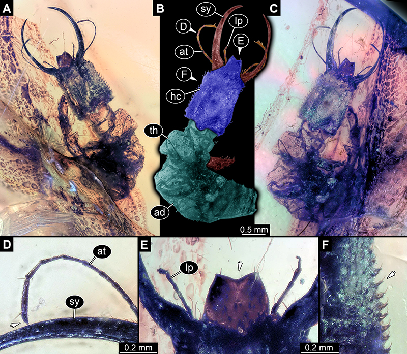
FIGURE 17. Specimen 39 (PED 0060); Burmese amber. A. Dorsal view. B. Dorsal view, colour marked. C. Ventral view. D. Close-up of antenna; arrow points to spine-like seta. E. Close-up of labrum (arrow) in dorsal view. F. Close-up of syn-inclusion, insect larva in lateral view. Abbreviations: ad = abdomen; at = antenna; hc = head capsule; ms = mesothorax; mt = metathorax; pt = prothorax; sy = stylet.
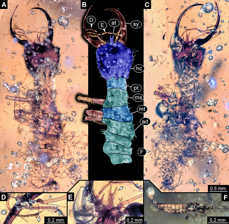
FIGURE 18. Specimen 40 (PED 0051); Burmese amber. A. Ventral view. B. Dorsal view, colour marked. C. Dorsal view. D. Close-up of labrum (arrow) in dorsal view. E. Close-up of empodium (arrow) of first walking appendage. F. Close-up of empodium (arrow) of second walking appendage. Abbreviations: ad = abdomen; at = antenna; cl = claw; hc = head capsule; lp = labial palp; sy = stylet; ta = tarsus; th = thorax; ti = tibia.
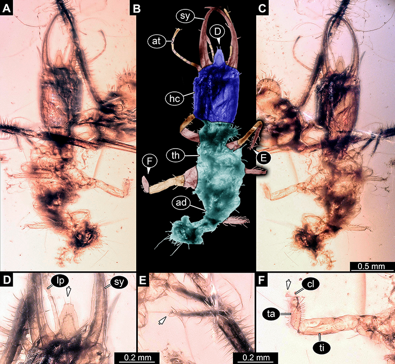
FIGURE 19. Specimens in Burmese amber, continued. A-C. Specimen 41 (PED 0127). A. Dorsal view. B. Dorsal view, colour marked. C. Close-up of labrum (arrow) in dorsal view. D-E. Specimen 42 (PED 0063). D. Dorsal view. E. Dorsal view, colour marked; arrows point to spine-like seta and labrum. Abbreviations: at = antenna; hc = head capsule; lp = labial palp; ms = mesothorax; mt = metathorax; pt = prothorax; sy = stylet.
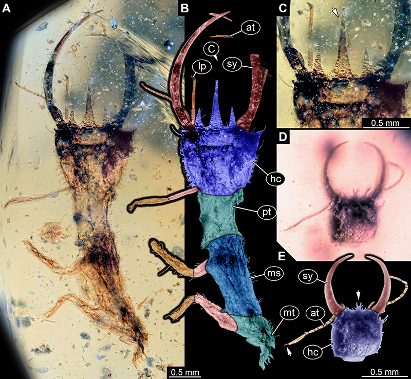
FIGURE 20. Specimens in Burmese amber, continued. A-D. Specimen 43 (PED 0153). A. Dorsal view. B. Dorsal view, colour marked. C. Close-up of labrum (arrow) in dorsal view. D. Close-up of empodium (arrow) of second walking appendage. E-F. Specimen 44 (BUB 3179). E. Dorsal view. F. Dorsal view, colour marked. Abbreviations: ad = abdomen; at = antenna; hc = head capsule; lp = labial palp; ms = mesothorax; mt = metathorax; pt = prothorax; sy = stylet.
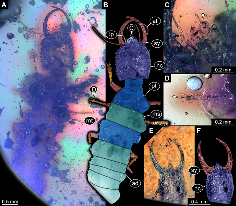
FIGURE 21. Specimen 45 (PED 0045); Burmese amber. A. Dorsal view. B. Dorsal view, colour marked. C. Ventral view. D. Close-up of labrum (arrow) in dorsal view. E. Close-up of labial palp. Abbreviations: at = antenna; hc = head capsule; lp = labial palp; sy = stylet; th = thorax.
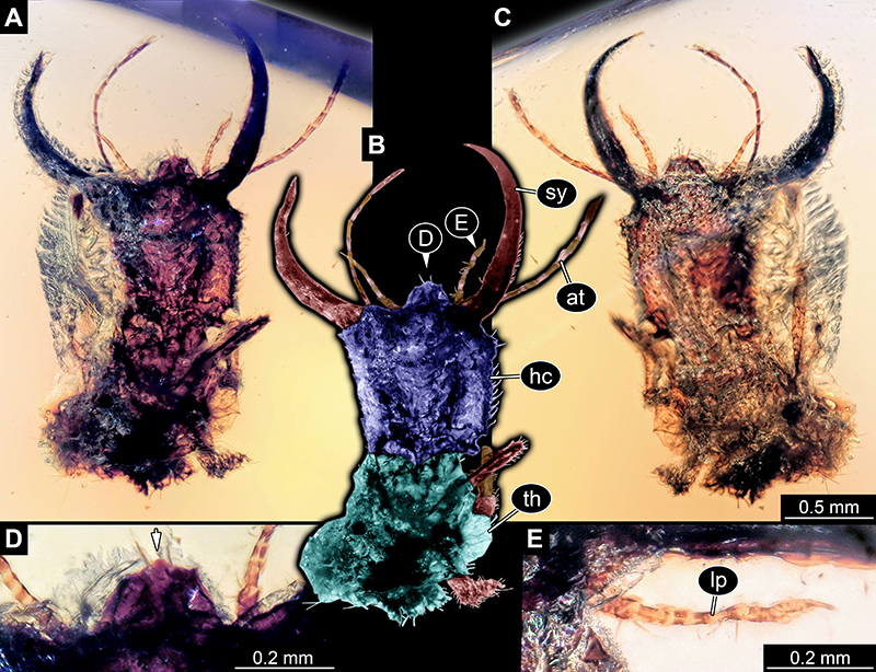
FIGURE 22. Specimens in Burmese amber, continued. A-C Specimen 46 (PED 0133). A. Ventral view. B. Dorsal view, colour marked; arrow points to labrum. C. Dorsal view. D-F. Specimen 47 (PED 0128). D. Ventral view. E. Dorsal view, colour marked; arrow points to labrum. F. Dorsal view. G-H. Specimen 48 (BUB 3386). G. Dorsal view. H. Dorsal view, slightly different angle; arrow points to labrum. Abbreviations: at = antenna; hc = head capsule; lp = labial palp; ms = mesothorax; mt = metathorax; pt = prothorax; sy = stylet.
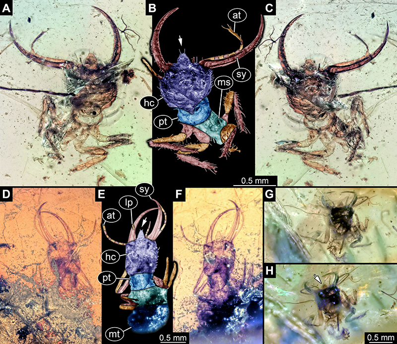
FIGURE 23. Specimen 49 (PED 0125); Burmese amber. A. Lateral view. B. Dorsal view. C. Lateral view, other side. D. Close-up of labrum (arrow) in dorsal view. E. Close-up of eyes in lateral view. F. Close-up of empodium (arrow) of a walking appendage. Abbreviations: at = antenna; hc = head capsule; lp = labial palp; sy = stylet; th = thorax.
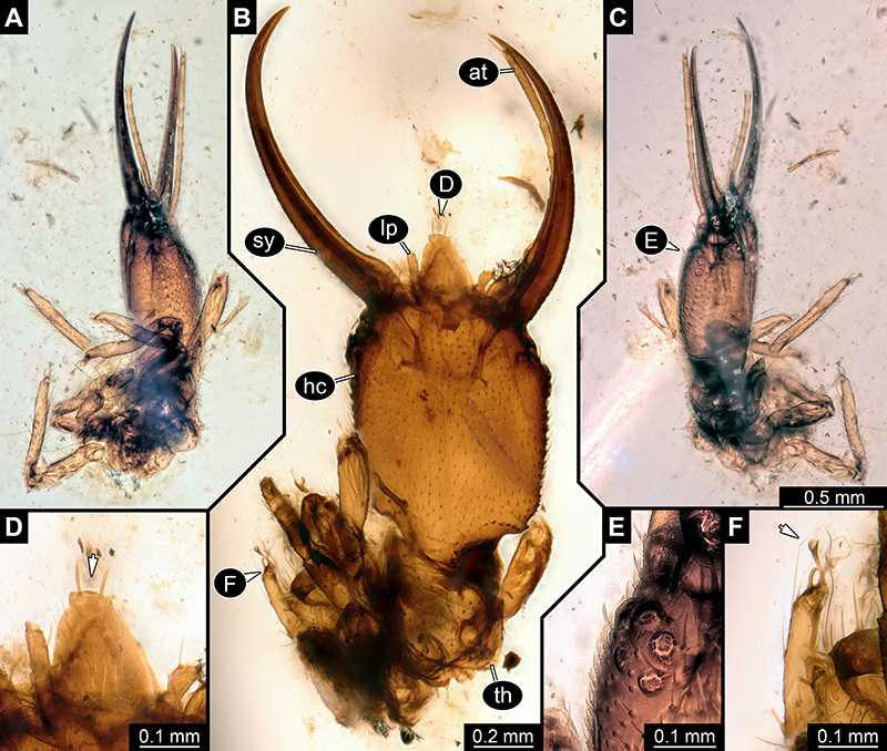
FIGURE 24. Specimens in Burmese amber, continued. A-C. Specimen 50 (PED 0039). A-C. Dorsal view. A. Ring light. B. Colour marked. C. Cross-polarised light. D. Specimen 51 (PED 0109). Abbreviations: at = antenna; hc = head capsule; sy = stylet; th = thorax.
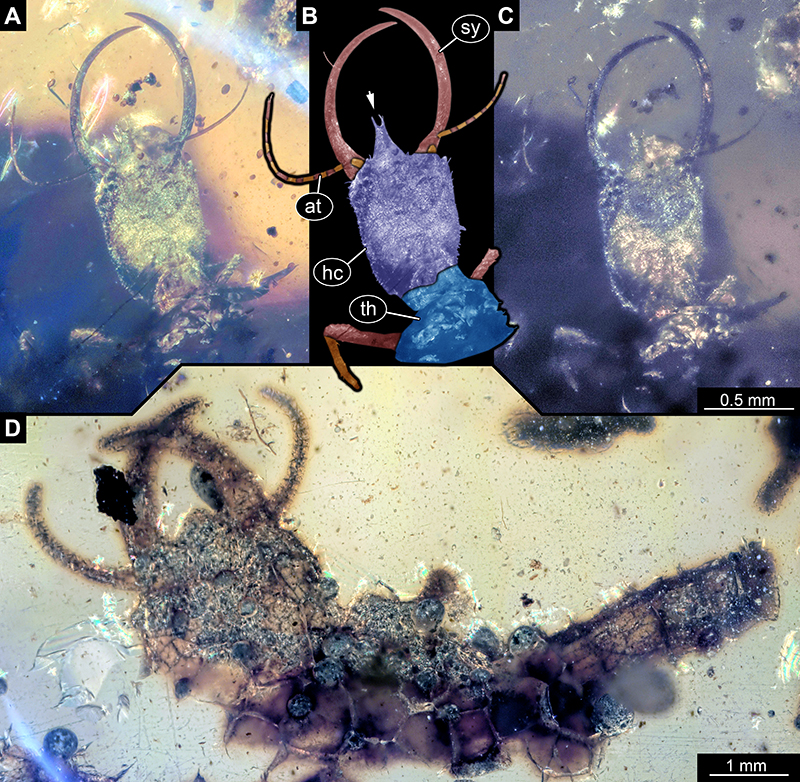
FIGURE 25. Specimen 52 (PED 0100); Burmese amber. A. White light composite image. B-C. Fluorescence images under blue light (GFP). B. Overview. C. Close-up of labrum; tip marked by arrow. Abbreviations: hc = head capsule; sy = stylet.
FIGURE 26. Scatterplot of PC1 and PC2 surrounded by all analysed heads. Numbers in the ellipses correspond to the numbers of the heads. Extant specimens as grey ellipses, Eocene specimens as white ellipses, Cretaceous specimens as black ellipses. The heads grouped together in circles cluster relatively closely together and possibly form relatively discrete types.
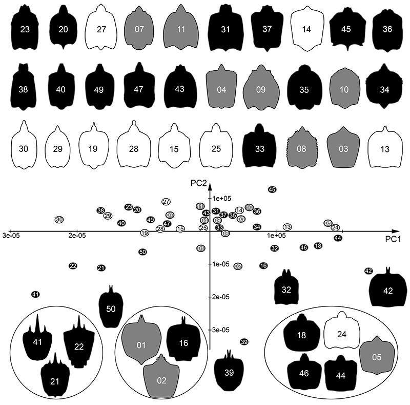
FIGURE 27. Different plots of the data of the analysed long-nosed antlion larvae. A. Range of PC1 and PC2 for specimens from different time slices. For Cretaceous specimens, the grey bars provide the sample-size-corrected ranges. B. Scatter plot of head capsule width w(h) vs. capsule length l(h) (without labrum). C. PC1 and PC2 plotted against head capsule length (without labrum). Note the low degree of correlation as well as low coefficient of determination: PC1: R2=0.039, PC2: R2=0.027.
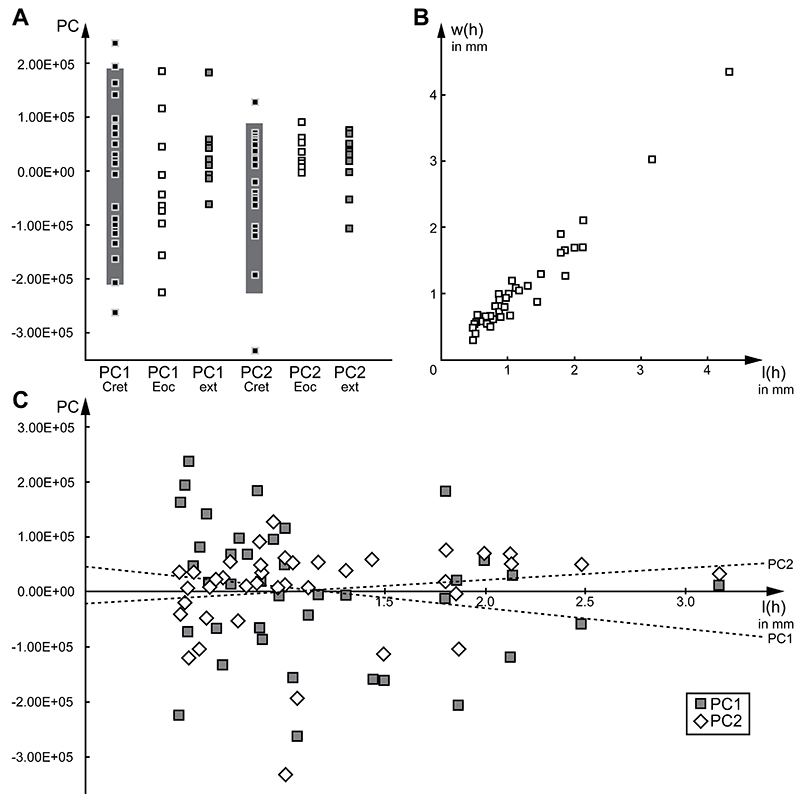
FIGURE 28. Comparison of different heads of lacewing larvae with labrum. A. Psychopsidae (MacLeod 1964, his fig. 66). B. Nevrorthidae (Beutel et al. 2010, their fig. 2). C. Myrmeleontidae ( Acanthaclisis occitanica; Badano 2012, image on p. 90; re-figured in Badano et al. 2017, their fig. 7C). D. Coniopterygidae (MacLeod 1964, his fig. 62). E. Dilaridae (MacLeod 1964, his fig. 33). F. Berothidae (MacLeod 1964, his fig. 36).