FIGURE 1. Topographic map (géoportail; Da Silva Pires, 2008) and geographical localization of the Palembert locality, Quercy, France.
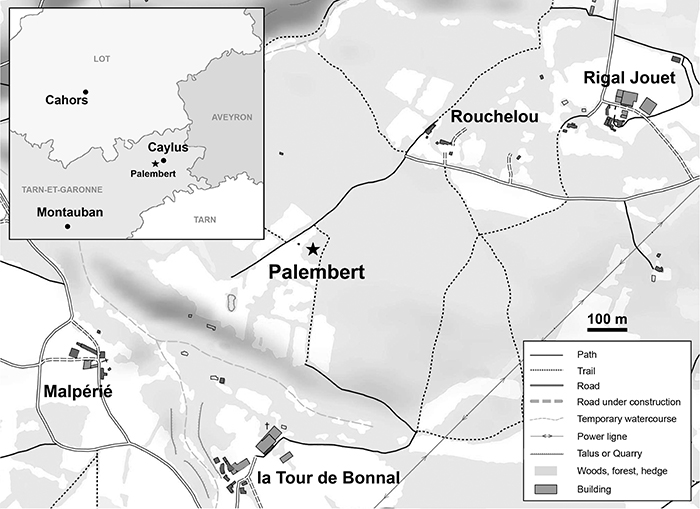
FIGURE 2. Material of the dentition of Oxacron courtoisii (A-D) and Palembertina deplasi gen. nov. sp. nov. (E-K). A, B, right dentary fragment with P/3-M/3 (PAL 59; mirror view); C, D, right dentary fragment with M/2-M/3 (PAL 61; mirror view); E, F, left dentary fragment with M/3 (PAL 60); G, H, left dentary fragment with P/4 and M/2 (PAL 62); I, J, left M2/ (PAL 21); K, right isolated M/3 (PAL 64; mirror view). In lingual view: J. In occlusal view: A, C, E, G, I, K. In buccal view: B, D, F, H. Scale bar equals 1 mm.
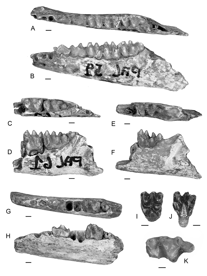
FIGURE 3. Dental diagrams of M2/ and M/3 in occlusal view. A-B, Robiacina; C-D, Palembertina deplasi gen. nov. sp. nov.; E-F, derived Cainotheriidae. In color, the diagnosis characters of interest: blue, protocone; light blue, paraconule; pink, metaconulid; red, entoconid; orange, hypoconulid; brown, neotrigonid (Sudre, 1977). Abbreviations: Centroulid., centroconulid; dist., distostyle; Entod., entoconid; Hypod., hypoconid; Hypoulid., hypoconulid; med., mediostyle; mes., mesostyle; Meta., metacone; Metad., metaconid; Metaul., metaconule; Metaulid., metaconulid; met., metastyle; Para., paracone; Parad., paraconid; Paraul., paraconule; par., parastyle; Proto., protocone; Protod., protoconid. Circles: cusps/cuspids/styles/conules/ conulids; full lines: crests; dashed lines: valleys/fossa; dotted lines: cingula/cingulids. Arrows indicate the mesio-lingual side. For more detailed dental labelling, see APPENDIX IV. Scale bar equals 1 mm.
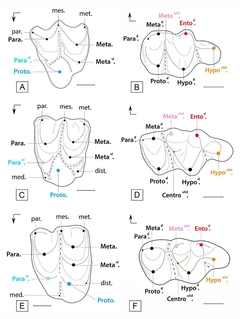
FIGURE 4. Phylogeny relationships of Palembertina deplasi gen. nov. sp. nov. and its position within Cainotherioidea. Most parsimonious tree at 183 steps, with CI = 0.48 and RI = 0.73. The Bremer Index (BI) is indicated in bold at each node. X • a - b: X, node number; a, number of synapomorphies; b, number of ambiguous characters (Acctran optimization). The stratigraphic extension of the species is determined from the collection of Montpellier University and literature: Emry (1978); Sudre (1978); Antunes (1986); Remy et al. (1987); Sudre and Erfurt (1996); Blondel (1997); Prothero (1998); Berthet (2003); Erfurt and Métais (2007); Hooker (2007); Ménouret (2014); Rincon et al. (2015); Weppe et al. (2020).
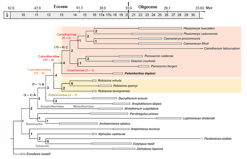
FIGURE 5. Dental occlusion diagrams between M2/-M3/ and M/3 in occlusal view. A, Robiacina; B, Palembertina deplasi gen. nov. sp. nov.; C, derived Cainotheriidae. Circles: cusps/cuspids/styles/conules/conulids; lines: crests. In blue: upper molars; in red: lower molars; light blue area: paraconule; pink area: metaconulid. Arrows indicate the mesio-lingual side. Scale bar equals 1 mm.
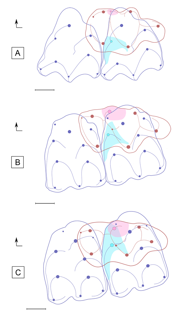
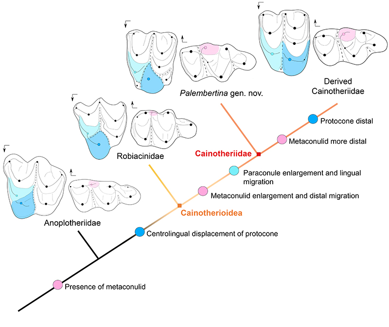
FIGURE 6. Evolutionary history of molar characters (metaconulid, protocone and paraconule) illustrating the steps towards the derived Cainotheriidae pattern ("Cainotherium plan"). Circles: cusps/cuspids/styles/conules/conulids; full lines: crest; dashed lines: valleys/fossa; dotted lines: cingula/cingulids; bold full lines: crown contour. Blue area: protocone; light blue area: paraconule; pink area: metaconulid. Arrows indicate the mesio-lingual side. Dental diagrams not to scale.