FIGURE 1. Diversity of fly larvae in Baltic amber. A, B, PED-230, Athericidae. Fringed lobes on the trunk end. B, Pseudopods with claws; C-E, Dip-00898, Chamaemyiidae, C, lateral view, head, anterior spiracle is marked with an arrow, render of SR-µCT scan; D, posterior spiracles openings are marked with arrows, render of SR-µCT scan; E, dorsal view, with clearly visible secondary annulation of the trunk; F, SMF-BE-10616, Chamaemyiidae, dorsal view; G, same, posterior spiracles, dorsal view; H, SMF-BE-10726, Phoridae representative puparium, dorsal view; I, same, head, dorsal view; J, same, trunk end, dorsal view.
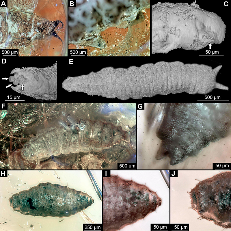
FIGURE 2. Diversity of fly larvae in Baltic amber. All volume renders based on SR-µCT scans A, PED-230, Athericidae, dorsal; B, same, lateral view; C, D, Chamaemyiidae; C, Dip-00898, dorsal view; D, Dip-00898, head in dorsal view; E, BI-2356, puparium Cyclorrhapha, morphotype 1, lateral view; F, Dip-00888, Syrphidae, Volucellini, dorsal view; G, same, ventral view; H-K, Cyclorrhapha. H-I, Morphotype 2; H, Dip-00892, sagittal slice; I, Dip-00892, lateral view; I, Morphotype 3; BI2354, lateral view; J, Morphotype 3; BI2354, lateral view; K, Dip-00893, lateral view.
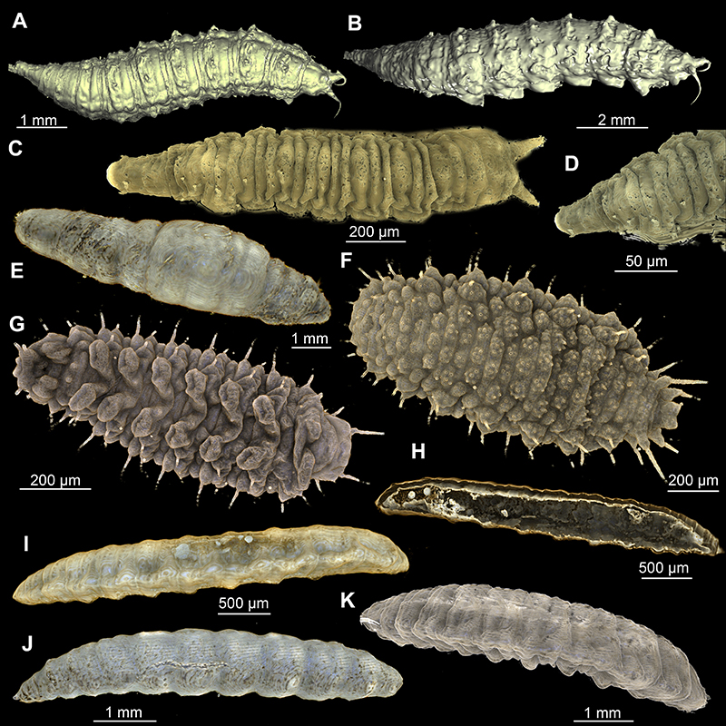
FIGURE 3. Diversity of fly larvae in Baltic amber. Optical images. A, AKBS-0030, full amber piece, arrow is pointing to the location of the inset from Fig. 3B; B, Cyclorrhapha, morphotype 1 close-up; C, PED-230, Athericidae, dorsal view; D, Heleomyzidae, puparium, Dip-00890,dorsal view; E, Volucellini, Dip-00889, lateral; F, Dip-00898, Chamaemyiidae, head in dorsal view; G, Dip-00892, Cyclorrhapha, morphotype 2, lateral view; H, Dip-00888, Syrphidae, Volucellini, lateral view; I, Dip-00896, Volucellini, lateral view.
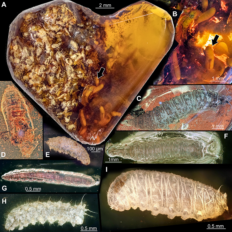
FIGURE 4. Representatives of Cyclorrhapha, morphotype 1, AKBS-0030. A, reconstruction of the Baltic amber forest: feces with larvae of Cyclorrhapha, morphotype 1 larvae at the front; the adult fly Gedanoleria eocenica Woźnica, 2019 (Heleomyzidae) at the feces; the early horse Eurohippus messelensis feeding at the background, representing hypothetical herbivores, which may have left feces, preserved as organic mass in the amber piece (Artist: Natalia Jagielska); B, SR-µCT scan render of the full amber piece, organic mass in light-grey and larvae in red; C, surface rendering on the SR-µCT scan, organic mass in violet and larvae in orange; D, surface renders of the individual larvae.
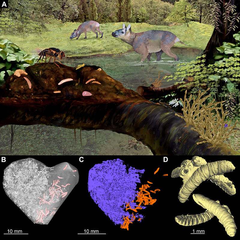
FIGURE 5. Volucella bombylans L. extant larva for comparison, from ZSM collection, collected at Ober-Bayern, Kiefersfelden, from wasp nest on the house leg. Seggmann. A, head, laterally; B, anterior spiracle; C, dorsal view; D, ventral view; E, lateral view; F, Posterior spiracle.
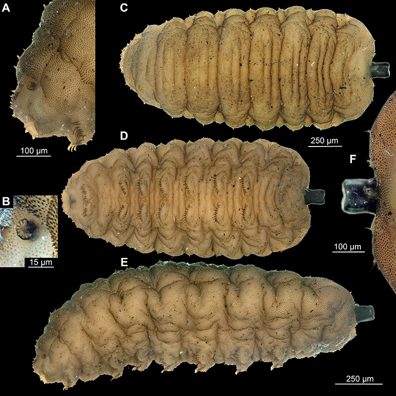
FIGURE 6. Larvae of the group Volucellini (Syrphidae) from the Baltic amber, renders of a SR-µCT scan. A-E, Dip-00897; F-I, Dip-00896. A, lateral view, render of a SR-µCT scan; B, Head, sagittal slice, with internal head skeleton marked in orange; C, Cephalo-pharyngeal skeleton, dorsal view; D, same, lateral view; E, same, ventral view; F, lateral view; G, frontal slice, through the head and thorax; H, Lateral slice, with well visible oesophagus, mandible marked in orange; I, lateral slice through the head; mandible marked in orange. Abbreviations: hp - hypopharynx; mp - metacephalic plate; md - mandibular hooks; hd - head; an - antennae, es - esophagus.

FIGURE 7. Diversity of fly larvae in Baltic amber. A, SMF-BE-10652, ventral view; B, same, trunk end, ventral view; C, Heleomyzidae, puparium, Dip-00890, dorsal; D, same, anterior spiracles; E, same, ventral view; F, same, posterior spiracles, dorsal view.
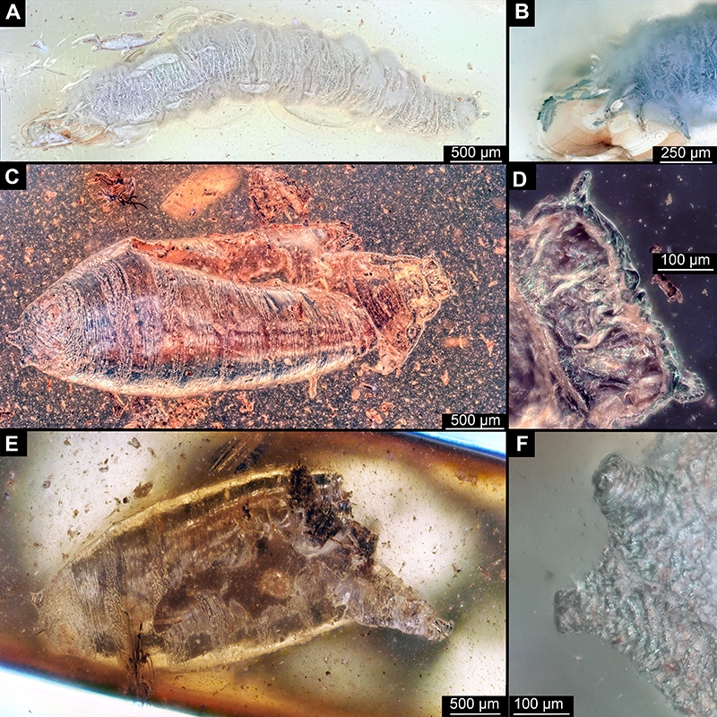
FIGURE 8. Diversity of fly larvae in Baltic amber. All representative of Cyclorrhapha, A, morphotype 1, AKBS-0030, lateral view, render of a SR-µCT scan; B, same, sagittal slice of the head, render of a SR-µCT scan; C, Dip-00893, morphotype 3, dorsal view, render of a SR-µCT scan; D, Dip-00893, morphotype 3, head, dorsal view, render of a SR-µCT scan; E, Dip-00893 morphotype 3, head, dorsal view, render of a SR-µCT scan; E, Dip-00893, morphotype 3, posterior spiracles, ventral view, render of a SR-µCT scan; F, Dip-00893, morphotype 3, lateral view, render of a SR-µCT scan; G, Dip-00892, morphotype 2, sagittal slice through the head, render of a synchrotron scan; H, same, lateral view, render of a synchrotron scan; I, morphotype 4, SMF-BE-10645, ventrolateral view; J, same, head.
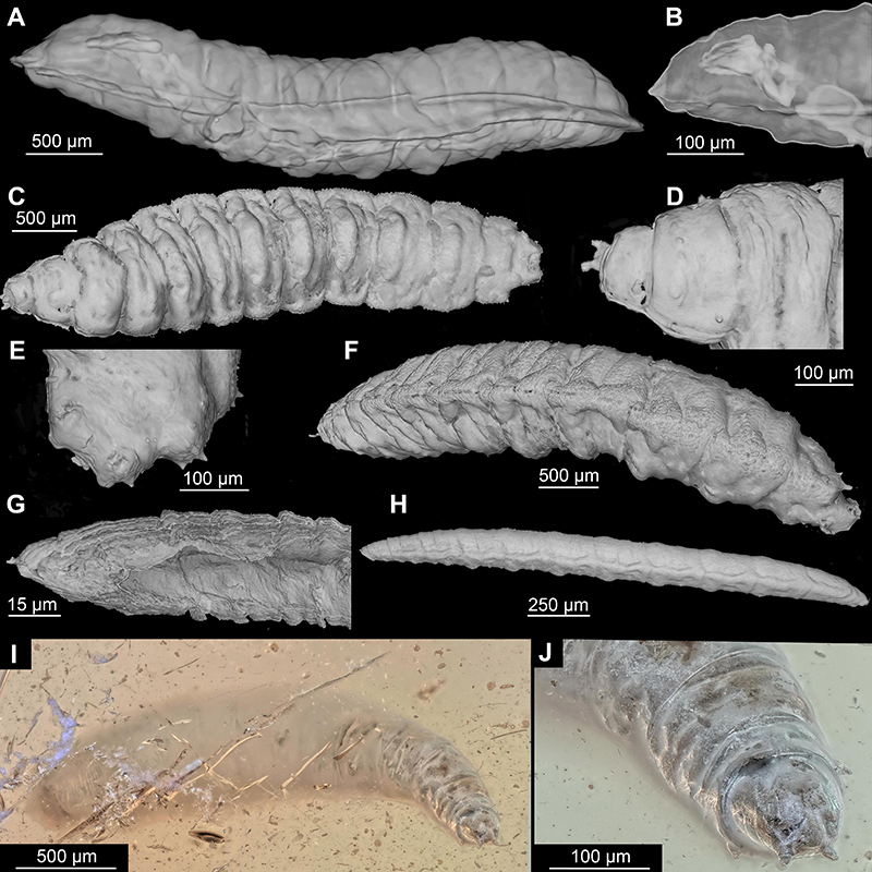
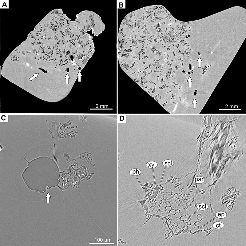
FIGURE 9. CT scans of the fecal matter in the AKBS-0030 amber piece. Fly larvae are marked with the arrows, while the rest objects in the matrix of the amber are the pieces of the fecal matter. A, lower part of amber piece; B, frontal view on the amber piece; C and D, close up on the plant remnants in the fecal matter. Abbreviations: ct - cuticle; ep - epiderma; ph – phloem; par- parenchyma; scl - sclerenchyma; xyl - xylem.