FIGURE 1. Diagrammatic transverse sections comparing the posterior portion of the ligament and nymphae of Acharax and Solemya. A, the upward-arching (parivincular) external ligament and nymphae, i.e., the plesiomorphic condition as seen in Acharax. (Drawing based on Carter, 1990, fig. 17D; Bailey, 2011, pl. 4, figs. 4-6; and Taviani et al., 2011, fig. 5). B, external nymphae of Acharax with the ligament removed. When the ligament is not preserved, the nymphae are visibly separated by an internymphal gap. C, hypothetical intermediate condition showing beginning stages of ligament depression and inward rotation of nymphae (arrows). D, submarginal ligament and nymphae - the apomorphic condition seen in Solemya. The depressed ligament is secondarily overgrown by a shelly outer prismatic layer. (After Carter, 1990, fig. 17A.) E, the submarginal nymphae of Solemya with the ligament removed. Notice the outer secondary layer that covers the former internymphal gap. Abbreviations: ne - external nymph, ns - submarginal nymph gr - ligament insertion groove, ig - internymphal gap, lgf - fibrous inner layer of ligament, lgl - lamellar outer layer of ligament, pe - periostracum, opl - outer (secondary) prismatic shell layer.
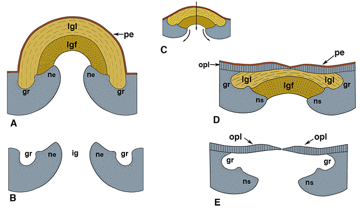
FIGURE 2. Comparative internal views of the posterior portion of the right valves of Acharax (A) and Solemya (B) showing character states of the nymphae, internal buttresses and posterior adductor scars. In Acharax the nymph (ne) is external, the condition of the internal buttress is simple (bts), and the condition of the posterior adductor muscle scar is entire (pae). In Solemya, depression of the nymph to a submarginal position (ns) results in a compound buttress (btc) and an occluded posterior adductor scar (pao).
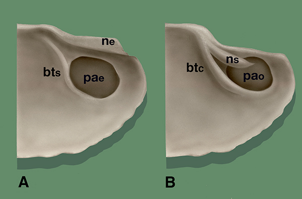
FIGURE 3. Internymphal gaps in fossil examples of Acharax. A, Acharax doderleini (Mayer, 1861), Pliocene of Italy; dorsal view showing external nymphae and ligament insertion grooves separated by internymphal gap that in life is covered by the ligament. In Solemya, the gap is secondarily closed off by the addition of a thin, outer prismatic shell layer. (Photo by permission, Taviani et al., 2011, fig. 5; yellow arrows with notations added here.) B-E, Solemya puzosiana de Koninck, 1842, Lower Carboniferous of Belgium. B, right lateral view of an incomplete articulated specimen (de Koninck, 1885, pl. 23, fig. 33). C, same specimen in dorsal view (de Koninck, 1880, pl. 23, fig. 34), reversed, anterior at left. D, right lateral view of an articulated specimen (de Koninck 1842, pl. 5, fig. 2b). E, same specimen in dorsal view (de Koninck 1842, pl. 5, fig. 2a). Hind (1900, p. 439) synonymized this specimen with Solemya primaeva Phillips, 1836, the type species of Janeia King, 1850. Internymphal gaps shown in de Koninck’s figures are consistent with Acharax but not Solemya. Red arrows with notations added here. Abbreviations: ne - external nymph; ig - internymphal gap; gr - ligament insertion groove.
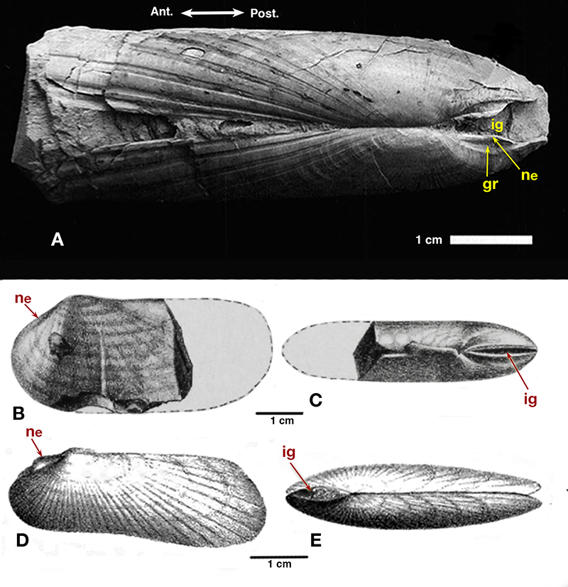
FIGURE 4. Phyletic schemes and biostratigraphic ranges (solid bars) of Paleozoic solemyoid genera. A, phyletic scheme of Pojeta (1988, fig. 3) postulating derivation of the Clinopisthinae and Solemyinae from nuculoid ancestors (i.e., Ctenodonta). B, proposed modifications (in red) of Pojeta’s scheme based on studies herein. Abbreviations of generic names: A. = Acharax; Cl. = Clinopistha; Ct. = Ctenodonta; D. = Dystactella; ‘J.’ = Janeia; and M. = Manzanella.
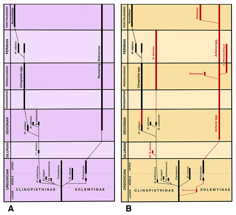
FIGURE 5. Classic exemplars of Janeia King of past authors. A-H, Solemya primaeva Phillips, 1836. A, putative holotype (lost) of Phillips, 1836, pl. 5, fig. 6 (= type species of Janeia by original designation of King, 1850, p. 177), Lower Carboniferous of Lowick, Northumberland, England. B, composite mold, left valve; a large hypotype identified by Portlock (1843, p. 441) as S. primaeva Phillips (figure of Hind 1900, pl. 50, fig. 1), Carboniferous shales of Aghaloo Parish, Co. Tyrone, N. Ireland. C, composite mold, left valve of a topotype (Hind, 1900, pl. 50, fig. 5); same figure used by Cox (1969, fig. B1.1a), Lower Carboniferous, Lowick, Northumberland. D, composite mold, right valve of a hypotype (Hind, 1900, pl. 50, fig. 6) (= type of Sanguinolites radiatus M’Coy, 1844, p. 50), Killymeal, Dungannon, Co. Tyrone. The simple buttresses and non-occluded posterior adductors in 5B-D are consistent with Acharax. E-F, Solemya primaeva Phillips as figured by M’Coy (1855), Lower Carboniferous, Lowick, Northumberland. E, left lateral view (M’Coy, 1855, pl. 3F, fig. 3). F, same specimen, dorsal view (M’Coy, 1855, pl. 3F, fig. 3a). G-H, actual specimen (Sedgwick Museum no. E1114) used by M’Coy as the basis for Figures 5E-F (photos by Matthew Riley). G, left lateral view. H, dorsal view. The details shown in M’Coy’s figures appear to be largely hypothetical. I, Janeia biarmica (de Verneuil, 1845), figure of King, (1850, p. 178, pl. 16, fig. 7) [= Wilkingia elegans (King)], Upper Permian, Humbledon Quarry, Durham, England. J, Solemya abnormis Howse, 1848, p. 244; 1857a, p. 309, pl. 4, fig. 8; Upper Permian “Shell-Limestone”, Silksworth, Durham; King (1850) accepted it as a synonym of J. biarmica; Logan (1967) called similar topotypic shells Stutchburia modioliformis (King). K-L, Solemya biarmica de Verneuil 1845, pl. 19, fig. 4a, 4b, Lower Permian, Nizhny, Novgorod Oblast, Russia; herein reassigned to Dystactella Hall and Whitfield. K, holotype, an internal mold, right lateral view. L, dorsal view of same showing anterior and posterior gapes; the infilling of the internymphal gap (ig) is evidence of an external parivincular ligament. M-N, Dystactella subnasuta (Hall and Whitfield, 1872), Lower Devonian (Upper Helderberg), Louisville, Kentucky. M, right lateral view of articulated specimen (= “Specimen A”, USNM 145650) of McAlester (1968, pl. 5, fig. 7). N, dorsal view of “Specimen A” of McAlester (1968, pl. 5, fig. 9) showing cylindrical external ligament (le) on the posterodorsum; the similarities to S. biarmica de Verneuil are conspicuous. O, Janeia phillipsiana King, 1848 (from King, 1850, p. 179, pl. 16, fig. 8); Howse (1857b, p. 9) called it a “mere fiction.” P, Solemya normalis Howse, 1848 as figured by Howse (1857a, pl. 4, fig. 7); placed in synonymy with J. phillipsiana by King (1850, p. 179), Upper Permian, Humbledon Hill, Durham. Q, Solemya phillipsiana King, figure of Schauroth (1854, p. 553, pl. 21, fig. 5), lower Zechstein (Upper Permian), Bucha, Germany. R, same specimen reinterpreted by Geintiz (1861, p. 60, pl. 12, fig. 19) and placed by him in synonymy with S. biarmica. Note the marked differences in shell shape and detail; both the cleft formed by the buttress and adjacent adductor scar are missing in Schauroth’s figure.
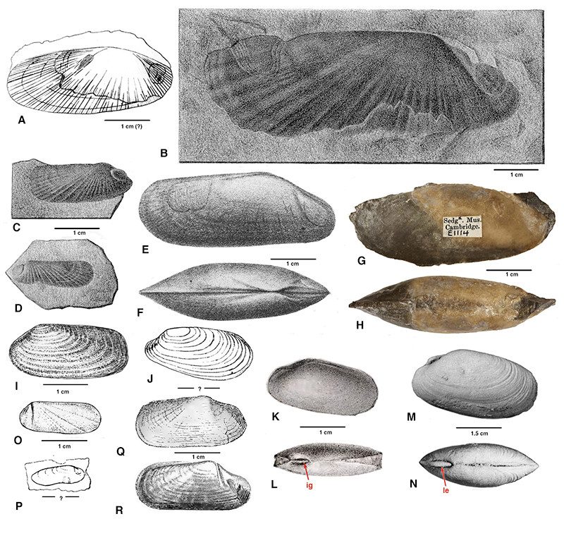
FIGURE 6. Classic exemplars of Janeia King of past authors. A-G, Solemya (Janeia) vetusta Meek, 1871 [= Dystactella Hall and Whitfield]. A, left lateral view, internal mold with partial shell (Meek, 1873, pl. 18, fig. 4), Devonian, Ohio. B-D, articulated specimen, Middle Devonian, Louisville, Kentucky. B, left lateral view (Hall, 1885, pl. 47, fig. 53). C, right lateral view (Hall, 1885, pl. 47, fig. 54). D, dorsal view, anterior to right (Hall, 1885, pl. 47, fig. 55). E, a left valve showing scalloped radii and cylindrical external ligament, Middle Devonian, Charlestown, Indiana (Hall, 1885, pl. 94, fig. 10). F-G, articulated specimen, Middle Devonian, Charlestown, Indiana. F, right lateral view (Kindle, 1901, pl. 16, fig. 1). G, dorsal view, posterior at right (Kindle, 1901, pl. 16, fig. 1a) showing external nymphae (ne) and internymphal gap (ig). H-J, Janeia? compressa (Goldfuss, 1840) of Beushausen (1895), an internal mold [= Pleurophorella ? aff. transversa (de Koninck)], ?Middle Devonian, Daleiden, Germany. H, right lateral view (Beushausen, 1895, pl. 26, fig. 1a). I, left lateral view (Beushausen, 1895, pl. 26, fig. 1b). J, dorsal view showing lunule and escutcheon (Beushausen, 1895, pl. 26, fig. 1c). K-P, Janeia laevigata (Goldfuss, 1840). K-L, an internal mold [= ? Pleurophorella cf. tricostata (Portlock)], Middle Devonian, Gerolstein, Germany; note prosoponal radii and short buttress bordering deeply impressed anterior adductor scar. K, left lateral view (Beushausen, 1895, pl. 26, fig. 3a). L, right lateral view (Beushausen, 1895, pl. 26, fig. 3b). M, lateral view of type (Goldfuss, 1840, pl.159, fig. 14), gen. indet., Devonian, Eifel; also figured in Beushausen (1895, pl. 26, fig. 8) N-P, an articulated specimen [= Pleurophorella ? sp. ], Devonian, Daleiden. N, right lateral view (Beushausen, 1895, pl. 26, fig. 2a). O, left lateral view (Beushausen, 1895, pl. 26, fig. 2b). P, dorsal view (Beushausen, 1895, pl. 26, fig. 2c); note strong similarities to Figure 6J. Q-V, Janeia truncata (Goldfuss, 1840). Q, internal mold with attached shell fragments, left lateral view [= Clinopistha Meek and Worthen], Middle Devonian, Stringocephalus Limestone, Gerolstein (Beushausen, 1895, pl. 26, fig. 5). R-T, internal mold with attached shell fragments [= Clinopistha ], Devonian, Eifel. R, right lateral view (Beushausen, 1895, pl. 26, fig. 4b). S, left lateral view (Beushausen, 1895, pl. 26, fig. 4a). T, dorsal view, anterior at right (Beushausen, 1895, pl. 26, fig. 4c). U-V, internal mold with partial shell [not a soleymid]; lateral views showing ciricular adductor scar (ad) and distinct pallial line (pl) (Beushausen, 1895, text-fig. 32, p. 297), Middle Devonian, Gerolstein. W-BB, Janeia phaseolina (Goldfuss, 1840). W, lateral view of type (Goldfuss, 1840, p. 279, pl. 159, fig. 15), gen. et sp. indet., Devonian, Eifel; also figured by Beushausen (1895, pl. 26, fig. 9). X, lateral view of an internal mold (Beushausen, 1895, pl. 26, fig. 6), Devonian, Eifel [= ? Edmondia de Koninck]. Y-AA, articulated specimen [= ? Edmondia ], Gerolstein (Beushausen, 1895, text fig. 31, p. 296). Y, right lateral view. Z, left lateral view. AA, end view. BB, lateral view of an internal or composite mold, lower Middle Devonian, Gerolstein (Beushausen, 1895, pl. 26, fig. 7) [possible sanguinolitid; = ? Myofossa Waterhouse].
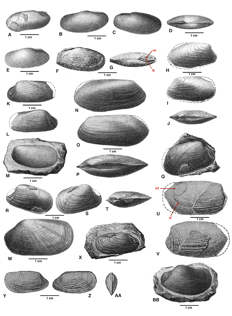
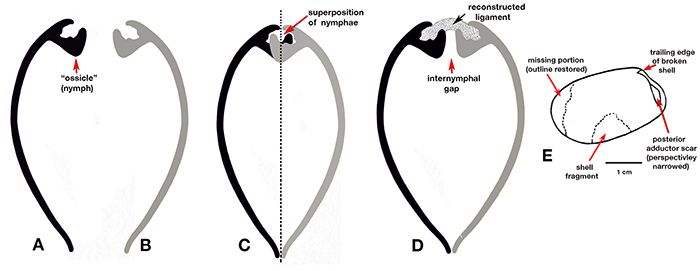
FIGURE 7. Interpretations of a transverse section through the beak of Janeia truncata, Middle Devonian of Gerolstein as figured by Quenstedt (1930, pl. 1, fig. 4) and reproduced in the Treatise by Cox (1969, N242, fig. B1,1b). A, Quenstedt’s original ink sketch, a left valve, showing nymph-like ossicle (arrow) alleged to be a chondrophore for attachment of an internal ligament. B, inferred transverse section of right valve. C, incompatibility of right and left valves when fitted together with closed dorsal margins. As shown, the ossicle and its inferred counterpart irreconcilably overlap (arrow). D, a workable solution achieved by addition of an internymphal gap (arrow) separating the valves posterodorsally. By inference, the ossicles functioned as nymphae supporting an external ligament (stippled pattern, here reconstructed). E, Quenstedt’s (1930, pl. 1, fig. 5) diagrammatic reconstruction of Janeia truncata based on the internal mold of a left valve figured by Beushausen (1895, pl. 26, fig. 5). For clarification, arrows and explanatory labels are added here.