FIGURE 1. Cladograms showing the various proposed placements of turtles (Testudines) relative to other amniotes. 1. Eunotosaurus as the ancestor of turtles (Watson, 1914). 2. Turtles as the sister group to the Dicynodontia Gardiner (1982). 3. Turtles as the sister group to the Captorhinidae (Gauthier et al., 1988). 4. Turtles as the sister group to Owenetta (Reisz and Laurin, 1991). 5. Turtles as the sister group to the Pareiasauridae (Lee, 1997). 6. Turtles as the sister group to the Sauropterygia (Rieppel and Reisz, 1999). 7. Turtles as a sister group to archosaurs (Hugal et al., 2007; Crawford et al., 2012; and Chiari et al., 2012).
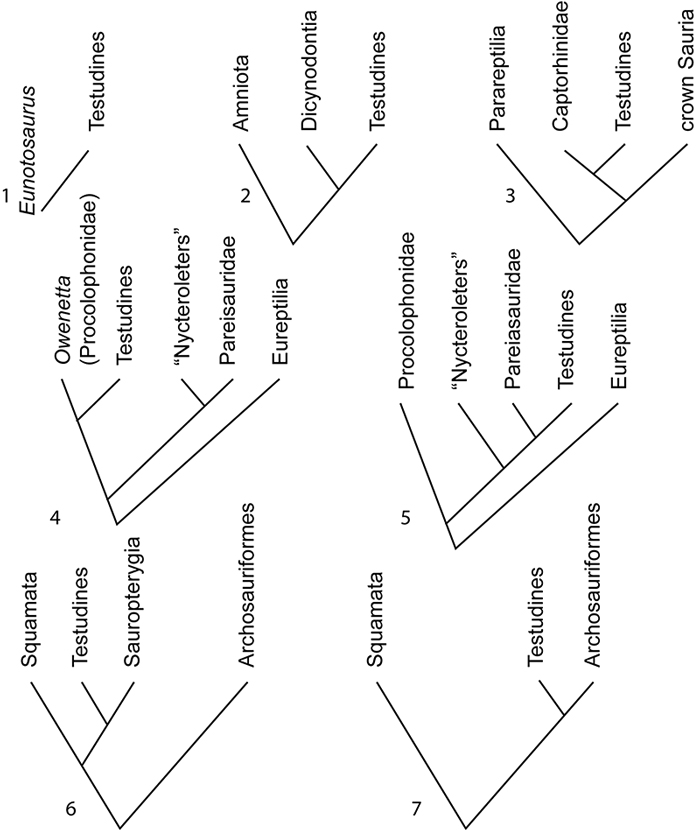
FIGURE 2. Paleogeographic location of genera of early turtles and other tetrapods discussed in this paper on maps of the Permian (1) and Triassic (2). Maps based on Wing and Sues (1992).
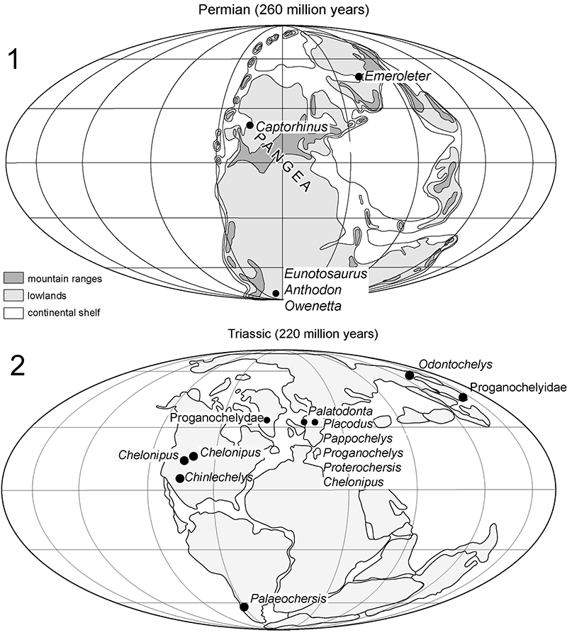
FIGURE 3. Temporal distribution of early turtles and other genera discussed in this paper.
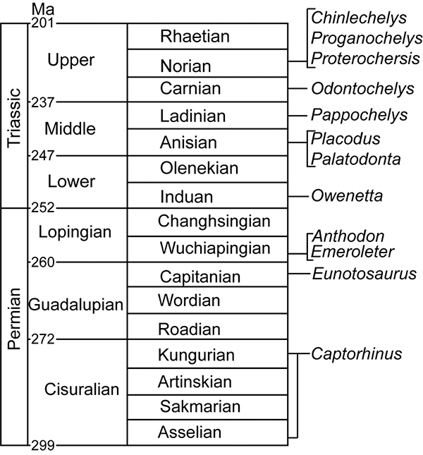
FIGURE 4. Entoplastron of Kayentachelys, Texas Memorial Museum 43687-109, showing sutures dorsally on either lateral margin of the entoplastron.
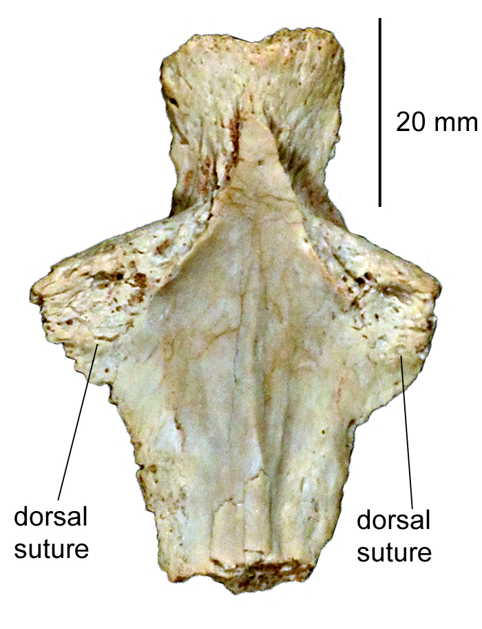
FIGURE 5. Locality and stratigraphic position of Chinlechelys tenertesta type locality. After Lucas et al. (2000).
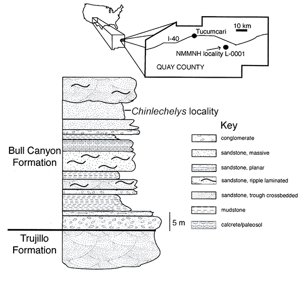
FIGURE 6. Outline drawing of Proganochelys quenstedti showing as shaded areas the approximate preserved portions of the shell and skull of Chinlechelys tenertesta . Numbers correspond to the suffix added to the specimen numbers in the text to identify specific fragments.
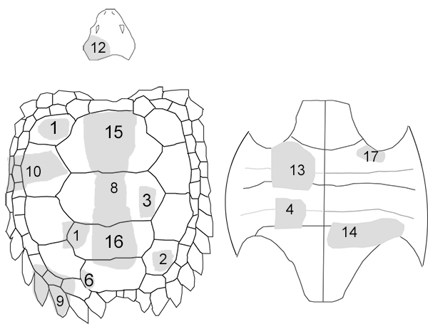
FIGURE 7. Part of skull of Chinlechelys tenertesta, NMMNH P-16697-12: 1, stereograph of the left posterior portion of Chinlechelys skull in dorsal view; 2, dorsal view of same with labels. Abbreviations: po, jugal; qj, quadratojugal; ri, ridge (a and b indicate the two separate ridges referred to in the text); sq, squamosal; te, temporal emargination.
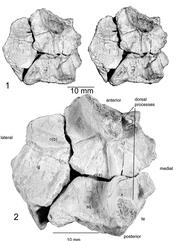
FIGURE 8. Part of skull of Chinlechelys tenertesta, NMMNH P-16697-12: 1, stereograph of the left posterior portion of Chinlechelys skull in ventral view; 2, line drawing highlighting major features of the skull fragment; 3, prootic and opistotic viewed from opposite sides, orientation unknown; 4, skull of Anthodon serrarius modified from Lee (1993) with the bones corresponding to those identified in Chinlechelys highlighted. Abbreviations: po, jugal; qj, quadratojugal; sq, squamosal; te, temporal emargination.
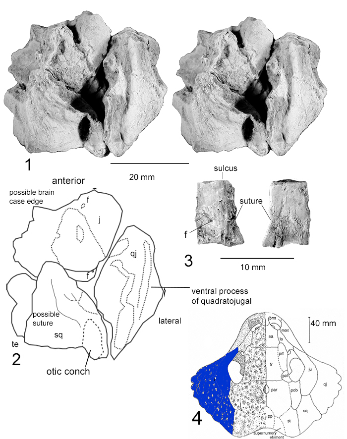
FIGURE 9. Chinlechelys tenertesta NMMNH P-16697-8,15,16, Neural plates and thoracic vertebrae 1-8 in 1, dorsal; and 2, ventral views.
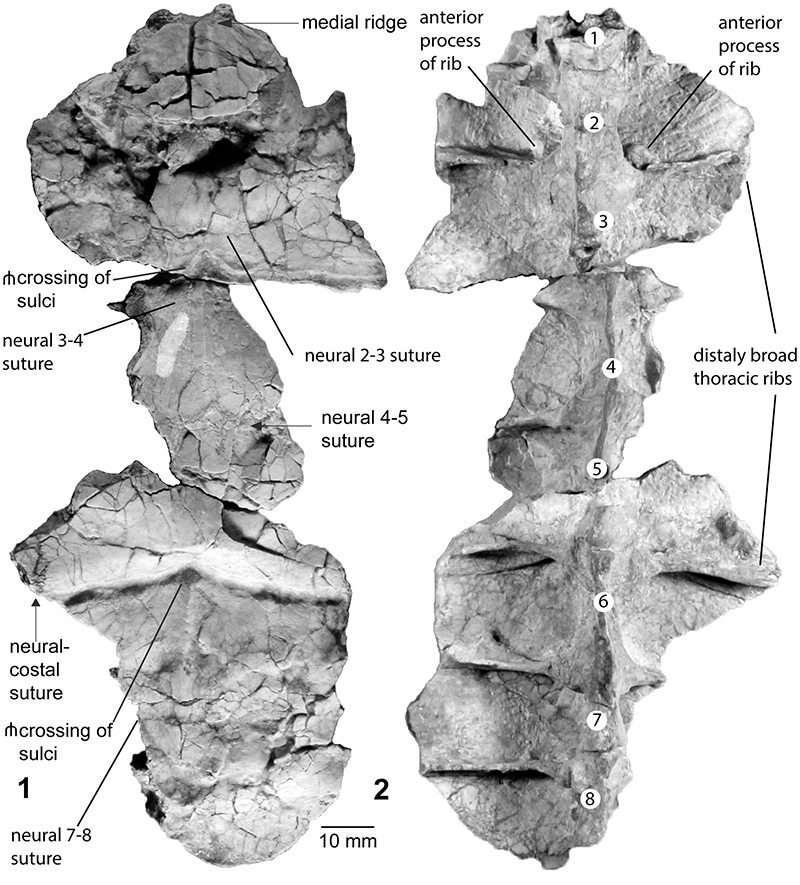
FIGURE 10. Chinlechelys tenertesta NMMNH P-16697-15, Neural plates and thoracic vertebrae 1-3 in 1, anterior; 2, right lateral; 3, posterior views; and 4, a close up of the anterior process of the right rib 3.
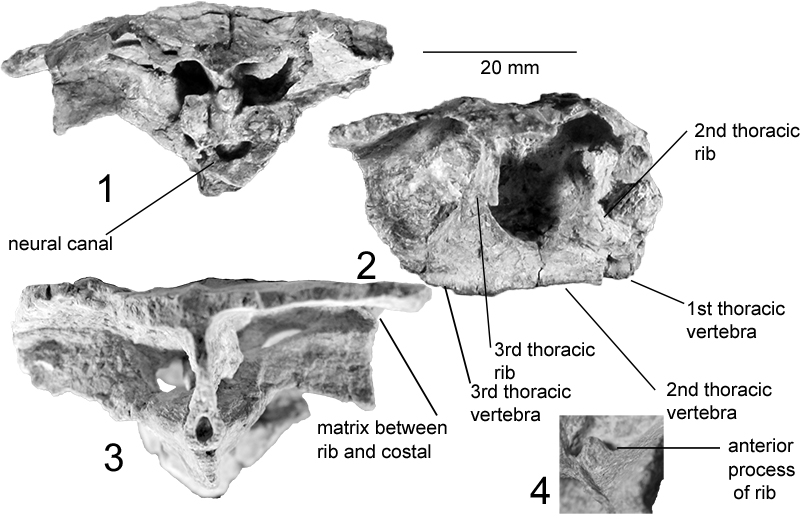
FIGURE 11. A-D, Chinlechelys tenertesta NMMNH P-16697-8: Two neural plates and vertebrae in 1, lateral; 2, dorsal views; 3, anterior; 4, posterior; and 5, dorsal outline drawing. Abbreviation: n1, first anterior neural; and n2 second neural; r, rib; and rid, medial ridge.
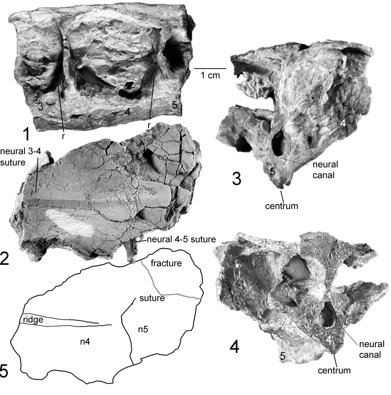
FIGURE 12. Chinlechelys tenertesta NMMNH P-16697-16 Neural plates and thoracic vertebrae 5-8 in 1, anterior; 2, dorsal; 3, left lateral views; 4, close up of thoracic rib highlighting the separation from the overlying costal plate. Numbers indicate the thoracic vertebrae positions.
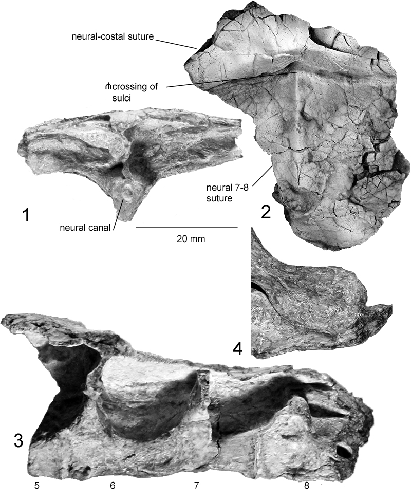
FIGURE 13. Chinlechelys tenertesta costal, NMMNH P-16697-1 in 1, Dorsal and 2, ventral views; 3-5, Costals and ribs P-16697-3: 3, Ventral view and 4, line drawing; 5, Close up of dorsal side of rib in F where the costal plate has been broken away; and 6, Lateral view. Abbreviations: r, rib and c, costal.
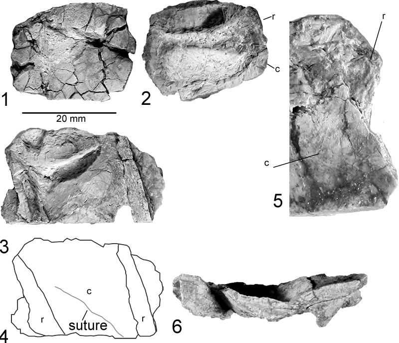
FIGURE 14. Chinlechelys tenertesta costal, NMMNH P-16697-3: 1, Close up of a cross section across the rib of NMMNH P-16697-1 on the right in Figure 12.6; and 2, cross section of rib on the left in Figure 12.6. 3, color close up of a cross section across the same rib of NMMNH P-16697-1 seen in 14.1; 4, color close up of a cross section across the same rib of NMMNH P-16697-1 seen in 14.2; 5, progressive ct scan slices through the rib in figure 14.1; and 6, progressive ct scan slices through the rib in figure 14.2.
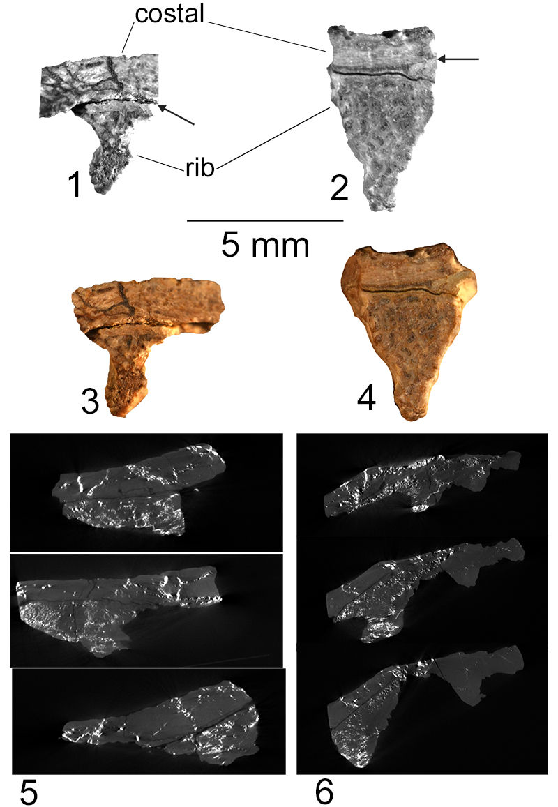
FIGURE 15. Carapace fragments of Chinlechelys tenertesta : 1-2, NMMNH P-16697-2 costal bones in dorsal view; 3-4, NMMNH P-16697-6, costal fragment in dorsal and ventral views; 5, NMMNH P-16697-7, costal fragment in dorsal view; and 6-7, NMMNH P-16697-5 costal fragment in dorsal view. Abbreviations: pr, dorsal process; r, rib; and s, suture.
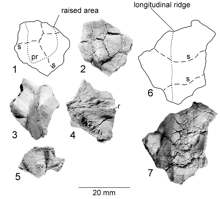
FIGURE 16. Chinlechelys tenertesta, NMMNH P-16697-10, Costals and peripherals: 1, Dorsal view; 2, line drawing of dorsal view; and 3-4, ventral view. Abbreviations: c, costal; p, peripheral; and r, rib.
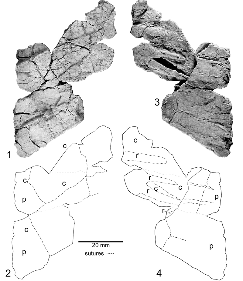
FIGURE 17. Chinlechelys tenertesta, NMMNH P-16697-9: Peripherals and supraperipherals in 1, lateral; 2, dorsal; and 4, ventral views. 3, dorsal line drawing; and 5, ventral line drawing. The ossifications labeled 1, 4, 5, 7, 8, 9, 10, and 11 forma a continuous line we consider equivalent to peripherals. The ossifications labeled as 2, 3, 6, and 7 are additional ossifications distal to these which we term supraperipherals.
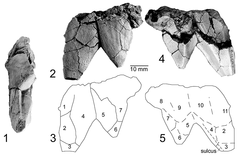
FIGURE 18. Chinlechelys tenertesta, NMMNH P-16697-13, hyoplastron and mesoplastron in 1, external view; 2, external view line drawing; and 3, internal view. 4, left axillary notch in dorsal view. Short dashes indicate sutures and longer dashes sulci and raised surface structures.
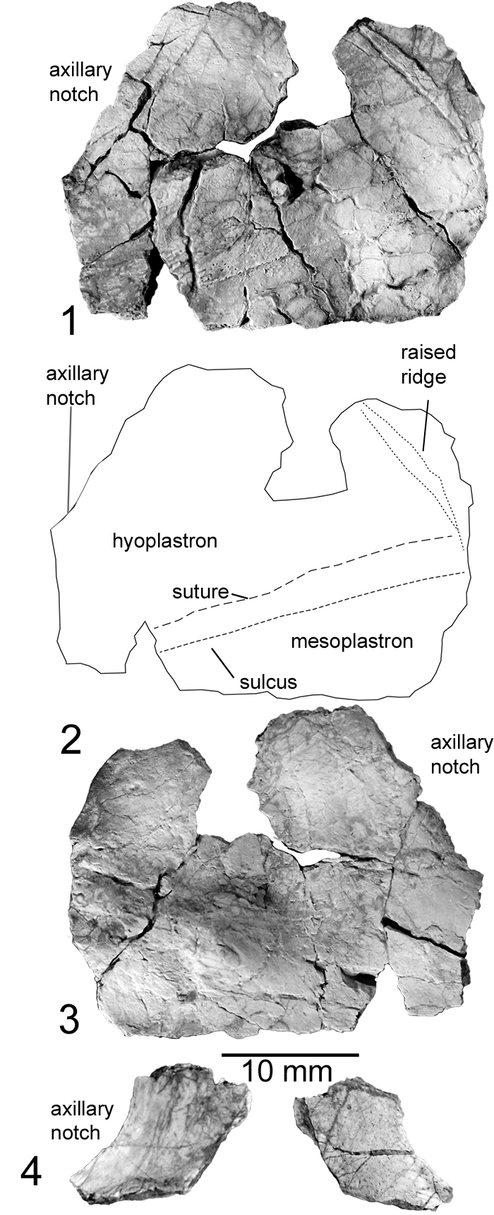
FIGURE 19. Chinlechelys tenertesta, NMMNH P-16697-4, mesoplastra in 1, external view; 2, external view line drawing; 3, internal view; 4, dorsal side of carapace fragment; and 5, plastron fragment. Short dashes indicate sutures and longer dashes indicate sulci and raised surface structures.
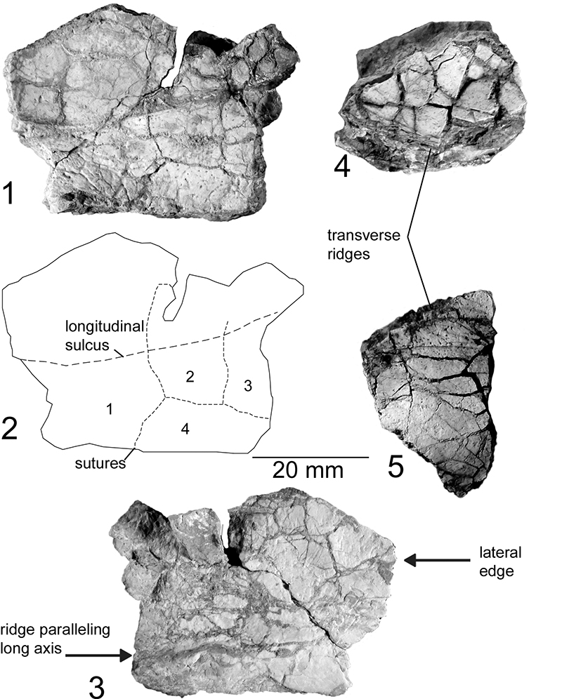
FIGURE 20. Chinlechelys tenertesta, NMMNH P-16697-14, Hypoplastron in dorsal (1) and ventral (3) views. Line drawings of dorsal view (2) and ventral views (4). Note the suture to the pelvis in the lower right of the dorsal view and the longitudinal sulcus crossing the hypoplastron in the ventral view near the edge of the bone. Abbreviation: in, inguinal scute.
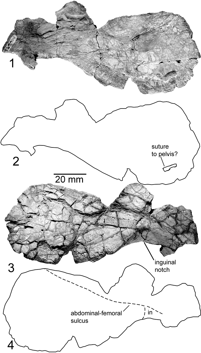
FIGURE 21. Chinlechelys tenertesta innominates: 1. Holotype innominate (P-16697-11) in distal view. 2. Proximal view. 3. Referred innominate (P-16621) distal view. 4. proximal view. Sutures are marked at the free edges of the innominate with a “s.”
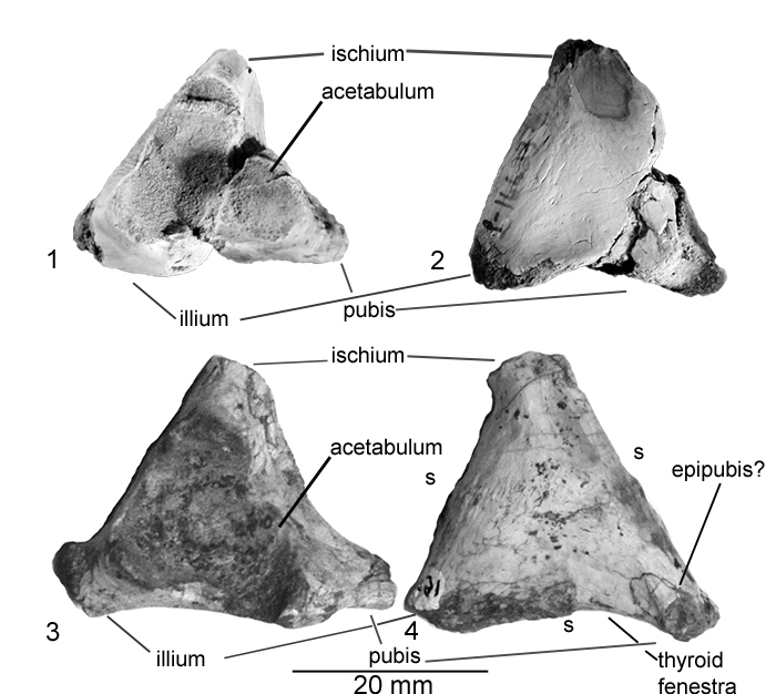
FIGURE 22. Chinlechelys tenertesta, NMMNH P- 4315, referred femoral head: in dorsal (1), posterior (2), anterior (3) and lateral views (4). Abbreviations: hh, head height; hw, head width; i, intertrochanteric fossa; ma, trochanter major; and mi, trochanter minor.
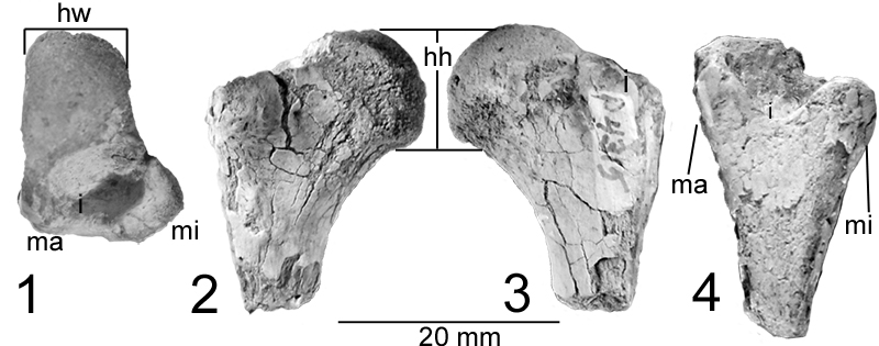
FIGURE 23. Final phylogeny resulting from our cladistic analysis excluding Eunotosaurus, and testing the position of Testudines within the Reptilia. This was based on the modified version of the Szczygielski matrix presented in this paper and run through the program TNT with multistate characters ordered. Bremer support values above one are listed above nodes, and bootstrap values above 50% are listed below nodes.

FIGURE 24. Phylogeny resulting from our cladistic analysis, excluding Eunotosaurus, adding in the two autapomorphies of the Pareiasauridae and testing the position of Testudines within the Reptilia. This was based on the modified version of the Szczygielski matrix presented in this paper and run through the program TNT with multistate characters ordered. Bremer support values above one are listed above nodes, and bootstrap values above 50% are listed below nodes.
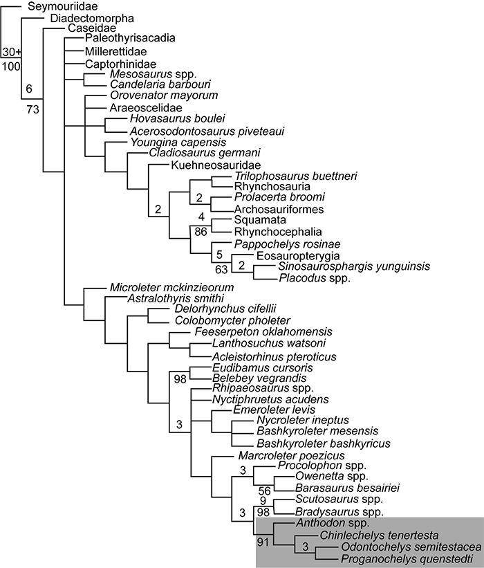
FIGURE 25. Phylogeny resulting from our cladistic analysis, excluding Eunotosaurus as well as all Chinlechelys skull characters, adding in the two autapomorphies of the Pareiasauridae and testing the position of Testudines within the Reptilia. This was based on the modified version of the Szczygielski matrix presented in this paper and run through the program TNT with multistate characters ordered. Bremer support values above one are listed above nodes and bootstrap values above 50% are listed below nodes.
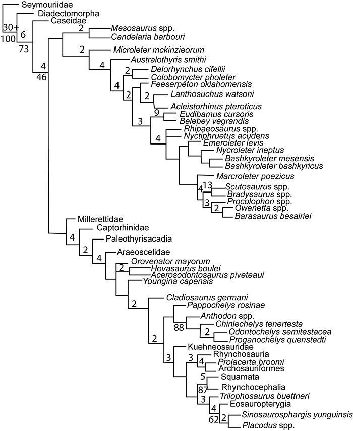
FIGURE 26. Phylogeny resulting from our initial analysis with Eunotosaurus and two caseids included. Bremer support values above one are listed above nodes and bootstrap values above 50% are listed below nodes.
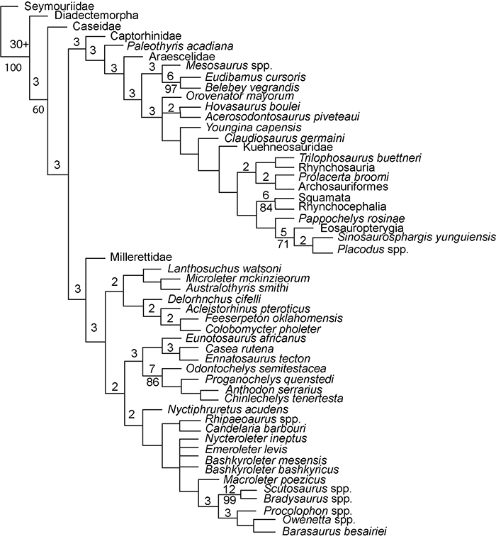
FIGURE 27. Side-by-side comparison of left lateral views of the skulls of: 1. Casea . 2. Eunotosaurus . 3. Milleretta . Not to scale. Casea based on Lee (1993), Eunotosaurus based on Keysler and Gow (1981), and Milleretta based on Romer (1956).
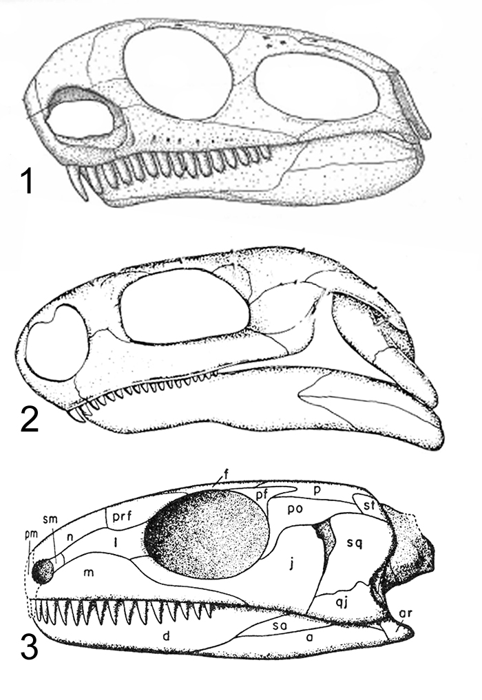
FIGURE 28. Phylogeny resulting from our cladistic analysis of Eunotosaurus using the matrix of Benson (2012) with no taxa pruned to assess the possible position of Eunotosaurus within the Synapsida. Bremer support values above one, followed by Jacknife values over 50% are listed below the nodes, and bootstrap values above 50% are listed above nodes.
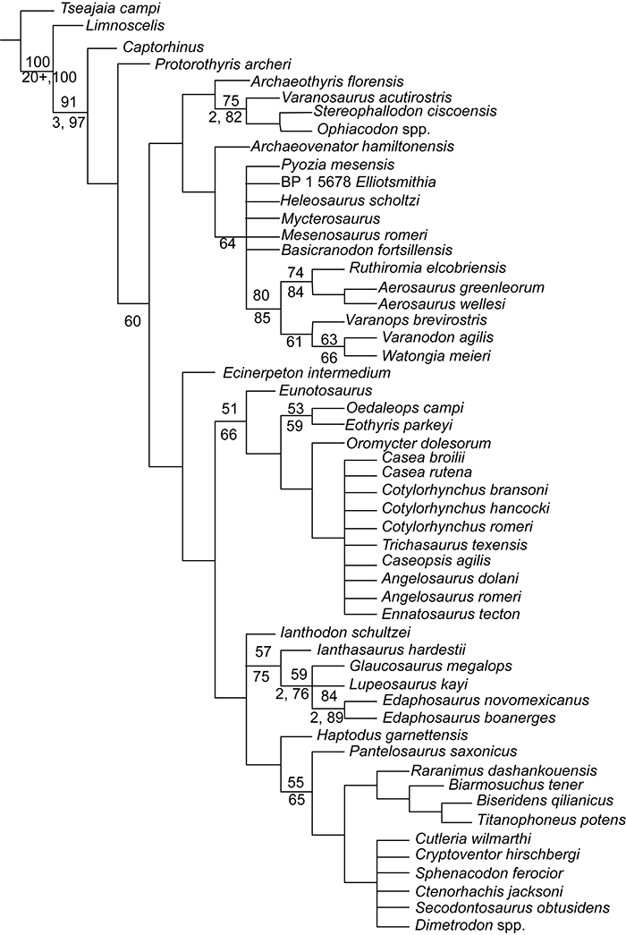
FIGURE 29. Plot showing the forelimb proportions of several putative basal turtles and the synapsid Edaphosaurus as a comparison relative to a variety of extant turtles. 1. comparison relative to various turtle taxa; and 2. comparison to different habitats of extant turtles. Figure is based on Joyce and Gauthier (2004).
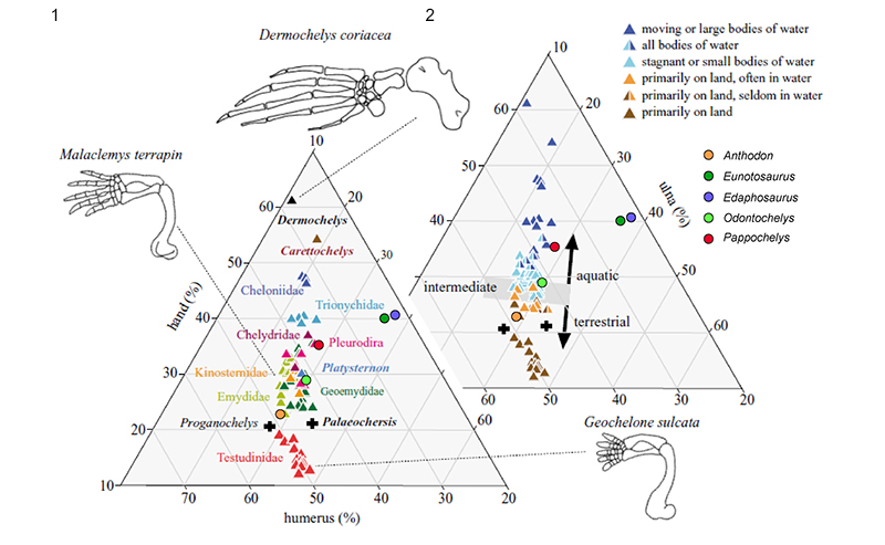
FIGURE 30. Drawings of proposed origin of turtles from left to right: Scutosaurus, modified from Lee (1997), Anthodon, modified from Lee (1997), Chinlechelys tenertesta, new reconstruction, Proganochelys quenstedti, modified from Joyce et al., (2009), Kayentachelys, modified from Joyce et al., (2009). Drawings by Matt Celeskey.
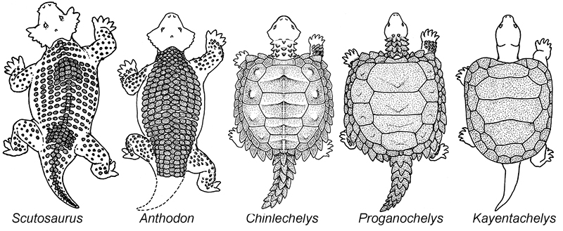
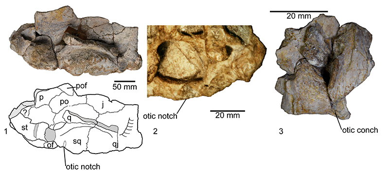
FIGURE 31. Otic structures of the Pareiasauridae and Chinlechelys : 1, Embrithosaurus schwarzi skull fragment in ventral view (from Van Den Brant et al., 2019), showing the simple otic notch; 2, Anthodon pricei skull in ventral view (image from Mike S.Y. Lee) showing the conical shape in one dimension of the notch in this pareiasaur, previously hypothesized to be close to Testudines; 3, ventral view of the skull fragment of Chinlechelys showing the cone-shaped (in two dimensions) otic conch.