FIGURE 1. Locality map showing plesiosaur-bearing fossil localities in Australia (modified from Kear, 2003). Specimens analysed here are from the Richmond area, Queensland and Coober Pedy, South Australia.
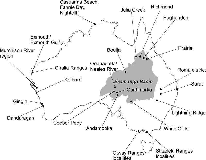
FIGURE 2. Measured morphometric parameters of vertebrae (specimen QM F12719); CW: centrum width; CH: centrum height; CL: centrum length; H of NC - height of neural canal; W of NC - width of neural canal; W of ZYG. - width of zygapophyses; H of NS - height of neural spine; angle - angle of zygapophysis with centre of centrum.
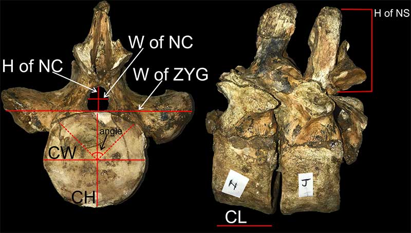
FIGURE 3. Specimen QM F12719. A. Dorsal vertebra, anterior view. B. Dorsal vertebra, lateral view. C. Cervical vertebra, anterior view. D. Cervical vertebra, ventral view showing foramina subcentralia (f.s.). E. Cervical vertebra, dorsal view showing foramen on neural arches. Scales shown on figure.
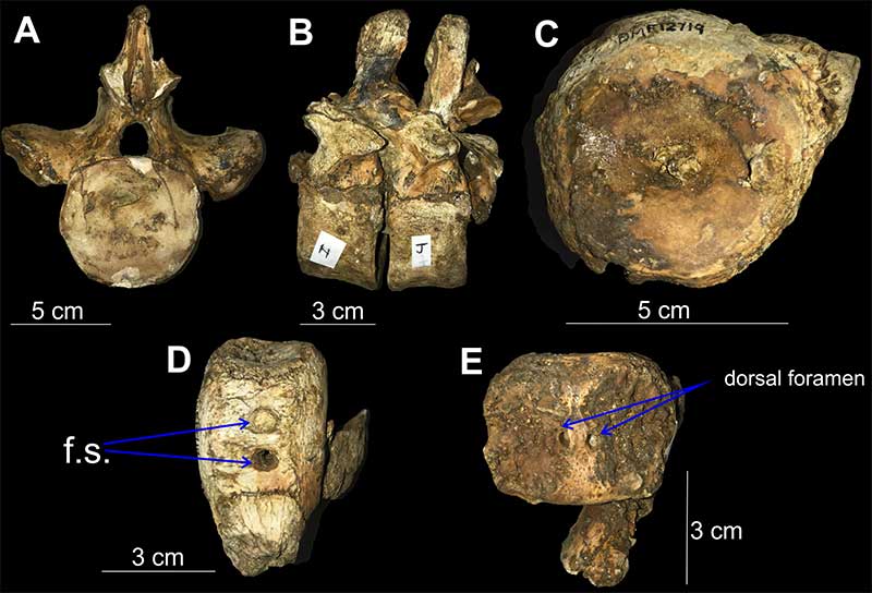
FIGURE 4. Vertebrae associated with specimen QM F11050 - Eromangasaurus australis holotype. A. QM F12216, lateral view showing lateral ridge (red arrow). B. Cojoined vertebrae QM F12217, anterior view. C. QM F12217, lateral view showing lateral ridges (red arrows). D. QM F12219a, anterior view. E. QM F12219a, lateral view showing lateral ridge (red arrow). F. QM F12219b, lateral view showing lateral ridge (long red arrow) and rib facet borne wholly on centrum (short red arrow). G. QM F12219b, ventral view showing foramina subcentralia (white arrows) separated by a mid-ventral keel. Scales shown on figure.
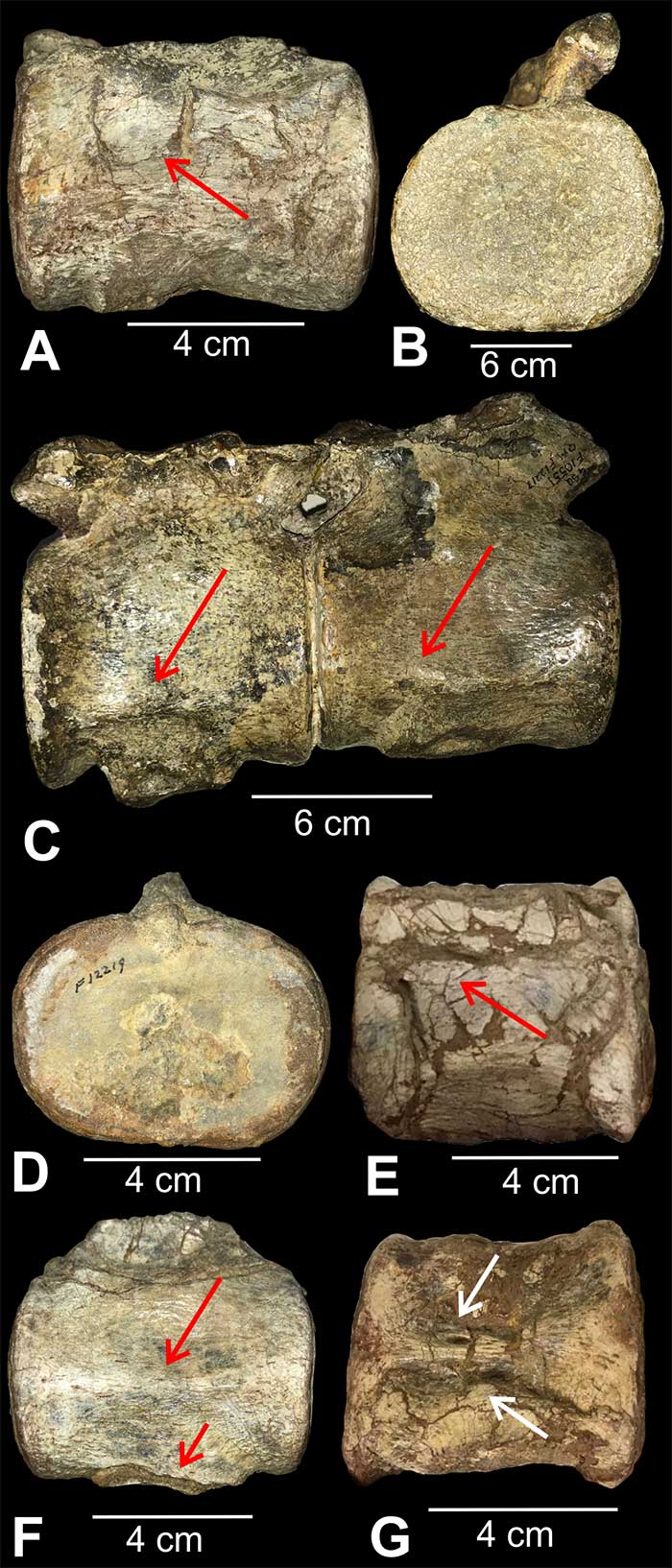
FIGURE 5. Specimen QM F12934. A. Caudal vertebra showing chevron facets; B. Caudal vertebra showing rib facet, lateral view. C. Sacral vertebra showing rib facets borne partly on centrum and partly on neural arch, lateral view; D. Sacral vertebra, anterior view. E. Posterior cervical showing rib facet, lateral view. F. Anterior cervical showing foramina subcentralia (f.s.), ventral view. G. Anterior cervical, lateral view. H. Dorsal vertebra, anterior view. Scales shown on figure.
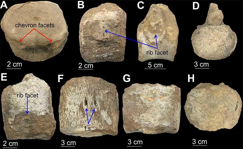
FIGURE 6. Specimen RM FR269. A. Cervical vertebra, anterior view with weakly fused neural arches and neural spine. B. Cervical vertebrae, lateral view with rib facets borne wholly on the centrum. C. Cervical vertebra, ventral view showing paired foramina subcentralia (f.s.). D. Dorsal vertebra, anterior view with rib facets (diapophyses) borne wholly on neural arches. Scales shown on figure.
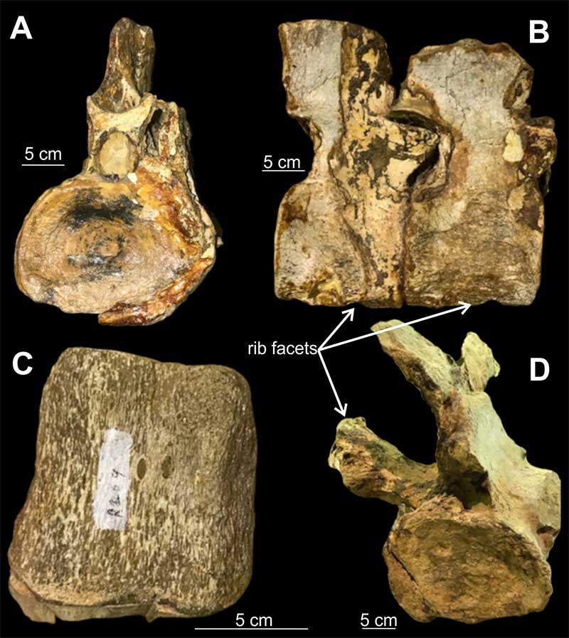
FIGURE 7. Specimen RM FR269. A. Transition from dorsals to sacrals, right lateral view. B. Transition from pectorals to dorsals, right lateral view. Scales shown on figure.
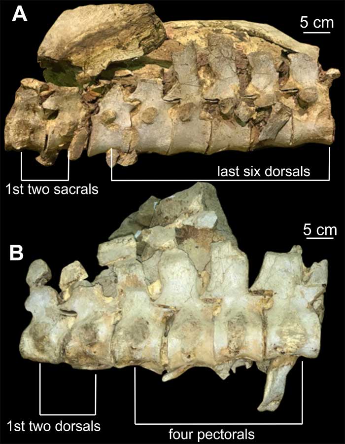
FIGURE 8. Specimen QM F2085. A. Pectoral vertebra, anterior view. B. Pectoral vertebra, lateral view. C. Sacral vertebra, posterior view. D. Sacral vertebrae, lateral view. E. Dorsal vertebra, lateral view. F. Dorsal vertebra, anterior view. Scales shown on figure.
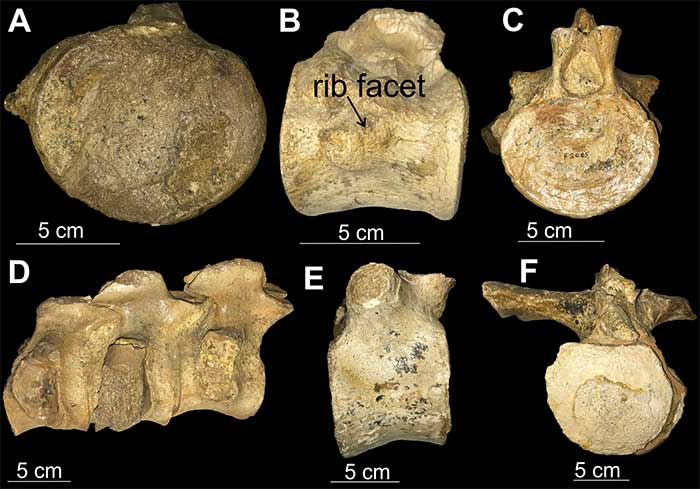
FIGURE 9. Specimen QM L39 - anterior cervicals. A. Lateral view with prominent ridge. B. Anterior view. C. Ventral view with foramina subcentralia. D. Anterior view. E. Lateral view showing rib facet. F. Ventral view showing foramina subcentralia. Scales shown on figure.
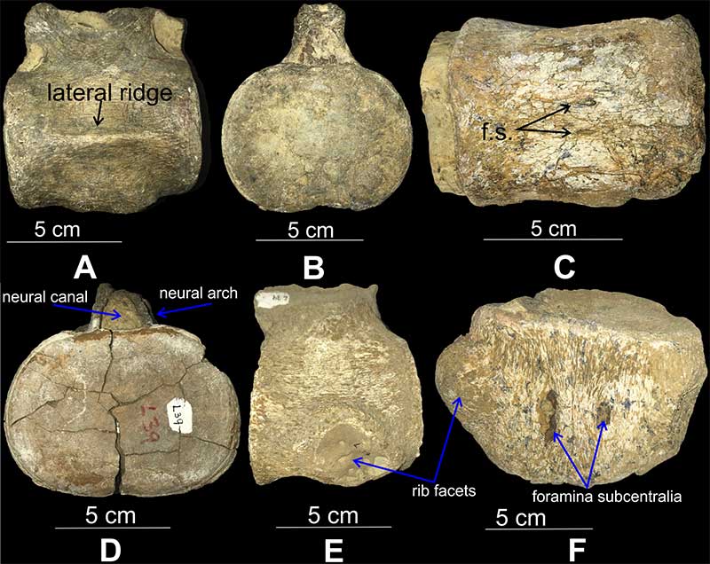
FIGURE 10. Specimen RM FR436. A. 12 anterior cervical vertebrae. B. Ventral view. C. Anterior view. Note the skewed vertebra. Scales shown on figure.
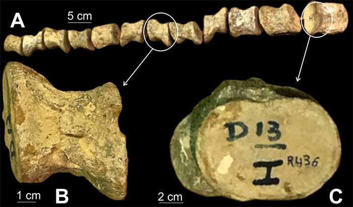
FIGURE 11. A-C. Specimen F171282/QM ISO. A. Lateral view showing lateral ridge. B. Anterior view, showing neural arch fused to the centrum. C. Ventral view showing paired foramina subcentralia. D-F. QM Specimen PL (unregistered). D. Anterior cervical, lateral view showing ridge. E. Anterior cervical, ventral view showing paired foramina subcentralia. F. Anterior cervical, anterior view, showing part of neural arches fused to the centrum. Scales shown on figure.
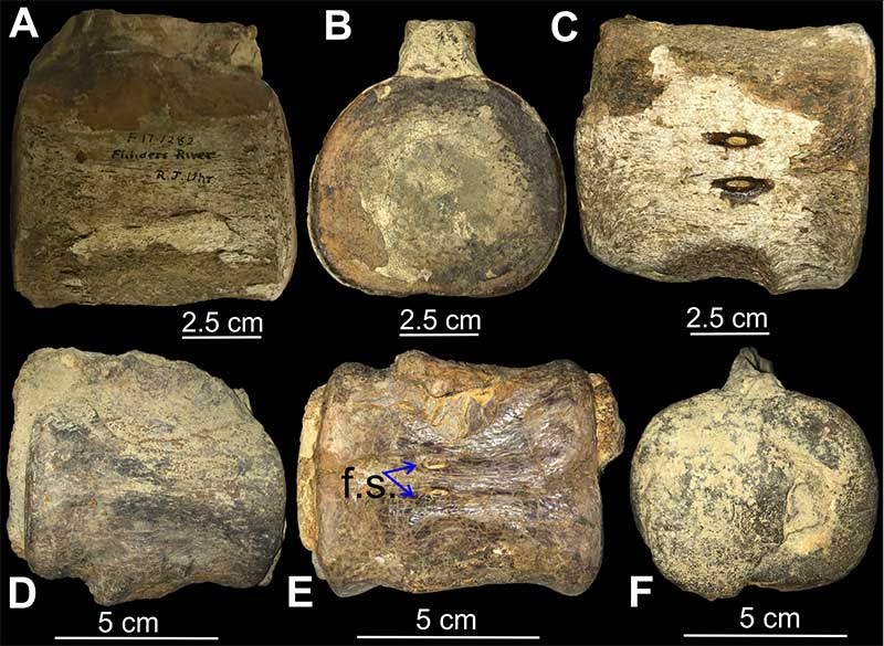
FIGURE 12. Normalised vertebral position (cervicals 0-1; dorsals 1-2; caudals 2-3) plotted against vertebral length index (VLI) for Australian plesiosaurians and non-Australian elasmosaurids. Data for QM F3567 and RM FR271 from Sachs (2004); Opallionectes andamookaensis from Kear (2005a); Elamosaurus platyurus, Thalassomedon haningtoni, Callawayasaurus colombiensis, and Cm Zfr 115 from O’Keefe and Hiller (2006); Vegasaurus molyi from O’Gorman el. (2015); AMNH FARB 1495, AMNH FARB 5835, and AMNH FARB 2554 from Otero (2016); Aristonectes parvidens from O'Gorman (2016a); Kawanectes lafquenianus from O’Gorman (2016b); Lagenanectes richterae from Sachs et al. (2017), and Jucha squalea from Fischer et al. (2020).
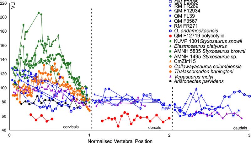
FIGURE 13. Normalised cervical vertebral position plotted against vertebral length index (VLI) for Australian plesiosaurians and non-Australian elasmosaurids. Data for QM F3567 and RM FR271 from Sachs (2004); Opallionectes andamookaensis from Kear (2005a); Elamosaurus platyurus, Thalassomedon haningtoni, Callawayasaurus colombiensis, and Cm Zfr 115 from O'Keefe and Hiller (2006); Vegasaurus molyi from O’Gorman el. (2015); AMNH 1495, AMNH 5835, and AMNH 2554 from Otero (2016); Aristonectes parvidens from O’Gorman (2016a); Kawanectes lafquenianus from O’Gorman (2016b); Lagenanectes richterae from Sachs et al. (2017) and Jucha squalea from Fischer et al. (2020).
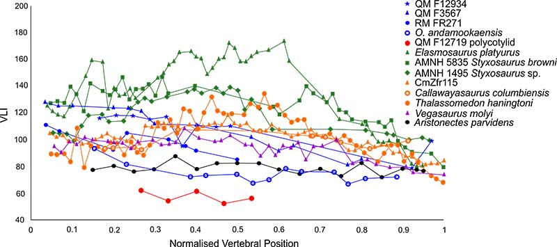
FIGURE 14. Plot for vertebral length index (VLI) against breadth index (BI) for Australian plesiosaurians and non-Australian elasmosaurids. Data for QM F3567 and RM FR271 from Sachs (2004); Opallionectes andamookaensis from Kear (2005a); Elamosaurus platyurus, Thalassomedon haningtoni, Callawayasaurus colombiensis, and Cm Zfr 115 from O'Keefe and Hiller (2006); Aristonectes quiriquinensis from Otero et al. (2014); Albertonectes vanderveldei from Kubo et al. (2012); Vegasaurus molyi from O'Gorman el. (2015); Tuarangisaurus keyesi from Hiller et al. (2017); AMNH FARB 1495, AMNH FARB 5835, Styxosaurus snowii, and AMNH FARB 2554 from Otero (2016); Aristonectes parvidens from O'Gorman (2016a); Kawanectes lafquenianus from O’Gorman (2016b); Lagenanectes richterae from Sachs et al. (2017) and Jucha squalea from Fischer et al. (2020).
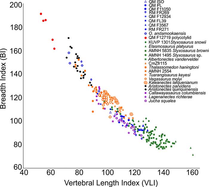
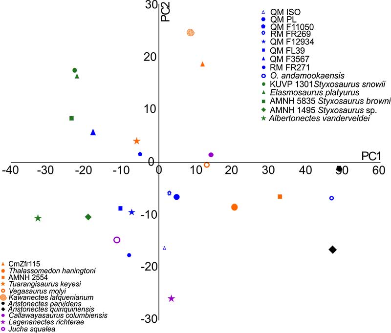
FIGURE 15. Principal Components Analysis for anterior cervicals of Australian xenopsarian specimens (including previously described QM F3567 from Sachs (2004) and Opallionectes andamookaensis from Kear (2006), but excluding polycotylid QM F12719) and non-Australian elasmosaurids using shape variables (HI, BI, BHI). Data for QM F3567 and RM FR271 from Sachs (2004); Opallionectes andamookaensis from Kear (2005a); Elamosaurus platyurus, Thalassomedon haningtoni, Callawayasaurus colombiensis and Cm Zfr 115 from O’Keefe and Hiller (2006); Aristonectes quiriquinensis from Otero et al. (2014); Albertonectes vanderveldei from Kubo et al. (2012); Vegasaurus molyi from O’Gorman el. (2015); Tuarangisaurus keyesi from Hiller et al. (2017); AMNH FARB 1495, AMNH FARB 5835, Styxosaurus snowii, and AMNH FARB 2554 from Otero (2016); Aristonectes parvidens from O'Gorman (2016a); Kawanectes lafquenianus from O’Gorman (2016b); Lagenanectes richterae from Sachs et al. (2017) and Jucha squalea from Fischer et al. (2020).
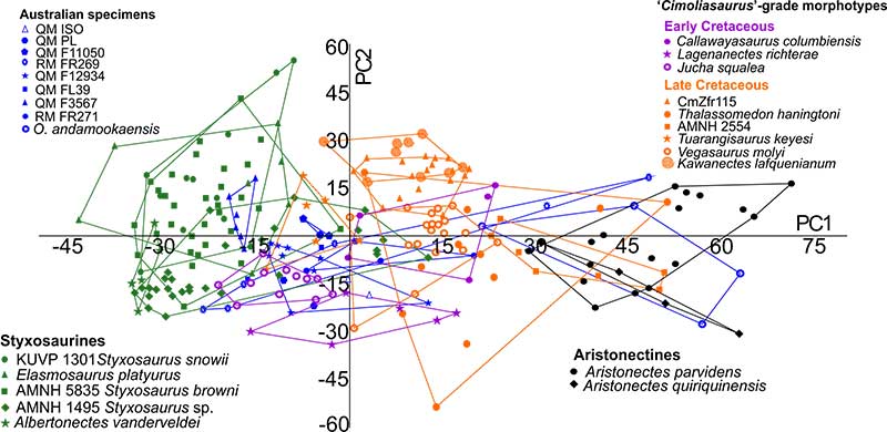
FIGURE 16. Taxon/specimen average plot for anterior cervicals of Australian plesiosauromorph specimens and non-Australian elasmosaurids. Data for QM F3567 and RM FR271 from Sachs (2004); Opallionectes andamookaensis from Kear (2005a); Elamosaurus platyurus, Thalassomedon haningtoni, Callawayasaurus colombiensis and Cm Zfr 115 from O’Keefe and Hiller (2006); Aristonectes quiriquinensis from Otero et al. (2014); Albertonectes vanderveldei from Kubo et al. (2012); Vegasaurus molyi from O’Gorman el. (2015); Tuarangisaurus keyesi from Hiller et al. (2017); AMNH FARB 1495, AMNH FARB 5835, Styxosaurus snowii, and AMNH FARB 2554 from Otero (2016); Aristonectes parvidens from O’Gorman (2016a); Kawanectes lafquenianus from O’Gorman (2016b); Lagenanectes richterae from Sachs et al. (2017) and Jucha squalea from Fischer et al. (2020).