FIGURE 1. Locations of Sloveniolimulus rudkini specimens and the geological context of the Strelovec Formation. A: Map of Europe. Box shows close up in B. B: Map of Slovenia. Box shows close up in C. C: Close up of specimen localities. Red star indicates holotype locality. Blue star indicates location of new material from the Kalška gora Mountain. D: Stratigraphic position of Strelovec Formation.
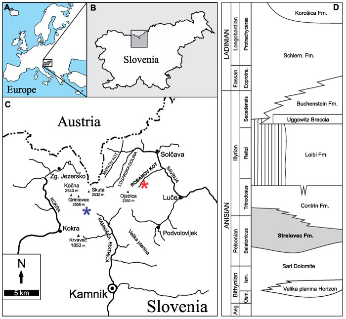
FIGURE 2. Holotype of Sloveniolimulus rudkini. A: PMSL T-993. B: Interpretative drawing of PMSL T-993, colour coded for main body sections. Specimen from the Section S1 of the Slatinski Plaz, Robanov Kot Valley. A coated in ammonium chloride sublimate and images converted to greyscale. Abbreviations: Car: cardiac lobe; Fla: thoracetronic flange; In: genal spine indentation; Med: medial thoracetronic lobe; Oph: ophthalmic ridge; PDub: prosomal doublure; Pro: prosoma; TDub: thoracetronic doublure; Thr: thoracetron; Tel: telson; Tk: telson keel. Image credit: Russell Bicknell.
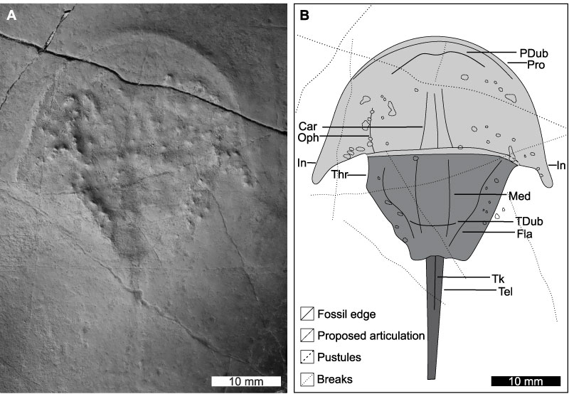
FIGURE 3. New material referred to Sloveniolimulus rudkini. A, B, E: PMSL T-2071. A: Specimen showing prosomal doublure and possible occipital lobe on left genal spine. B: Interpretative drawing of PMSL T-2071. E: Close up of phosphatized spheres in A. C, D: PMSL T-2072. C: Specimen showing injured right genal spine and hypertrophied left genal spine. D: Interpretative drawing of PMSL T-2072. Specimens from the Western slopes of the Kalška gora Mountain, within the Kamniška Bistrica Valley. B and D colour coded for main body sections. A and C coated in ammonium chloride sublimate. All images converted to greyscale. Abbreviations: Car: cardiac lobe; In: genal spine indentation; Oc: possible occipital lobe; Pro: prosoma; Thr: thoracetron. Image credit: A, C: Russell Bicknell; E: Tomaž Hitij.
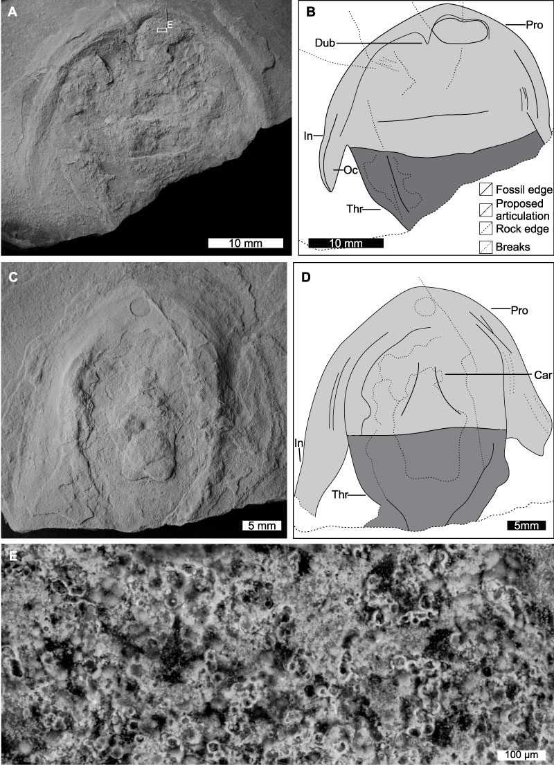
FIGURE 4. Additional xiphosurids with genal spine indentations. Arrows indicate the structure on all taxa. A: Franconiolimulus pochankei from the Bayreuth Formation, (Early Jurassic, Hettangian) Pechgraben, Germany. SSN 8PG35, holotype. B, C: Panduralimulus babcocki from the Maybelle Limestone, Lueders Formation (Permian, Cisuralian, Kungurian), Texas, USA. B: USNM 520723, holotype. C: USNM 520724, paratype. D: Tasmaniolimulus patersoni from the Jackey Shale (Permian, Lopingian), Tasmania, Australia. UTGD 123979, holotype. E: Dubbolimulus peetae from the Ballimore Formation (Middle Triassic, Ladinian), New South Wales, Australia. MMF 27693, holotype. D coated in ammonium chloride sublimate. D and E converted to greyscale. Image credit: A: Andreas Hecker; B-D: Russell Bicknell; E: David Barnes.
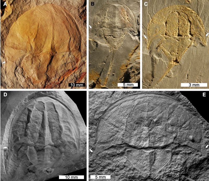
FIGURE 5. Molluscs and brachiopods from the Strelovec Formation. A: An ammonoid. PMSL T-1805. B: Lingulid brachiopod. PMSL T-865. C: Modiolus sp. PMSL T-836. D: Worthenia sp. PMSL T-1772. A coated in ammonium chloride sublimate. All converted to greyscale. Image credit: A: Tomaž Hitij; B, C, D: Jure Žalohar.
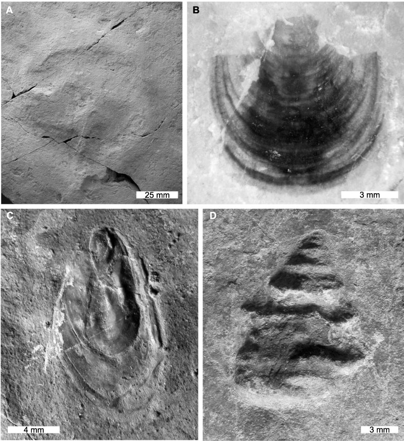
FIGURE 6. Non-xiphosurid euarthropods from the Strelovec Formation. A, E: Aeger sp. A: PMSL T-1270. E: PMSL T-1238 showing exceptional appendage detail. B: An as-of-yet undescribed thylacocephalan. PMSL T-1742. C, D: Exceptionally well-preserved indeterminate decapod crustaceans. C: PMSL T-1262 showing appendage segmentation and ocular structures. D: PMSL T-1263. F: Schimperella sp. PMSL T-1236. Image credit: Jure Žalohar.
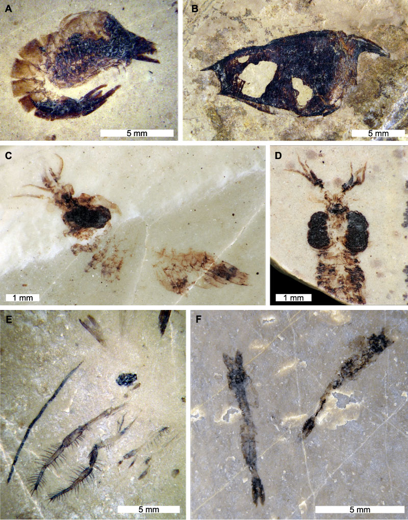
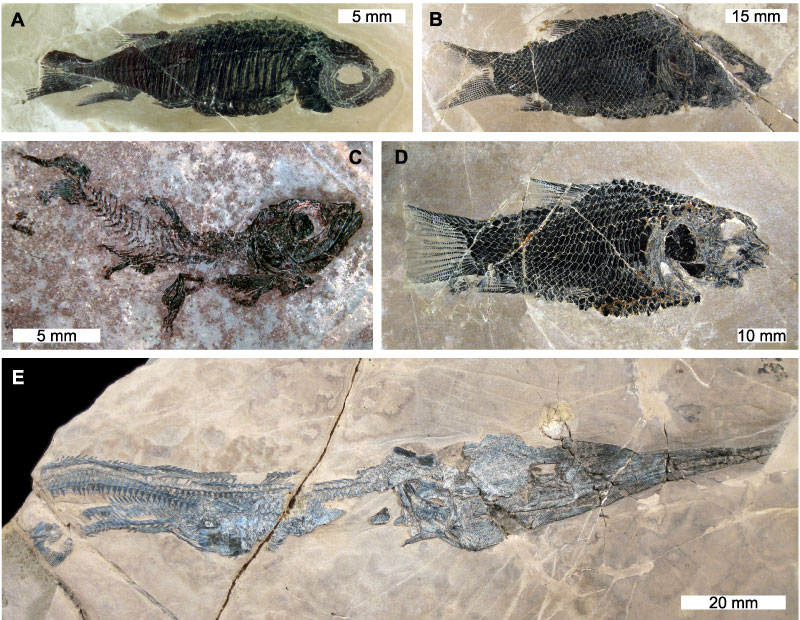
FIGURE 7. Marine vertebrates from the Strelovec Formation. A: Placopleurus sp. PMSL T-899. B: Sangiorgioichthys sp. PMSL T-900. C: Marcopoloichthys sp. preserving ocular structures. PMSL T-1745. D: Eosemionotus sp. PMSL T-1262. E Saurichthys sp. with stomach contents containing Eosemionotus sp. A and E reflected to align with other images. Image credit: Jure Žalohar.