FIGURE 1. Schematic drawing of a pygocephalomorphan shield in dorsal aspect, showing the measurements taken for the morphometric analysis. 1: Length of rostrum, 2: Length of shield, 3: Width of anterior (outer) margin, 4: Medial width, 5: Width of posterior margin, 6: Length of antero-lateral spine.
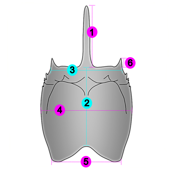
FIGURE 2. Anthracaris gracilis (Meek and Worthen, 1865) from the Carboniferous of Germany. Digital microscopy image of part (A-C) and counterpart (D-F) (specimen 1.529), in ventral view. (A, D) Ring light. (B, D) non-polarized co-axial light. (C, F) polarized co-axial light.
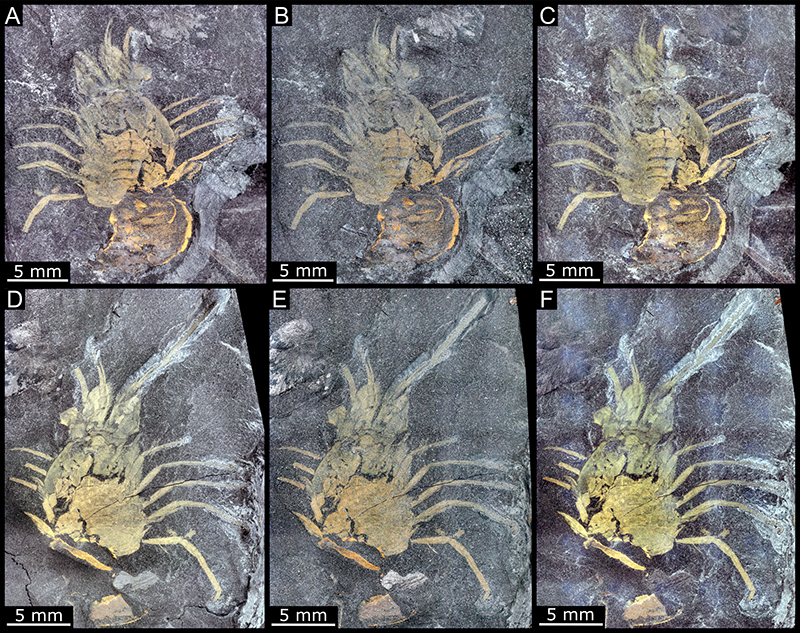
FIGURE 3. Anthracaris gracilis (Meek and Worthen, 1865) from the Carboniferous of Germany. Digital microscopy images of shields. (A-D) Dorsal view of specimen 1.228. (A, B) Part. (C, D) Counterpart. (E, F) Dorsal view of specimen Pal590-2. (A, C, E) Non-polarized co-axial light. (B, D, F) Polarized co-axial light. Images of the counterparts are mirrored. Abbreviations: als = antero-lateral spine; ls = lateral spine; r = rostrum.

FIGURE 4. Anthracaris gracilis (Meek and Worthen, 1865) from the Carboniferous of Germany. Digital microscopy images of shields. (A-G) Specimen 1.587 in dorsal view. (A-C) Part. (D, E) Counterpart. (A, D) Non-polarized co-axial light. (B, E) Polarized co-axial light. (C) Ring light. (F) Detail of B, antero-lateral shield corner. (G) Detail of B, rostrum. (H) Detail of Pal590-2, antero-lateral shield corner, mirrored. Images of the counterpart are mirrored. Abbreviations: als = antero-lateral spine; g = groove; ls = lateral spine; p = punctuations; r = rostrum.
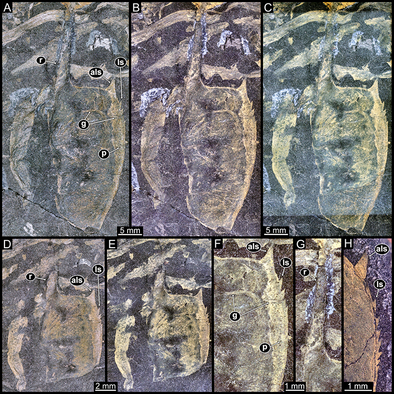
FIGURE 5. Anthracaris gracilis (Meek and Worthen, 1865) from the Carboniferous of Germany. (A) Virtually overlaid part and counterpart of specimen 1.529. (B) Same as A, but structures colour marked. Abbreviations: ab = antenna basipod; ant = antenna; atl = antennula; ec = eye cornea; es = eye stalk; lb = labrum; md = mandible; ml = maxillula; mx = maxilla; pl = pleon; sc = scaphocerite; tp1-8 = thoracopod 1-8; ts1-8 = thoracic segment 1-8.
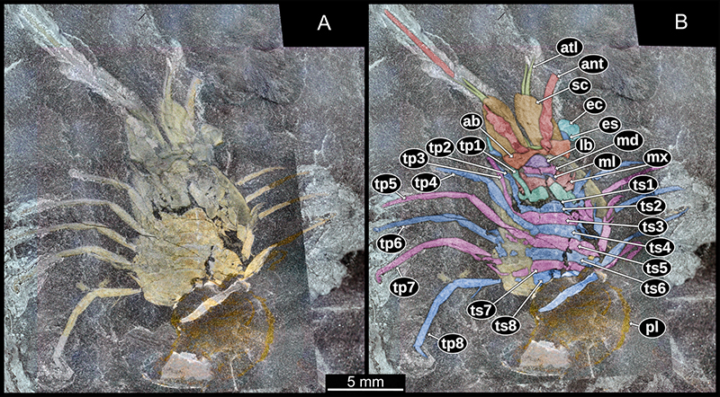
FIGURE 6. Anthracaris gracilis (Meek and Worthen, 1865) from the Carboniferous of Germany. (A-E) Digital microscopy image of isolated pleon in dorsal view of specimen 1.756. (A, B) Part. (C) Same as B, but structures colour marked. (D, E) Counterpart. (A, D) Non-polarized co-axial light. (B, E) Polarized co-axial light. Images of the counterpart are mirrored. (F-H) Details of specimen 1.529. (F) Detail of exopods of thoracic appendage. (G) Same as F, but colour marked. (H) Detail of the distal portion of an endopod of thoracic appendage, colour marked. Abbreviations: b = basipod; c = carpus; cr = caudal ramus; d = dactylus; en = endopod; ex = exopod; pp = pleopod; ps1-6 = pleon segment 1-6; pr = propodus; s = setae; t = telson; ts = telson spine.
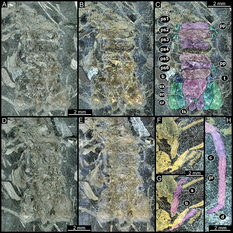
FIGURE 7. Plot of the eumalacostracan shields of Anthracaris gracilis (Meek and Worthen, 1865) from the Carboniferous of Germany (1.587A, Pal.590-2, 1.228A) and of A. gracilis from Mazon Creek, USA. Values for A. gracilis from Brooks (1962).
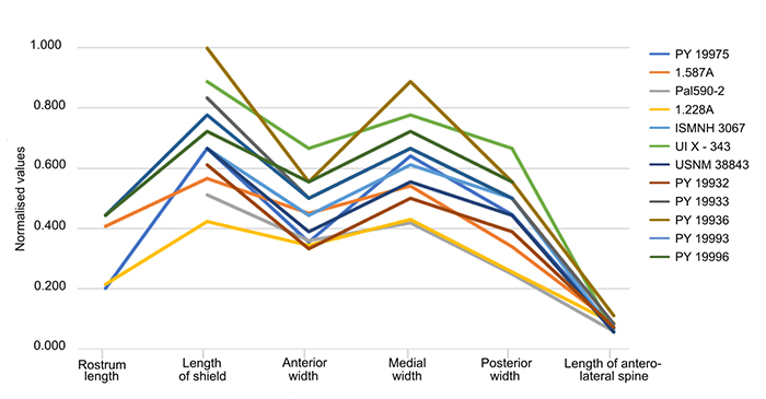
FIGURE 8. Restoration drawings of shields of pygocephalmorphans. (A) Specimen 1.587, (B) 1.228 and (C) Pal.590-2 of Anthracaris gracilis (Meek and Worthen, 1865) from the Carboniferous of Germany. (D) Anthracaris gracilis, based on Brooks (1962, pl. 32, fig. 2). (E) Pygocephalus cooperi Huxley, 1857, based on Schram (1979, fig. 39a). (F) Pygocephalus dubius (Prestwich, 1840), based on Schram (1979, fig. 39c).
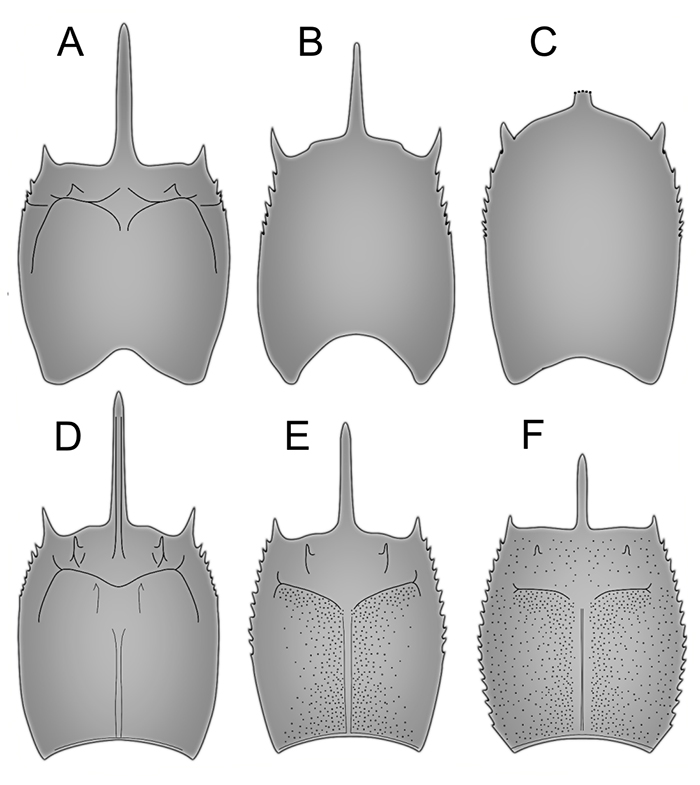
FIGURE 9. Restoration drawings of Anthracaris gracilis (Meek and Worthen, 1865) from the Carboniferous of Germany with the three different shields recovered. (A, C, E) Dorsal aspect. (B, D, F) Ventral aspect. (A, B) Specimen 1.587. (C, D) Specimen Pal.590-2, the rostrum is broken off. (E, F) Specimen 1.228.
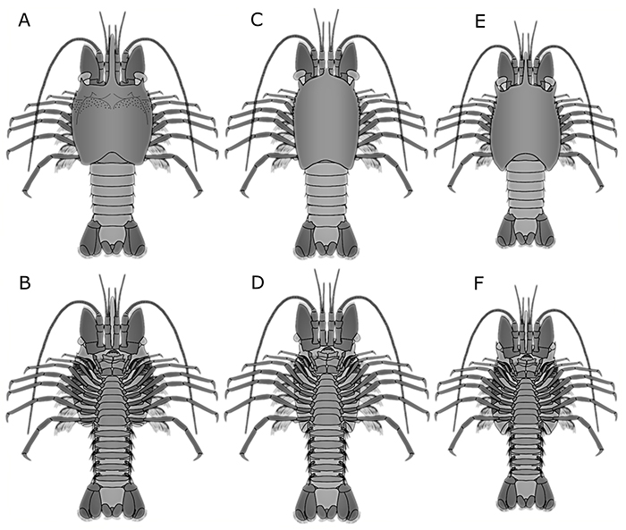
FIGURE 10. Restoration drawings of the pygocephalmorphan Anthracaris gracilis (Meek and Worthen, 1865) and Pygocephalus cooperi Huxley, 1857. (A-C) A. gracilis. (A) Dorsal aspect. (B) Ventral aspect of putative female, note the pouch on the last thorax segment. (C) Ventral aspect of putative male. (D-F) P. cooperi. (D) Dorsal aspect. (E) Ventral aspect of female with thorax sternites hidden by oostegites. (F) Putative male in ventral aspect.
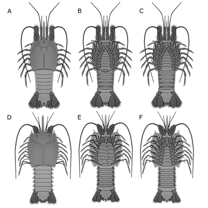
FIGURE 11. Frequency plot of the distribution of ingroups of Eumalacostraca and of its sister-group Phyllocarida, during the Carboniferous (A) of North America, the UK and continental Europe, the Mississippian (B) and the Pennsylvanian (C).
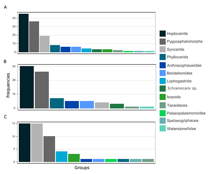
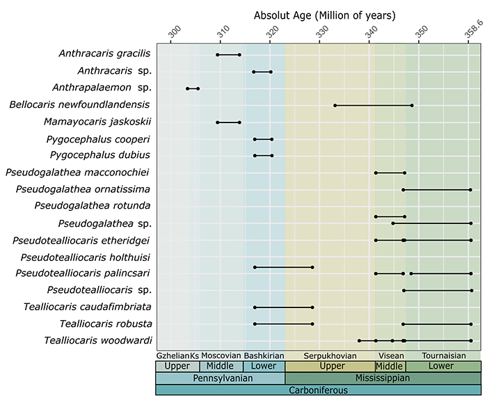
FIGURE 12. Time span of the species of Pygocephalomorpha during the Carboniferous of North America, the UK and continental Europe. Abbreviation: Ks, Kasimovian.