FIGURE 1. Covidifructus multicarpellatus gen. et sp. nov.; specimen No. NMP F3200; scale bars equal 100 µm in all figures. (A) Small premature capsular fruit in lateral view, semi-globose in overall shape; SEM. (B) Fruit seen in apical view; note preformed dorsal lines of fruit dehiscence; SEM. (C) Close-up of fruit apex showing styles and stigmatic areas (asterisks); note irregular closure of ovary in the very centre; SEM. (D) MicroCT volume rendering, lateral view, showing 10 elongate seeds (green), one seed per carpel. (E) MicroCT volume rendering, apical view, showing regular arrangement of carpels and seeds. (F) Detail of central ovary closure (dashed line); note that some of the carpel flanks (arrowheads) do not extend to the very centre of the closure zone; SEM.
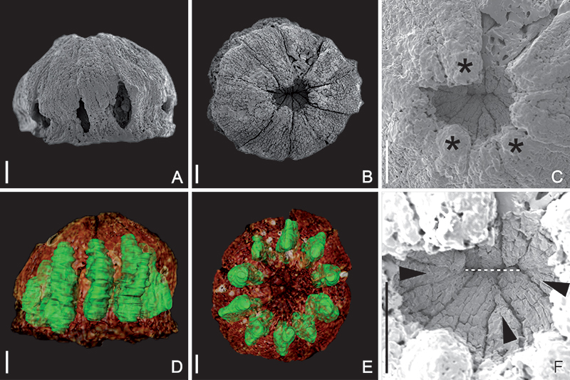
FIGURE 2. Covidifructus multicarpellatus gen. et sp. nov. specimen No. NMP F3200, scale bars equal 300 µm in all figures, series of microCT sections of premature capsular fruit. (A) Volume rendering of fruit in lateral view; lines B-F indicate approximate levels of transverse sections shown in the following images. (B) Transverse section at the level of styles and stigmas. (C) Transverse section at the level of the symplicate zone of the gynoecium where the carpels are postgenitally united in the centre of the ovary; distalmost parts of locules and seeds are visible. (D) Transverse section at the level of the empty space (asterisk) where carpels do not meet in the centre of the ovary. (E) Transverse section at the level of the synascidiate zone of the gynoecium, i.e., below the enclosed floral apex and the empty space. (F) Transverse section through the very base of the fruit showing the basal-most parts of the locules. (G) Longitudinal median section showing empty space in the centre of the ovary (asterisk) and axile ovule/seed attachment (arrow) in the distalmost part of the ovary. (H) Longitudinal tangential section with one seed rendered and coloured in green. (I) Transverse section at the level of seed attachment in the distal part of the ovary, with one seed rendered and coloured in green.
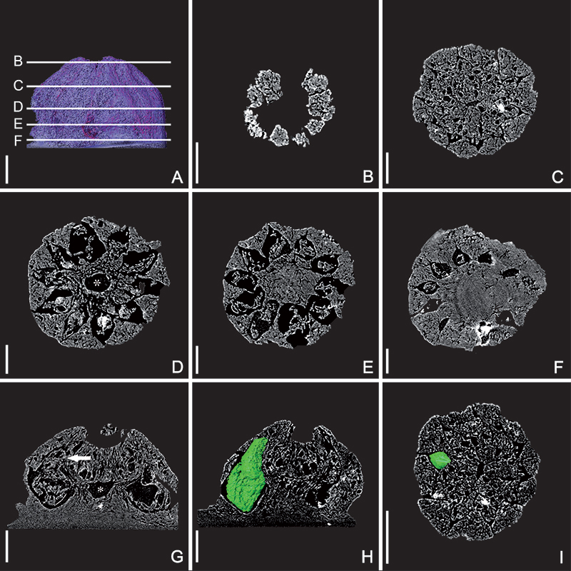
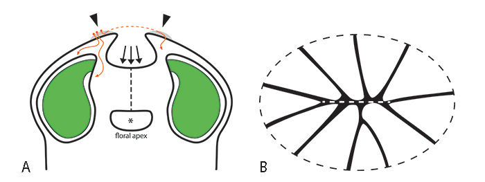
FIGURE 3. Schematic line drawings of the gynoecium of Covidifructus multicarpellatus. (A) Longitudinal median section through the gynoecium showing its complex internal morphology with a remaining floral apex and an empty space (asterisk) in the centre of the ovary; arrowheads indicate stigma positions; grey shaded areas indicate potential stigmatic secretion forming an extra-gynoecial compitum across neighbouring stigmas; pollen grains and hypothetical pathways of pollen tubes are given in orange; dashed orange line indicates hypothetical pathway of pollen tube reaching a stigma via growth through the extra-gynoecial compitum; dashed black line indicates area of postgenital carpel union in the centre of the ovary (symplicate region); arrows indicate area of irregular ovary closure shown in (B); placentation is axile with the seeds (green) attached in the distalmost part of the ovary. (B) Line drawing showing zone ovary closure (see also Figure 1C, F) as seen from above, radial lines correspond to ventral slits of individual carpels; carpel flanks meet in an irregular pattern in the centre of the gynoecium; the area of closure is flattened (compressed; indicated by dashed ellipse), and the 10 carpels are roughly arranged in a double row facing each other (dashed line in centre of figure) rather than in a smooth circle.