FIGURE 1. Fossil larva of Coniopterygidae from Baltic amber, CCHH 540-1, specimen 6301. A. Ventral view. B. Dorsal view. C. Colour-marked version of B. D. Close-up of head in dorsal view. E. Close-up of head in ventral view. F. Close-up on right hind leg; arrows mark claws. G. Close-up of hind leg; arrows mark claws. Abbreviations: a1-a8 = abdomen segment 1-8; at = antenna; cx = coxa; e1-3 = element 1-3; fe = femur; hc = head capsule; lp = labial palp; ms = mesothorax; mt = metathorax; pt = prothorax; st = stemmata; ta = tarsus; te = trunk end; ti = tibia; tr = trochanter.
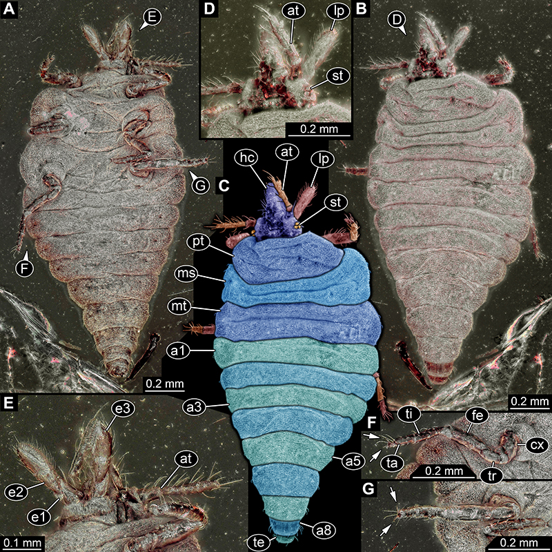
FIGURE 2. Fossil larva of Sisyridae from Baltic amber, CCGG 1383, specimen 6501. A. Dorsal view. B. Ventral view. C. Colour-marked version of B. D. Close-up of head in dorsal view. E. Colour-marked version of D; arrows mark stemmata. F. Close-up on gills. G. Close-up on processes on trunk segments. Abbreviations: a3-a7 = abdomen segment 3-7; at = antenna; gi = gills; hc = head capsule; ms = mesothorax; mt = metathorax; pt = prothorax; sy = stylet; te = trunk end.
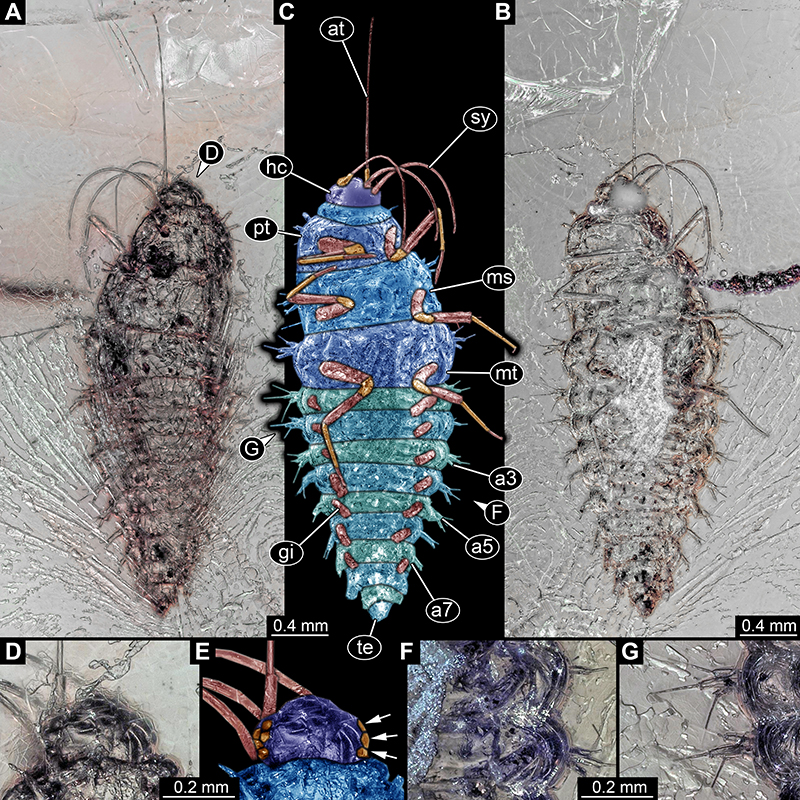
FIGURE 3. Fossil larva of Sisyridae from Baltic amber, CCGG 7122, specimen 6502. A. Dorsal view. B. Ventral view. C. Colour-marked version of B. D. Close-up of head in dorsal view. E. Colour-marked version of D. F. Close-up on gills. G. Close-up on trunk end. Abbreviations: at = antenna; hc = head capsule; ms = mesothorax; mt = metathorax; pt = prothorax; sy = stylet.
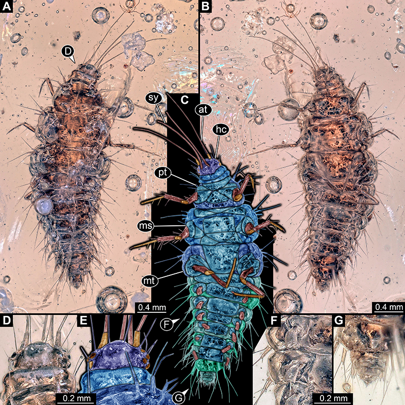
FIGURE 4. Fossil larva, “beak larva” type 2, from Myanmar amber, PED 0596, specimen 6803. A. Dorsal view; posterior part not well accessible. B. Colour-marked version of A. Abbreviations: 1t = trunk appendage 1; at = antenna; hc = head capsule; ms = mesothorax; pl = palp; pt = prothorax.
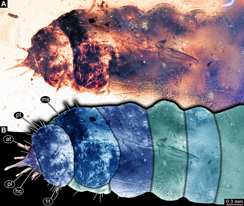
FIGURE 5. Fossil larva, “beak larva” type 2, from Myanmar amber, PED 0596, specimen 6803, continued. A. Ventral view; details not well accessible; posterior end not inside the amber. B. Close-up on head in dorsal view. C. Close up on trunk appendage 1 in dorsal view. D. Same as C; estimated outline of appendage, visible through tergite outlined in red. Abbreviations: at = antenna; “b” = beak; pl = palp.
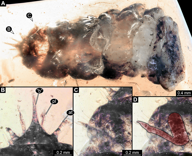
FIGURE 6. Additional specimens of larvae of Sisyridae preserved in Baltic amber. Images kindly provided by Jonas Damzen (JD) and Marius Veta (RMV). A. JD 6345, specimen 6503, in ventral view, 3.5 mm long. B, C. RMV 2380, specimen 6504, 3 mm long. B. Dorsal view. C. Ventral view. D, E. JD 4198, specimen 6505, 4 mm long. D. Dorsal view. E. Ventral view. F, G. RMV 2793, specimen 6506, 2 mm long. F. Left lateral view. G. Right lateral view. H, I. RMV 2794, specimen 6507, 3.2 mm long. H. Lateral view. I. Dorsal view.
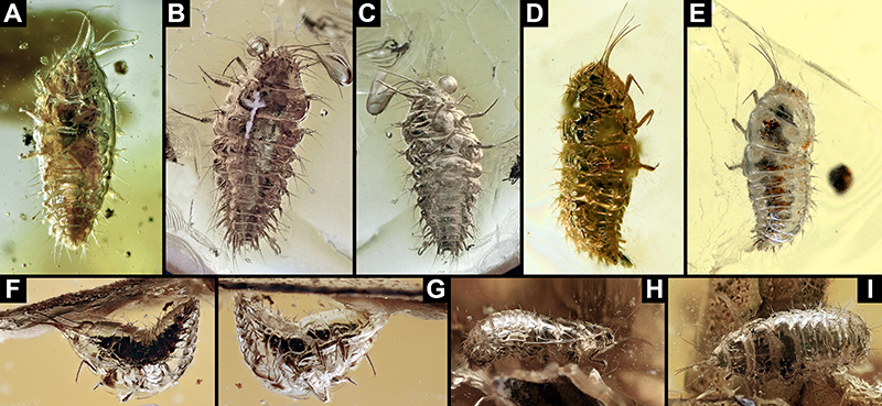
FIGURE 7. Scatterplot of PC2 vs. PC1. Note how the two beak larvae (specimens 6802 + 6803) plot together with larvae of Coniopterygidae.
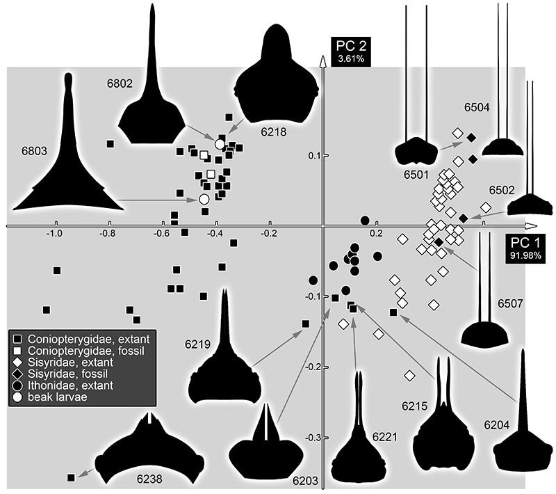
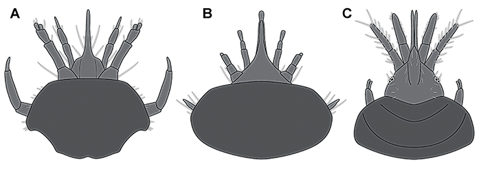
FIGURE 8. Comparison of the two beak larvae to a larva of Coniopterygidae. A. Larva (specimen 6802) from Haug et al. (2020b). B. New beak larva (specimen 6803, based on Figure 5B). C. Larva of Aleuropteryx loewi (based on several figures from Rousset, 1966).