FIGURE 1. A. Depositional area of the Upper Jurassic Morrison Formation, and location of the Nail Quarry in southeastern Wyoming, and B. the stratigraphic location of Nail Quarry in the Morrison Formation.
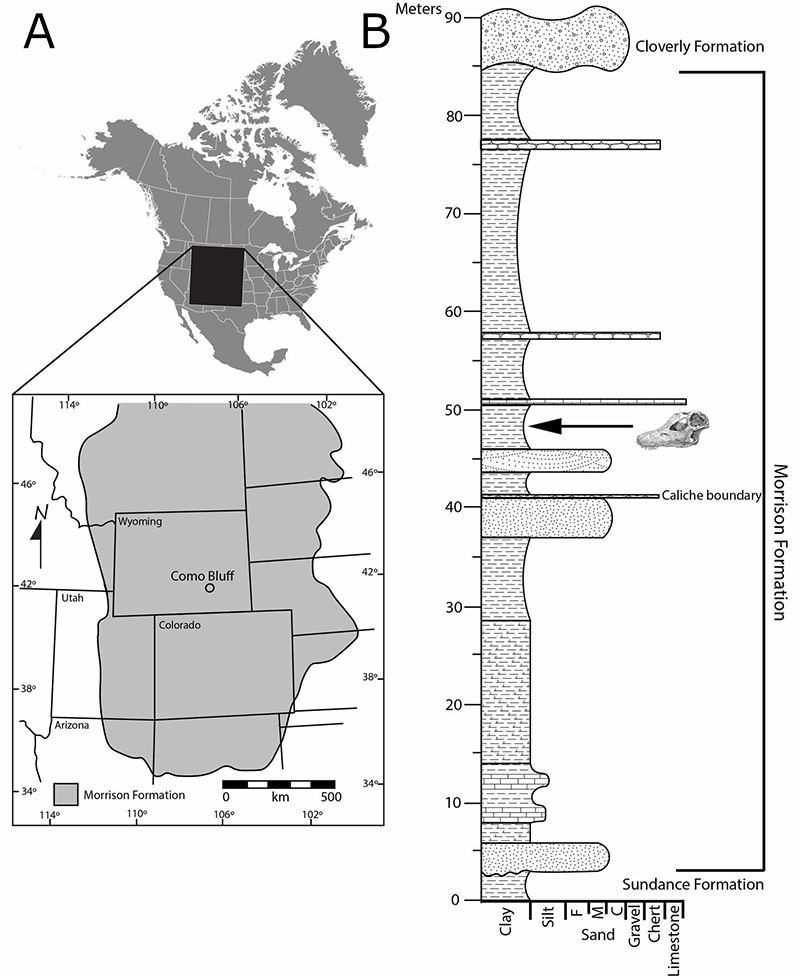
FIGURE 2. Left (A, B) and right (C, D) premaxillae in dorsal ventral views, respectively. Scale bar equals 10 cm. Artwork by Ryan Steiskal.
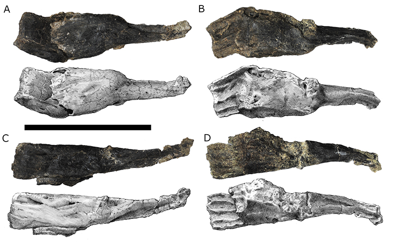
FIGURE 3. Right maxilla in (A) dorsal and (B) ventral views with the left ectopterygoid (i) and left palatine (ii) adhered to the ventral surface. Scale bar equals 10 cm. Artwork by Ryan Steiskal. Abb: aof, antorbital fenestra; paof, preantorbital fenestra.
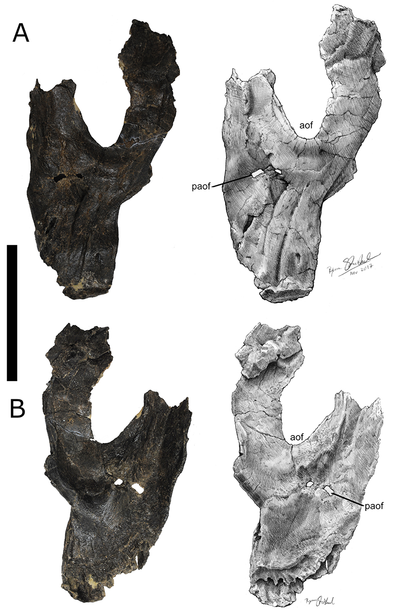
FIGURE 4. Left maxilla in (A) dorsal and (B) ventral views. Scale bar = 10cm. Artwork by Ryan Steiskal. Abb: aof, antorbital fenestra; paof, preantorbital fenestra.
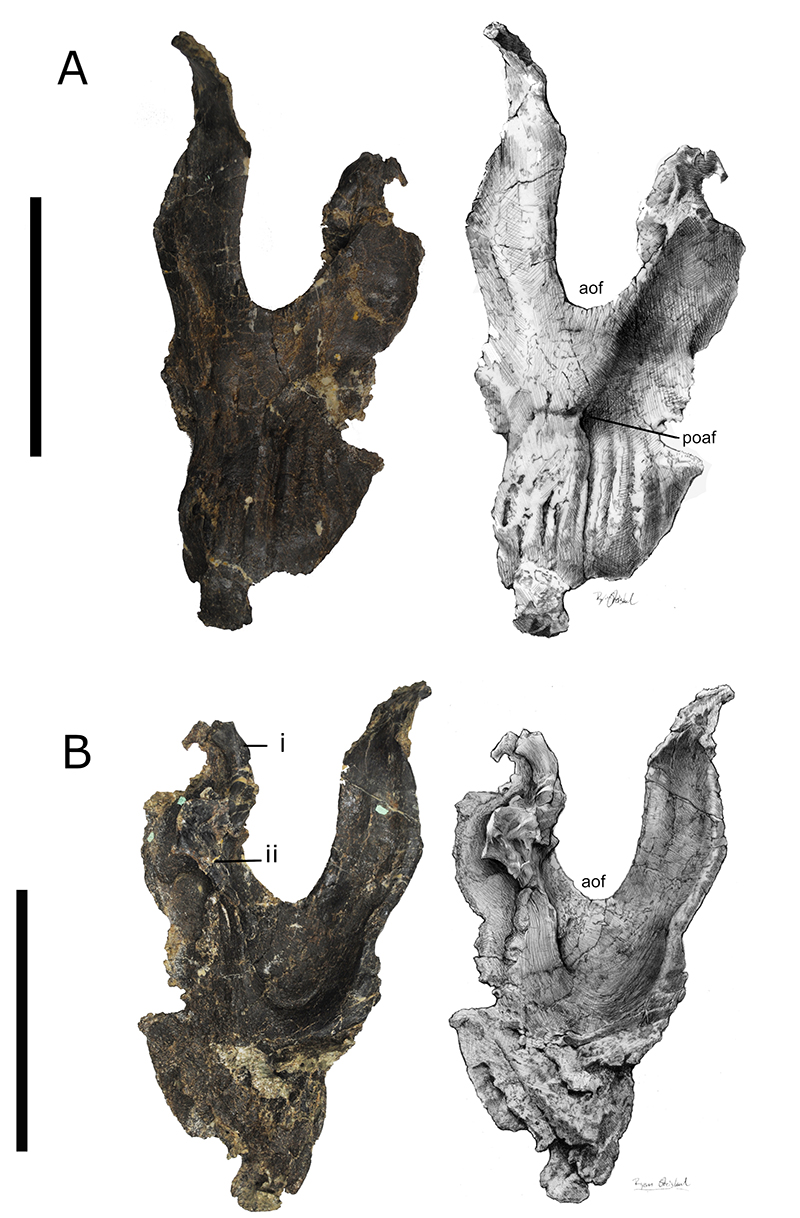
FIGURE 5. Left pterygoid (i, iv), quadrate (ii, v), and quadratojugal (iii, vi) in A) dorsal and B) ventral views. Scale bar equals 10 cm. Artwork by Ryan Steiskal. Abb: pt, pterygoid; q, quadrate; qj, quadratojugal.
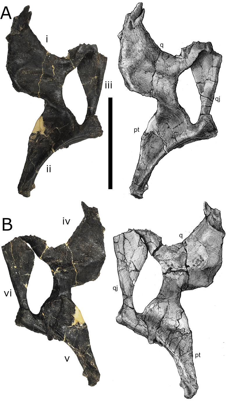
FIGURE 6. Reconstruction of the skull of TATE-099 in A) left lateral view, B) right lateral view, C) dorsal view, and D) ventral view. Scale bar equals 10 cm. Artwork by Ryan Steiskal.
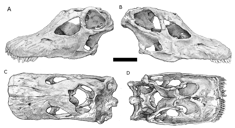
FIGURE 7. Left (A) and right (B) ectopterygoid right and left lateral views (C, D). Scale bar equals 10 cm. Artwork by Ryan Steiskal.
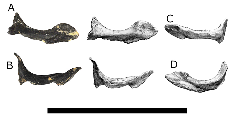
FIGURE 8. Reconstructed cast and 3D model of the braincase of TATE-099 in dorsal view (A, B), ventral view (C, D), posterior view (E, F), right lateral view (G, H), and left lateral view (I, J). Scale bar equals 10 cm. Abb: bo, basoccipital; bpr, basipterygoid process; bs, basisphenoid; bt, basal tuber; cpr, crista prootica; eo, exoccipital-opithsotic; f, frontal; fm, foramen magnum; p, parietal; pas, parasphenoid; pft, posttemporal fenestra; po, postorbital; popr, paroccipital process; so, supraoccipital; snc, sagittal nuchal crest; stf, supratemporal fenestra.
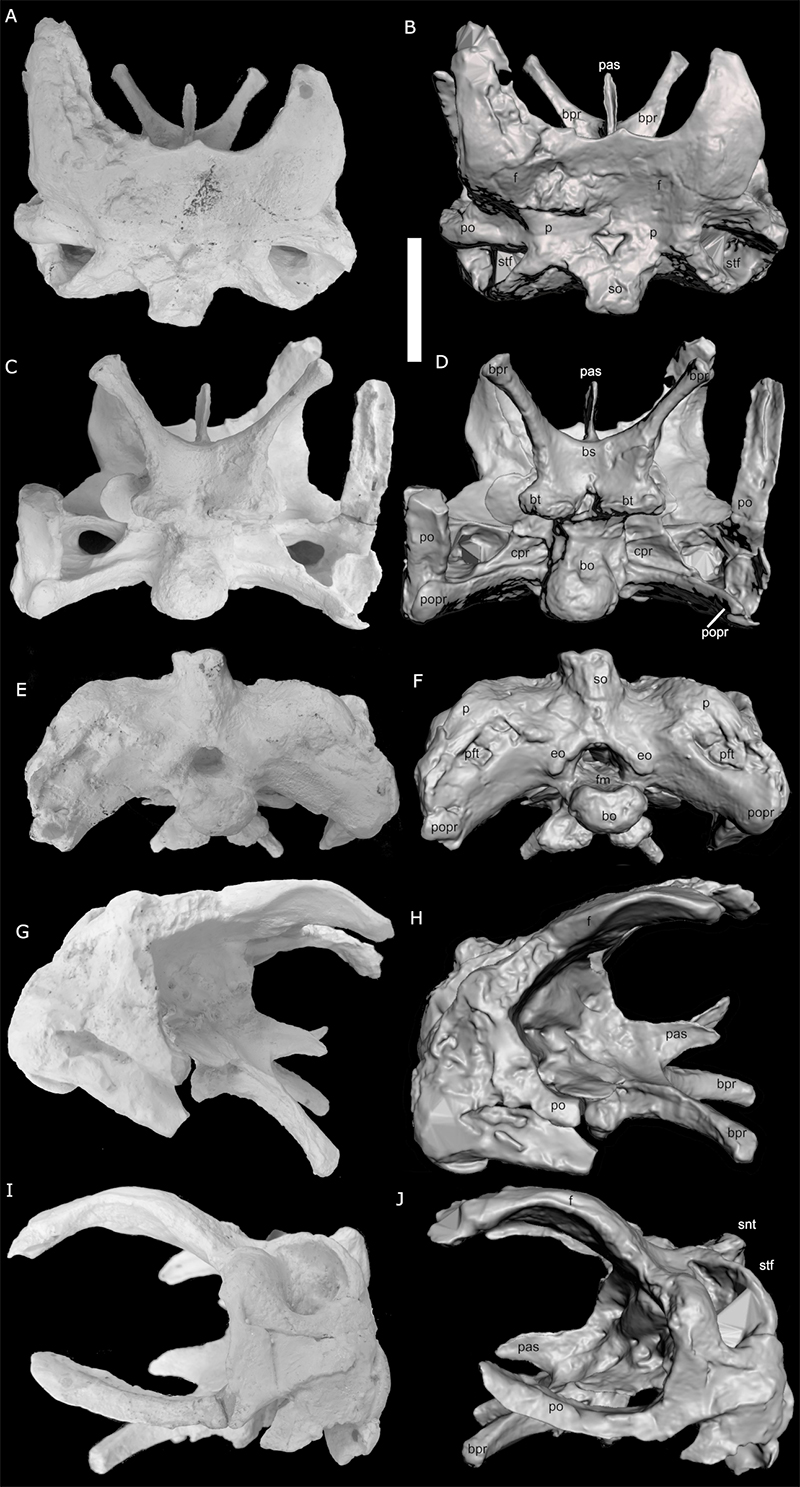
FIGURE 9. Right dentarty and surangular in A) left lateral view, and B) right lateral view. Scale bar equals 10 cm. Artwork by Ryan Steiskal.
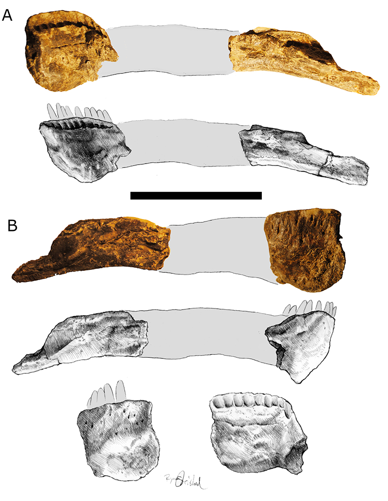
FIGURE 10. Computed tomography of the right and left maxillae (A-B, respectively) are displayed in coronal (upper) and sagittal (lower) cross sections; the left premaxilla (C) is shown in sagittal (left) and coronal (right) cross sections. The unerupted teeth (ii=first unerupted tooth) of the maxilla (D; left) and premaxilla (D; right) were segmented for 3D rapid prototyping. Note: proceeding caudally, the number of unerupted teeth in the maxilla declines from 5 (ii-vi) to 2 (ii-iii).
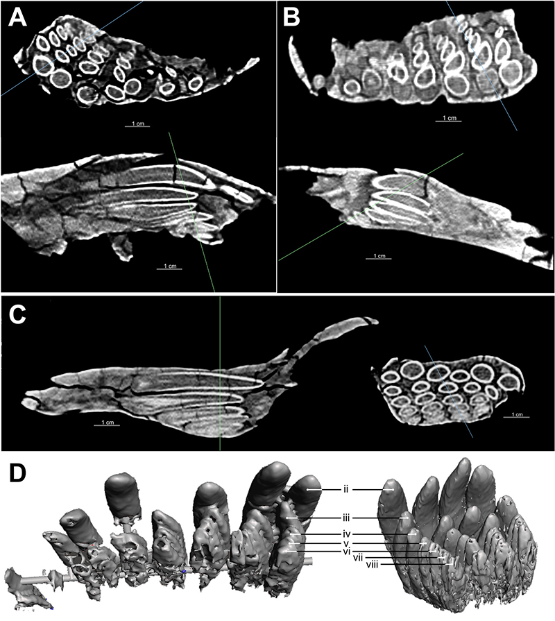
FIGURE 11. Computed tomography of the right dentary is displayed in coronal (left) and sagittal (right cross section, illustrating the presence of 1-2 unerupted teeth per alveolar position. The unerupted teeth were segmented for 3D rapid prototyping (lower left).
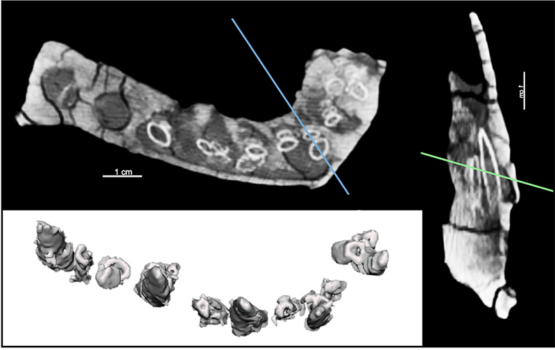
FIGURE 12. Strict consensus of 47 MPTs demonstrating the relative position of TATE-099 using the matrix of Tschopp and others (2015).
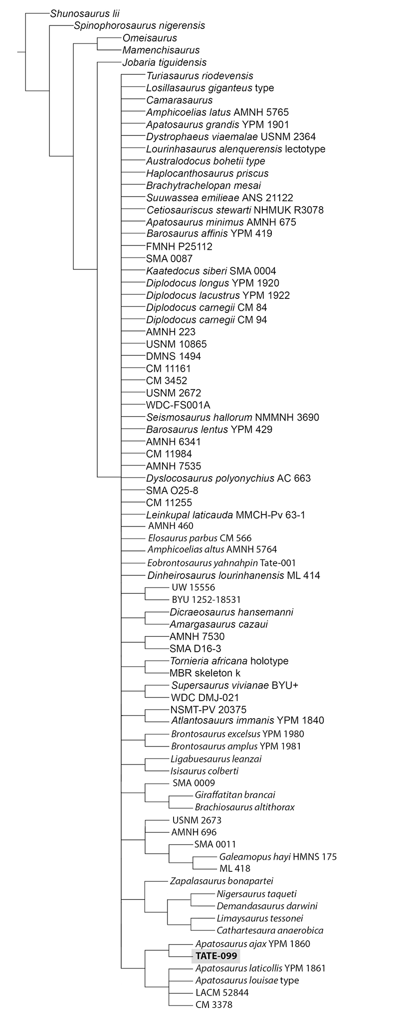
FIGURE 13. Strict consensus of 23 MPTs demonstrating the relative position of TATE-099 using the matrix of Whitlock and Wilson (2020).
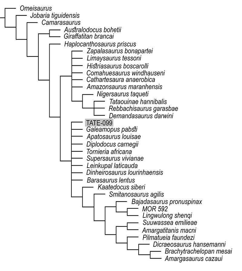
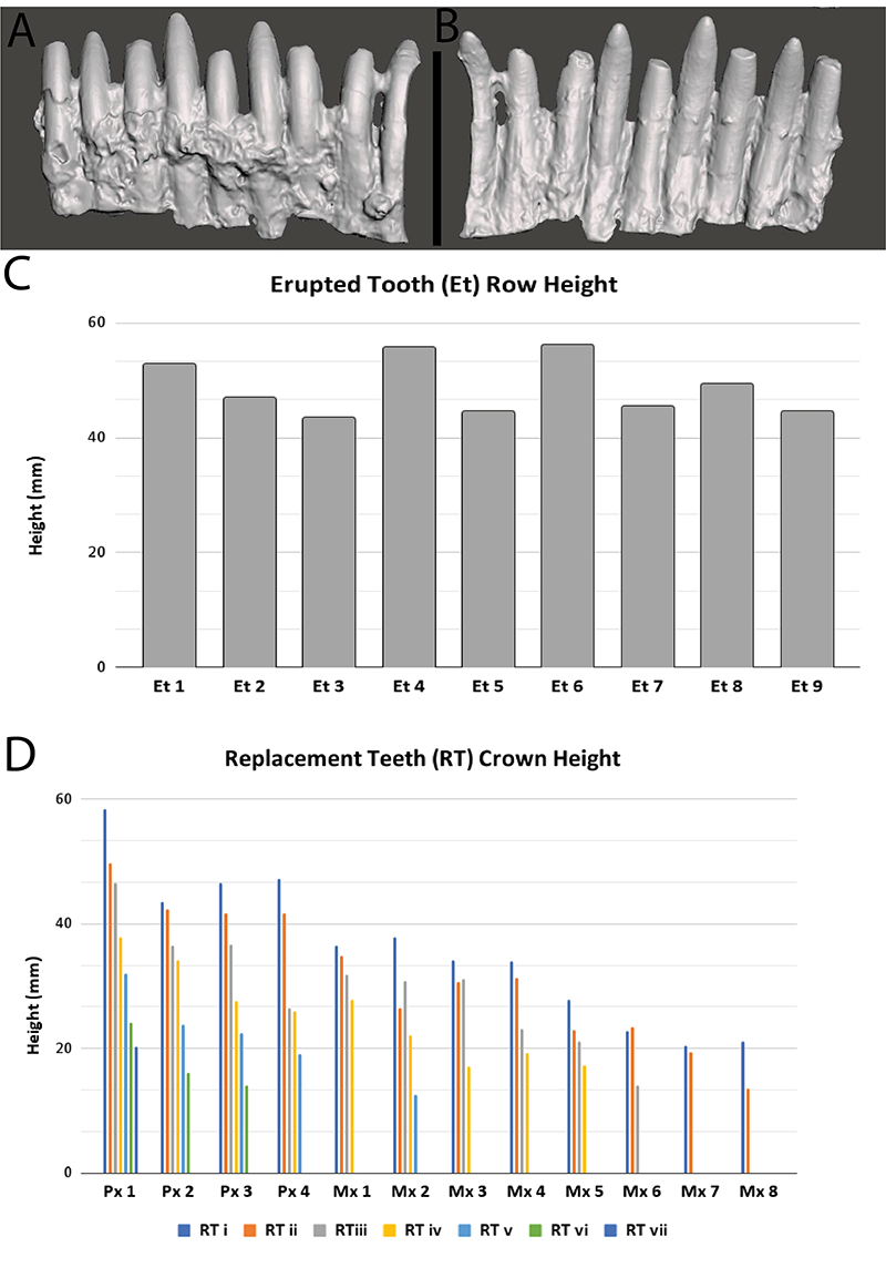
FIGURE 14. 3D model of a cast of the erupted tooth row of TATE-099 in (A) labial and (B) lingual views with crown heights (C) and comparative measurements of the unerupted replacement teeth (D). Scale bar equals 5 cm.