FIGURE 1. Area of study. A - Highlights the position of Wenlock Edge within the English Midlands and Welsh Marches. B - Shows the Off-Reef tract where thin samples studied in this paper originate from. C - Inset in A, highlights the exposures of the Much Wenlock Limestone Formation in the English Midlands and Welsh Marches.
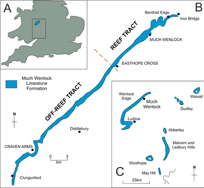
FIGURE 2. The original measurements taken by Munnecke et al. (2000) from each individual specimen of Ovummuridae based on Minourella gotlandica (left) and modified measurements for Ovummurus duoportius (right). The measurements recorded are: overall height (H) and width (w) of the specimen, height (H1 and H2) and width (w1 and w2) of both internal chambers, the thickness of the wall in three loci: overall thickness of the wall (th1), thickness of the septum separating the two chambers (th3) and the thickness between the wall and the chamber (th2) and finally the size of the aperture (ap). (After Munnecke et al., 2000).
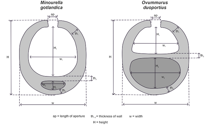
FIGURE 3. Photomicrographs of Ovummurus duoportius within the MWL Fm. showing this species typical morphology and characteristic. These specimens are from: Longville Stanway (A and B) and Moorwood Quarry (C and D). (A) = Ovummurus duoportius exhibiting two chambers (C1 and C2) separated by a septal structure (ss) with an apical aperture (ap) present in C2. All specimens here are found within a sparitic (sp) grainstone matrix. (B) = O. duoportius with thickened. (C) = Longitudinal cross section showing the equal-sized chambers present within O. duoportius. (D) = O. duoportius specimen with pyrite growth within C2. All scale bars equal 50 µm.
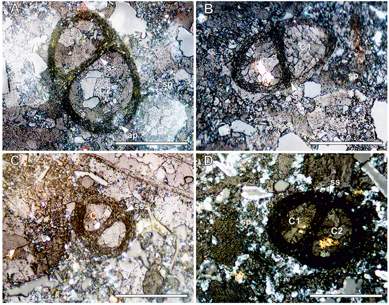
FIGURE 4. Photomicrographs of Minourella gotlandica (A-D), Minourella wenlockensis (E-G) and Minorella cameroni (H) within the MWL Fm., these specimens are from Eaton Top (A, B and C), Longville Stanway Track (D and F), Moorwood Quarry (E and G) and Harton Hollow (H). (A) = M. gotlandica (mg) specimen near a bryozoan (bry) within a spiritic matrix (sp) highlighting the similarities in wall structure, (B) = M. gotlandica with a larger globular dorsal chamber (DC) separated from the smaller globular ventral chamber (VC) by a septum like structure (ss). Sparitic (sp) matrix. Specimen has a pyritic wall. (C) = M. gotlandica with an aperture (ap) within the VC. (D) = Thickened walls of the dorsal chamber with an apical aperture (ap). (E) = Sectional cut through M. wenlockensis highlighting the conical ventral chamber (VC) in contrast to the globular dorsal chamber (DC) separated by septum like structure (ss) within a sparitic (sp). (F) = Apertures present in both DC and VC. Note pyritic growth within the specimen walls. (G) = M. wenlockensis with an apical aperture (ap) within the DC. Matrix comprised of sparite. (H) = M. cameroni Longitudinal transverse sectional cut through M. cameroni exhibiting the conical nature of the test. Lateral dorsal chamber (LDC) is separated from the ventral chamber (VC) by a septum like structure (ss). Sparitic infill of the LDC and VC from the surrounding sparitic (sp) matrix. All scale bars equal 50 µm.
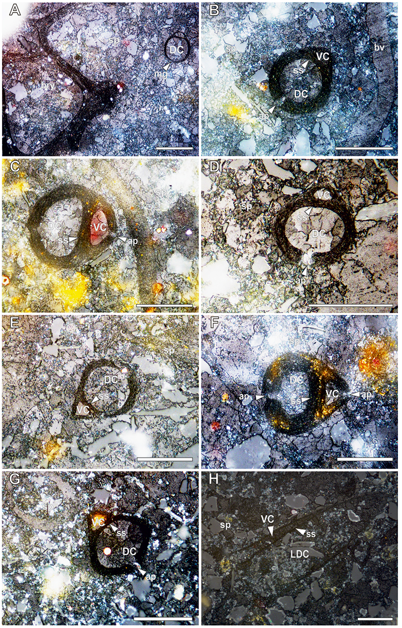
FIGURE 5. Photomicrographs of Arouxina pluricellata (A-F) and Samtlebenella circumcamerata (G-H) within the MWL Fm., these specimens are from Longville Stanway Track (A), Moorwood Quarry (B, C, E and H), Harton Hollow (D and G) and Strefford Quarry (H). (A) = Sectional cut through A. pluricellata highlighting the multiple hemispherical chambers (HC) surrounding the globular dorsal chamber (DC) and ventral chamber (VC). HC can form concentrically. Occasional apical apertures (ap) are observed within the DC.The bulbous growth are associated with several walls. (B) = Thickened A. pluricellata specimen with six HC surrounding the central DC and VC. Note the presence of pyrite within a few of the HC. Matrix comprised solely of sparite. (C) = An elongate A. pluricellata test with three HC. (D) = Specimen of A. pluricellata highlighting the disjointed nature of the surrounding HC. (E) = A. pluricellata with a single HC chamber but exhibits an ap within both VC and DC. (F) = A. pluricellata specimen with thinner walled HC compared to the walls surrounding the DC and VC. (G) = S. circumcamerata resembling Darth Vader’s helmet. Presence of HC within the walls surrounding the DC. (H) = Thick walled specimen of S. circumcamerata with a partially preserved VC and two chambers (c) are visible within the walls surrounding the DC. Sparitic matrix = (sp). All scale bars equal 50 µm.
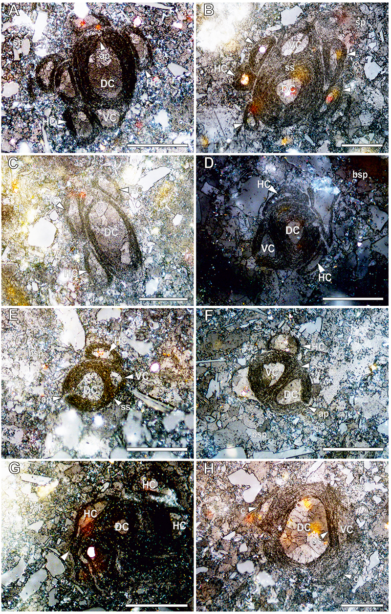
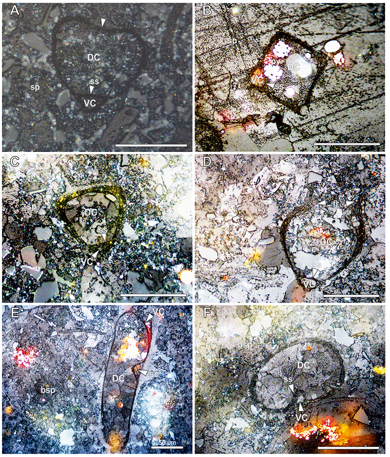
FIGURE 6. Photomicrographs of Natantesprifmata rogersi (A-D) and Hartonella oblonga (E-H) within the MWL Fm., these specimens are from Longville Stanway (A), Longville Stanway Track (B), Eaton Top (C), Moorwood Quarry (D) and Harton Hollow (E-H). (A) = N. rogersi with a prismatic dorsal chamber (DC) separated from the conical ventral chamber (VC) by a septum-like structure (ss). Apical aperture is present in the DC. (B) = N. rogersi with pyrite within the walls (pyw) of the test. Thickened walls (tw) are present around DC. Note quartz grains (qtz) within the DC infill. (C, D) = N. rogersi showing the typical kite-like shape. (E, F) = H. oblonga showing a large elongated dorsal chamber (DC) separated by a septum-like structure (ss) from its smaller globular ventral chamber. (G) H. oblonga with a pointed VC and a smoother, more oval DC. Bivalve fragment (bv) (top right). (H) An H. oblonga with a thicker than normal wall and elongated ss. Specimen of mg is neighbouring H. oblonga (bottom centre). Sparitic matrix (sp); Biosparitic matrix (bsp). All scale bars equal 50 µm.

FIGURE 7. (A) = Photomicrographs of Munneckella tribuscamera within the MWL Fm., these specimens are from Harton Hollow (A and B), Strefford Quarry (C) and Eaton Top (D). (A) = M. tribuscamera with two septum-like structures (ss) forming a three-chambered morphology consisting of a slightly larger central chamber (CC) and two smaller, often dome or cone-shaped peripheral chambers (PC). (B) = M. tribuscamera with three unequal sized chambers and varying wall thickness. An apical aperture is present in one of the peripheral chambers. (C) = Large specimen of M. tribuscamera with conical PC. (D) = M. tribuscamera. with slightly thickened thicker than normal walls. Sparitic matrix (sp); Biosparitic matrix (bsp). All scale bars equal 50 µm.
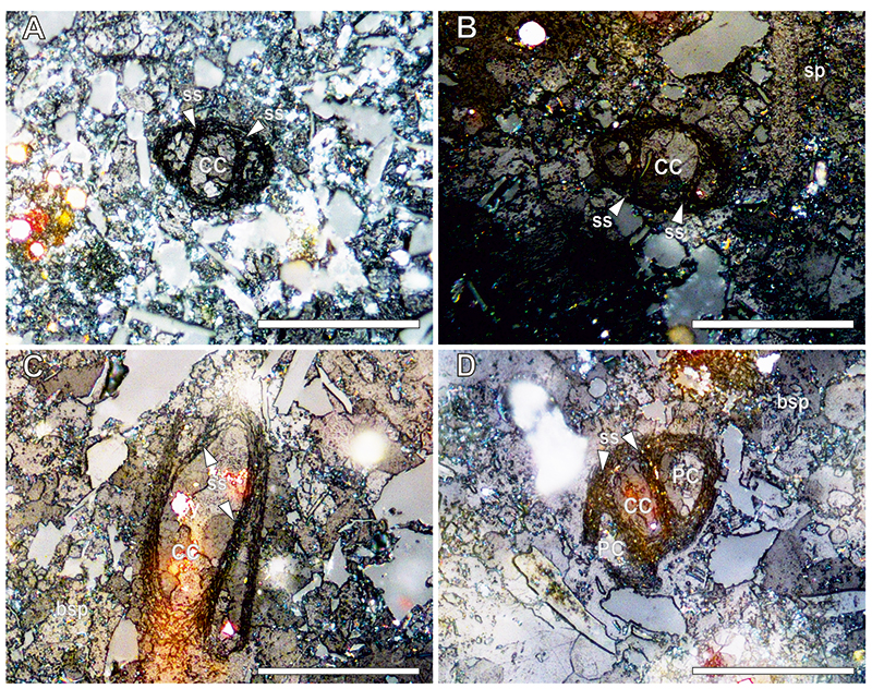
FIGURE 8. Photomicrographs of Ovummuridae left to open nomenclature within the MWL Fm. (A) = Open gen. 1, sunken apex of the dorsal chamber (DC) producing a ‘love heart’ shape. (B) = Open gen. 2, a square test shape. (C) = Open gen. 3, Triangular test shape. Dorsal chamber (DC) is separated from the ventral chamber (VC) by the septum like structure (ss). A possible transverse cross-sectional cut of M. cameroni. (D) = Open gen. 4, open DC of an elongate specimen. Possibly due to cross-sectional cuts. Flanges (fl) creating an opening at the apex of the DC. (E and F) = Open gen. 5 = Three-chambered variant (E) in which the dorsal chamber (DC) is fully split into a smaller chamber (sc) by the septum like structure (ss). A ventral chamber (VC) is still observable. A two-chambered variant (F) of this form showing various morphological differences. A third chamber is not observed. Sparitic matrix (sp); Biosparitic matrix (bsp). All scale bars equal 50 µm.