FIGURE 1. Map of Asia (A) showing the location of Russian and Kazakh late Miocene and early Pliocene sites (B): ANR, Andreevka 1A, 2A; BRK, Borki 1A-C; BTK, Biteke; CHR, Cherlak 1A; ISK, Isakovka 1A, 2A; KRN, Krasnokutsk; KRT, Kartashovo; LZN, Lezhanka 1A, 2B; NIL, Nizhneil’inka; NST, Novaya Stanitsa 1A-B; PPL, Petropavlovsk 1A; PSH, Peshnevo 1A-B; PVL, Pavlodar 1A, 2 ‘Quarry’; SLT, Selety 1A (type locality of Paranourosorex seletiensis Storch and Zazhigin, 1996). Age of the deposits is marked by colours, which corresponds to ELMA Neogene zones ‘MN 10, 12-14’; the redeposited fossiliferous layers are marked by ‘?’. Map data from resource ESRI (http://www.esri.com/) using SASPlanet software (v.160707.9476).
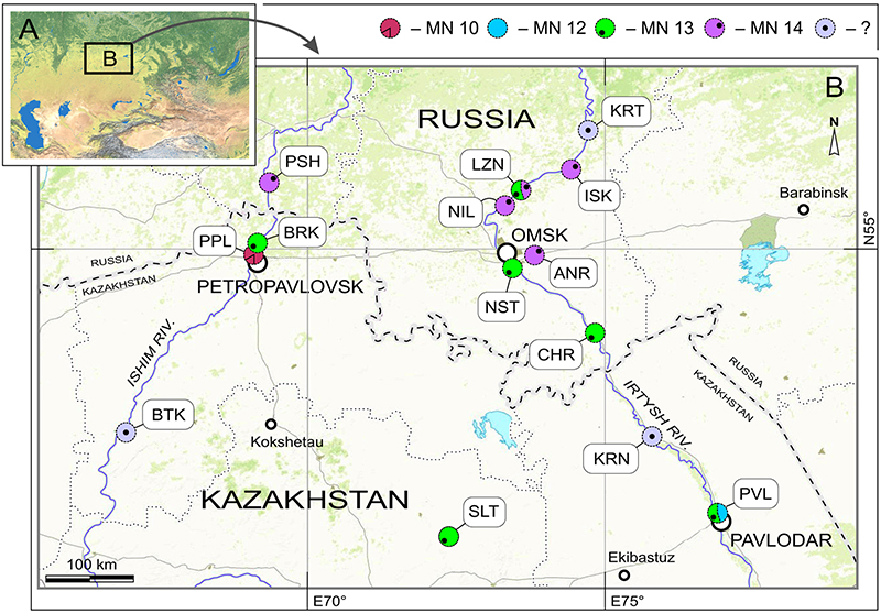
FIGURE 2. Teeth and bones of Ishimosorex ishimiensis gen. et sp. nov. from late Miocene Petropavlovsk 1A locality (A-E, G-K) and Borki 1A locality (F). A, GIN 952/1161, left I1 in lateral view (Figure 2A cf. Figure 7A); B, GIN 952/1160, right I1 in medial view; C, GIN 952/1152, left fragment of mandibular ramus in posterior view (image displays articular surface of the condylar process); D, ibid., diagrammatic image of mandibular ramus in medial view; E, GIN 952/1159, left i1 in lateral view; F, GIN 1115/1149, right i1 in medial view; G, GIN 952/1155, left M1 in occlusal view (G1, pure view; G2, augmented view) (Figure 2G cf. Figure 7H); H, GIN 952/1153, right dentary fragment with p4-m1 and anterior alveolus of m2; J, GIN 952/1158, right m1 in occlusal view; K, GIN 952/1154, left m2 in occlusal view. Star indicates reversed image. Scale bars equal 1 mm.
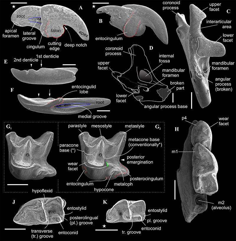
FIGURE 3. The characters of the first lower incisors of Paranourosorex gigas from ANR/1A (GIN 1112/1108; A1, lateral, A2, medial views), Ishimosorex ishimiensis gen. et sp. nov. from BRK/1A (GIN 1115/1149, paratype; B1, lateral, B2, medial views), Crusafontina endemica Spanish from Can Llobateres 1 locality (C, CL1 2217, see van Dam, 2004: 746; in lateral view), C. kormosi (D, Polgárdi 4, in lateral view) and A. oligodon (E, Polgárdi 4, in lateral view). Scale bars equal 1 mm; C, unscaled. Abbreviations see in Figure 1.
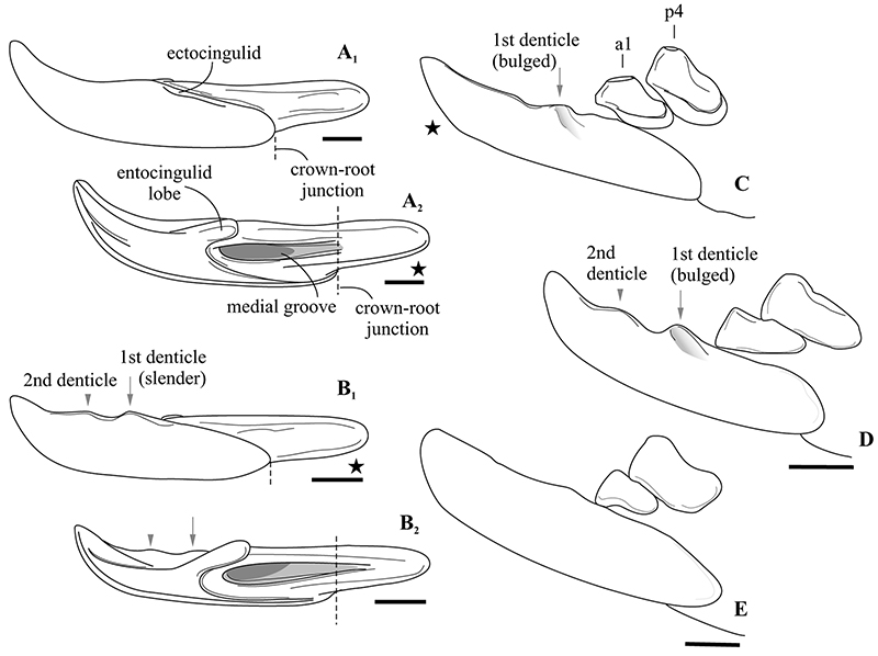
FIGURE 4. Schematic image of the fourth lower premolar of Ishimosorex ishimiensis gen. et sp. nov. (A , GIN 952/1153, PPL/1A), Crusafontina kormosi (B, Polgárdi 4), Paranourosorex intermedius sp. nov. (C, GIN 948/1051, NST/1A), Paranourosorex gigas (D, GIN 1118/1011, PSH/1B), Anourosorex squamipes (E, ZIN 98253, Recent), Crusafontina fastigata (F , AG5A from Los Aguanaces 5A, Teruel Basin, Spain by van Dam, 2004: figs. 4-1), Amblycoptus oligodon (G, Polgárdi 4) and Amblycoptus jessiae (H, KS 3157 from Las Casiones, Teruel Basin, Spain by van Dam, 2004: figs. 5-11). Abbreviations: a, concavity; add, additional crown elements (1, crest; 2, cusplet); ccr, central crest; eccd, ectocingulid; encd, entocingulid; pcd, postcingulid; plb, posterolingual basin (1, shallow, weak; 2, expressed); wen, wrinkled enamel; wft, wear facet (1, spot-like; 2, comma-like). Star indicates reversed image. Scale bars are 1 mm; F, H unscaled.
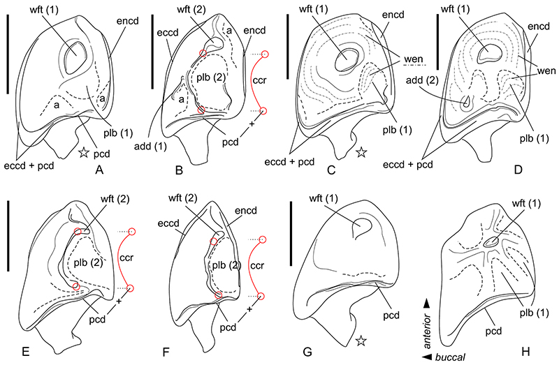
FIGURE 5. Bivariate plot of linear differences between five Asian species, Paranourosorex seletiensis Storch and Zazhigin, 1996 (PSt.), Paranourosorex gigas Rzebik-Kowalska, 1975 (P. gigas), Paranourosorex intermedius sp. nov. (PIm.) and Ishimosorex ishimiensis gen. et sp. nov. (PPl) based on the second lower molar measurements (L vs TRL), mm). Studied remains from Asian Neogene localities are marked by colours, which corresponds to ELMA (see Figure 1); numbers are given in the Appendix 2.
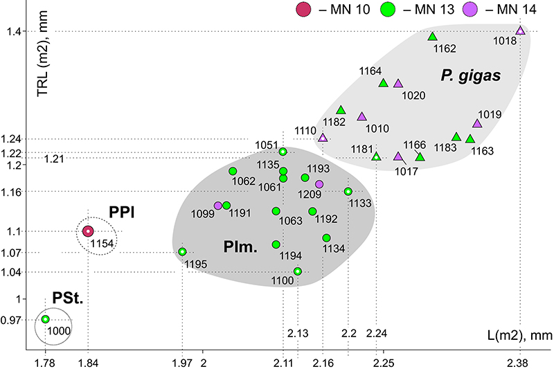
FIGURE 6. Teeth and bones of Paranourosorex seletiensis Storch and Zazhigin, 1996 (A, B), Crusafontina sp. 1, (C), Crusafontina sp. 2 (D) and Paranourosorex intermedius sp. nov. (E). A, GIN 951/1000 (holotype) from SLT/1A, left hemimandible fragment (cut image) with m1 and m2 in occlusal view (A2, magnified m2); B, GIN 951/1001 (paratype) from SLT/1A, left P4 in occlusal view; C, GIN 640/1004 from PVL/1A, left M1 in occlusal view; D, GIN 951/1003 SLT/1A, left P4 in occlusal view; E, GIN 948/1051 (holotype) from NST/1A, right dentary fragment with m1 and m2 (E1, occlusal view; E2, lateral view E3, medial view;). Scale bars equal 1 mm.
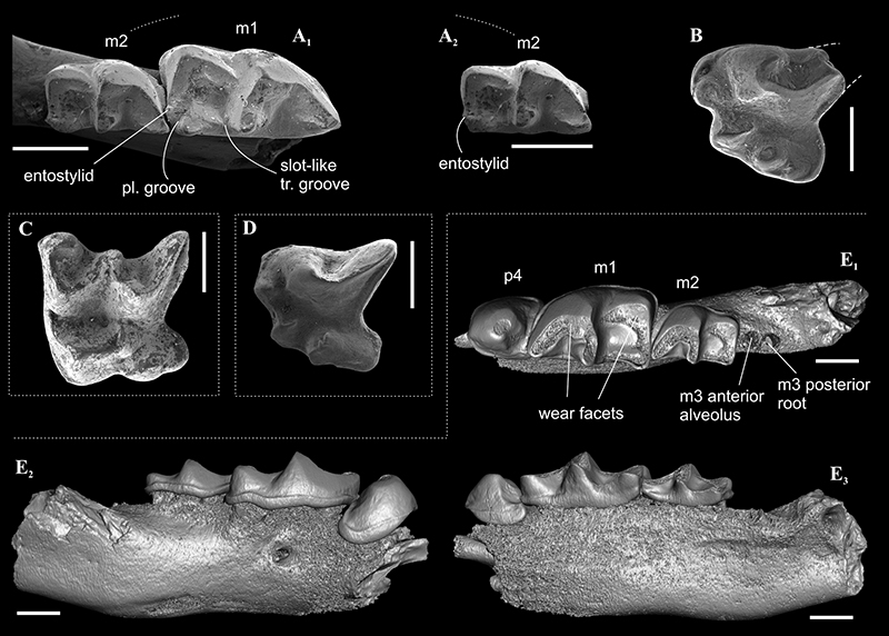
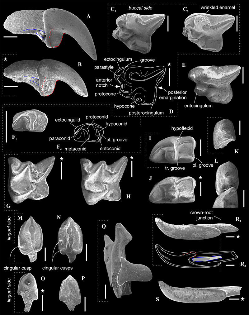
FIGURE 7. Teeth and bones of Paranourosorex intermedius sp. nov. (B, C-D, F-G, I, K, M, O, Q-R; with dotted frames) and Paranourosorex gigas Rzebik-Kowalska, 1975 (A, E, H, J, L, N, P, S). A, GIN 1118/1044 from PSH/1B, right I1 in lateral view; B, GIN 948/1073 (paratype) from NST/1A, left I1 in lateral view; C, GIN 948/1065 (paratype) from NST/1A, left P4 (C1 , occlusal view; C2, tilted occlusal view); D, GIN 1130/1104 from LZN/2B, diagrammatic image of right P4 in occlusal view; E, GIN 1118/1028 from PSH/1B, left P4 in occlusal view; F, GIN 948/1064 (paratype) from NST/1A, right m3 in occlusal view (F1, SEM image; F2, diagrammatic image); G , GIN 948/1069 (paratype) from NST/1A, right M1 in occlusal view; H , GIN 1118/1005 from PSH/1B, right M1 in occlusal view; I, GIN 948/1056 (paratype) from NST/1A, right m1 in occlusal view; J, GIN 1118/1013 from PSH/1B, left m1 in occlusal view; K, GIN 948/1078 (paratype) from NST/1A, left p4 in occlusal view; L, GIN 1118/1011 from PSH/1B, left p4 and m1 fragment (cut image) in occlusal view; M, GIN 948/1089 (paratype) from NST/1A, left A1 in occlusal view; N, GIN 11181036 from PSH/1B, left A1 in occlusal view; O, GIN 948/1081 (paratype) from NST/1A, left a1 in occlusal view; P, GIN 1118/1008 from PSH/1B, left a1 in occlusal view; Q, GIN 1115/1123 from BRK/1A, left fragment of mandibular ramus in posterior view; R, GIN 948/1075 (paratype) from NST/1A, right i1 (R1, SEM image in lateral view; R2, diagrammatic image in medial view); S, 1118 951/1021 from PSH/1B, right i1 in lateral view. Scale bars equal 1 mm.