FIGURE 1. Position and geology of the fossil locality. A, Map of USA with the position of Oklahoma (red area) and of Arbuckle Mountains (grey area). B, Map of Arbuckle mountains with the position of the type locality of Concavicaris woodfordi (Cooper, 1932). C, Section of the upper Woodford Shale at Interstate 35 road-cut section (I-35) (sec. 25, T2S, R2E, Arbuckle Mountains, Oklahoma, USA; redrawn after Over [1992]).
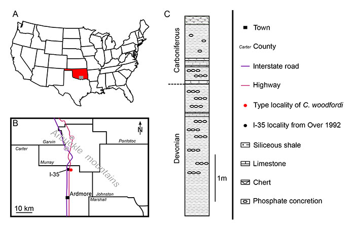
FIGURE 2. General view of Concavicaris woodfordi (Cooper, 1932). A, B, Right and left lateral views. C, line drawing (lateral view). D, location of virtual slices presented in the figures (dorsal view). Abbreviations: vld, ventro-lateral depression. Arrows indicate the anterior side of the specimen. Yellow doted lines indicate longitudinal sections. Green dotted lines indicate transversal sections. Scales: 10 mm. Photos: T. A. Hegna.
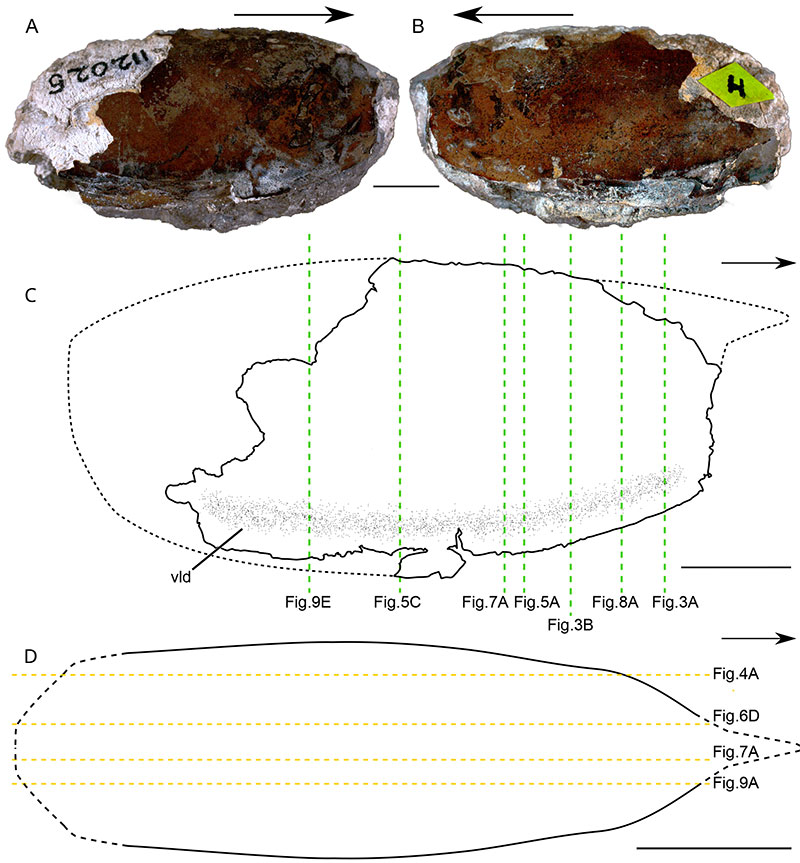
FIGURE 3. Marginal fold of Concavicaris woodfordi (Cooper, 1932). A, B, anterior part of the shield (tomogram and drawing). C, close-up of marginal fold in the anterior part of the shield (tomogram). D, E, middle part of the shield (tomogram and drawing). F, close-up of marginal fold in the middle part of the shield (tomogram). Abbreviations: il, inner layer; mf; marginal fold; so, shield outline. Scales: A, B, D, E, 5 mm; C, F, 1 mm.
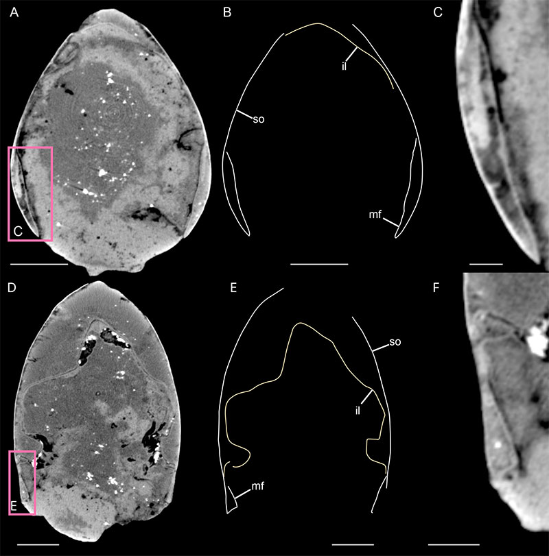
FIGURE 4. Anatomy of Concavicaris woodfordi (Cooper, 1932). A, longitudinal virtual section. B, longitudinal virtual section (colour-marked). C, dorsal view (3D rendering). D, lateral view (3D rendering). Abbreviations: am, adductor muscles; cs, cylindrical structure; g2-7, gills; gm, gastric muscles;ila, anterior part of the inner layer; ilp, posterior part of the inner layer; lvp, latero-ventral pouch; pta, posterior trunk appendages; ra1-3, raptorial appendages; so, shield outline; sto, stomach. Arrows indicate the anterior side of the specimen. Scales: 10 mm.
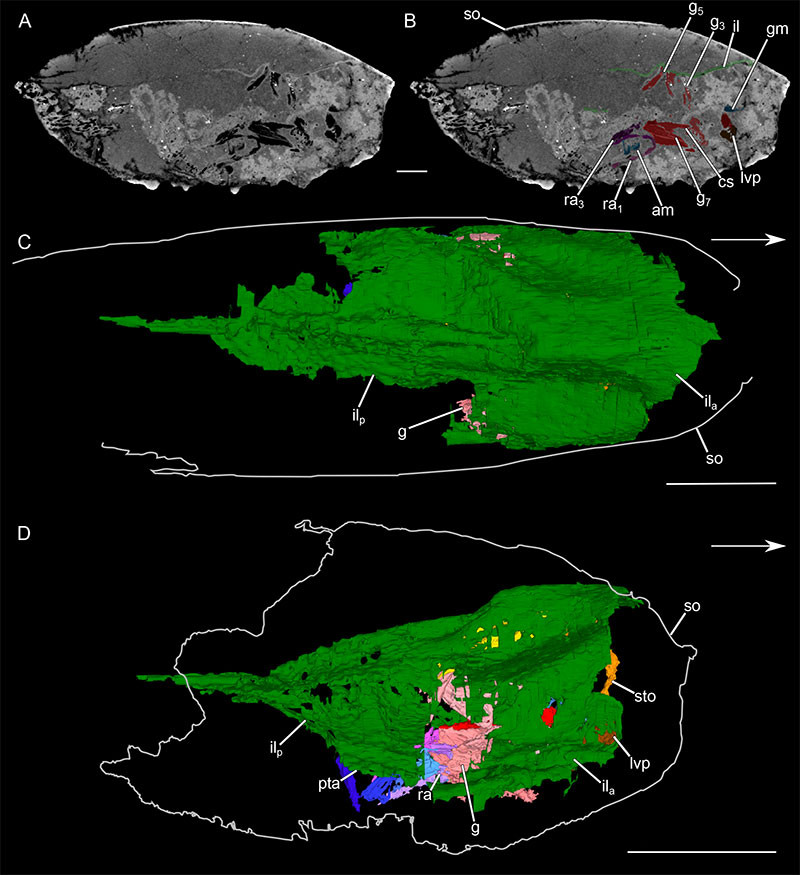
FIGURE 5. Inner layer of Concavicaris woodfordi (Cooper, 1932). A-C, anterior part of the specimen. A, tomogram (transversal slice). B, tomogram (transversal slice; colour-marked). C, anterior view (3D rendering). D-F, posterior part of the specimen. D, tomogram (transversal slice). E, tomogram (transversal slice; colour-marked). F, cross-section (3D rendering). G, cross-section of cephalothorax of a reptantian decapod (after Glaessner, 1969). H, I, cross-section of the carapace structure of a myodocopan (Euphilomedes japonica (Müller, 1890); after Yamada, 2019). H, attached region. I, duplicated region. Arrow indicates the rotation of the posterior part of the inner layer. Abbreviations: am, adductor muscles; app, appendage; cb, chitinous body; ef, epimeral fold; g, gills; hi, hinge; il, inner layer of the shield; ila, anterior part of the inner layer; ilp, posterior part of the inner layer; mua, attractor muscles; ol, outer layer of the shield; pl, pleural part; pta, posterior trunk appendages; so, shield outline; st, sternal part; te, tergal part. Scales: 5 mm.
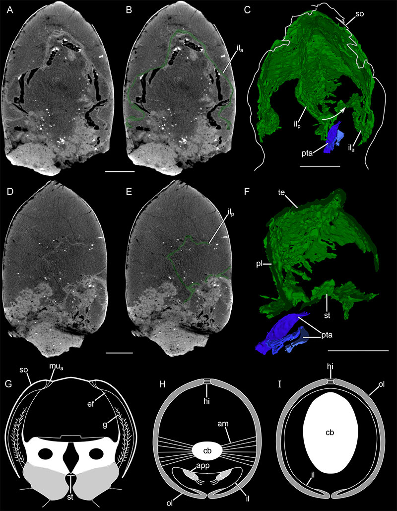
FIGURE 6. Internal anatomy of Concavicaris woodfordi (Cooper, 1932). A, right lateral view (3D rendering). B, anterior view (3D rendering). C, dorsal view (3D rendering). D, longitudinal section (3D rendering). Abbreviations: am, adductor muscles; cs, cylindrical structure; g1-8, gills; gm, gastric muscles; go, gonads; lvp, latero-ventral pouch; pta1-3, posterior trunk appendages; r, rostrum; ra1, 3, raptorial appendages; s, shield; so, shield outline; sto, stomach. Arrows indicate the anterior side of the specimen. Scales: A, C, D, 10 mm; B, 5 mm.
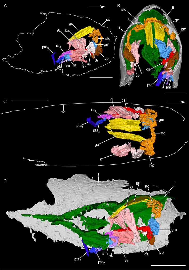
FIGURE 7. Digestive, reproductive, and circulatory systems of Concavicaris woodfordi (Cooper, 1932). A, longitudinal virtual section. B, longitudinal virtual section (colour-marked). C, D, digestive and reproductive systems (3D rendering). C, anterior view. D, right lateral view. E, F, G, circulatory system. E, tomogram. F, tomogram (colour-marked). G, 3D rendering of the left part. Abbreviations: cs, cylindrical structure; g1-8, gills; go, gonads; il, inner layer; lvp, latero-ventral pouch; sto, stomach. Arrows indicate the anterior side of the specimen. Scales: A, B, 10 mm; C-G, 5 mm.
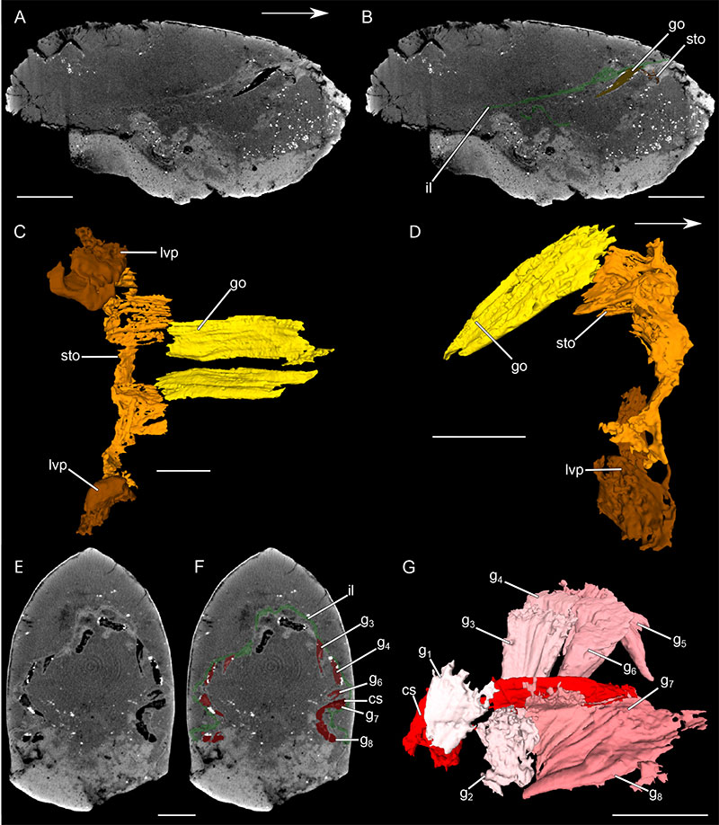
FIGURE 8. Muscular structures of Concavicaris woodfordi (Cooper, 1932). A, tomogram. B, tomogram (colour-marked). C, gastric muscles (3D rendering). D, cross-section (3D rendering). E, adductor muscles (3D rendering). Abbreviations: am, adductor muscle; gm, gastric muscles; il, inner layer; lvp, latero-ventral pouch; s, shield; sto, stomach. Scales: A, B, D, 5 mm; C, E, 2 mm.
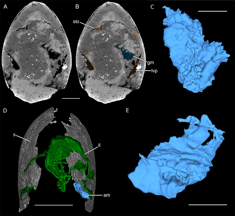
FIGURE 9. Appendages of Concavicaris woodfordi (Cooper, 1932). A, longitudinal section (3D rendering). B, raptorial appendages (3D rendering). C, tomogram. D, close-up of third posterior trunk appendage. E, posterior trunk appendages (3D rendering). Arrow indicates the third posterior trunk appendage. Abbreviations: il, inner layer; pta1-3, posterior trunk appendages; ra1-3, raptorial appendages; s, shield. Arrow indicates the anterior side of the specimen. Scales: A-C, 10 mm; D, G, 2 mm; E, 5 mm; F, 1 mm.
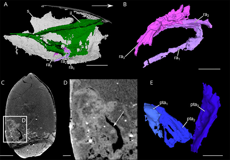
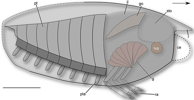
FIGURE 10. Hypothetical reconstruction of Concavicaris woodfordi (Cooper, 1932). Morphology of anterior and posterior sides of the shield, eyes and number of posterior trunk appendages are reconstructed based on Concavicaris submarinus (Jobbins et al., 2020). Abbreviations: ce, compound eyes; g, gills; go, gonads; il, inner layer: lvp, latero-ventral pouch; pt, posterior trunk; pta, posterior trunk appendages; r, rostrum; ra, raptorial appendages; s, shield; sto, stomach. Arrow indicates the anterior side of the specimen. Scales: 10 mm.