FIGURE 1. Generalized map of the Orcadian Basin (modified from Burrow et al., 2016, fig. 2) with a few key sites indicated and their relative stratigraphical horizon indicated. Circles indicate the Achanarras Fish Bed Member horizon, crosses the upper Mey Flagstone Formation horizon, and stars the lower Mey Flagstone Formation horizon and their equivalents.
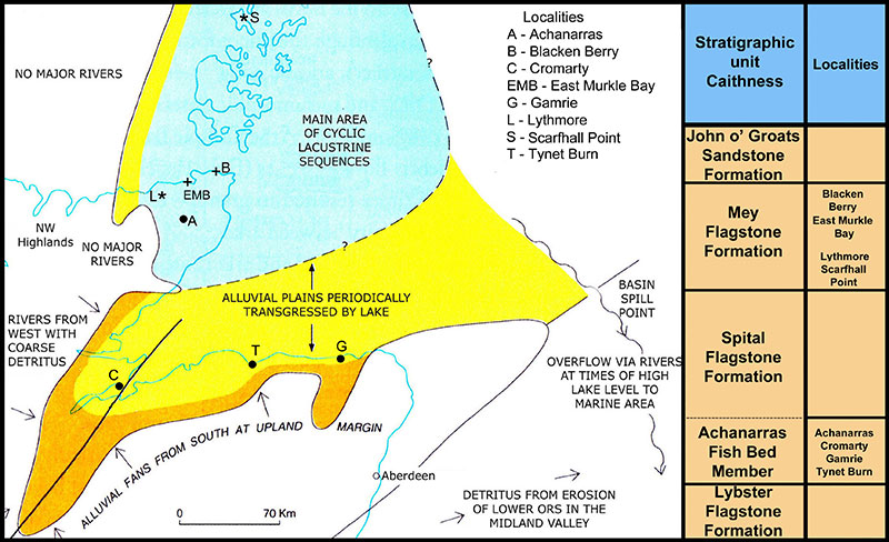
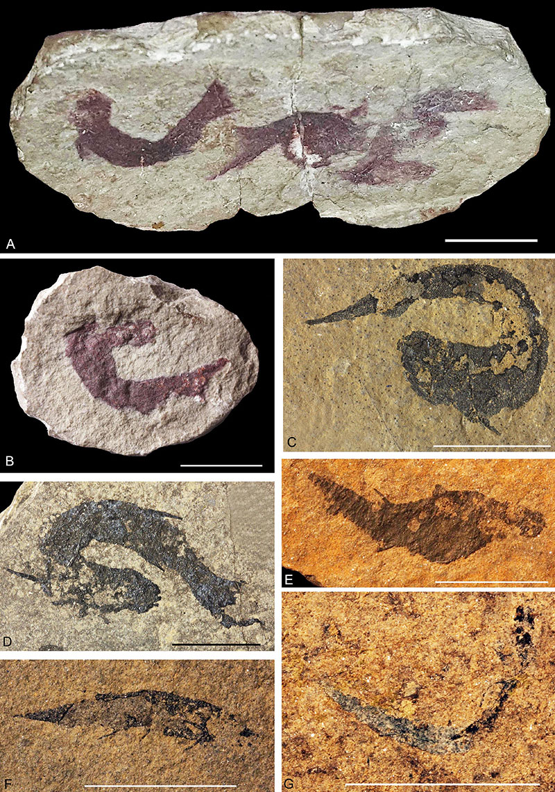
FIGURE 2. Orcadacanthus pusillus specimens from Tynet Burn, Scotland and type specimens of junior synonyms Acanthodes peachi and A. coriaceus from Caithness. A, ELGNM.1978.191.1, electronically flipped horizontally to match B. B, Agassiz type specimen (1844-5, plate 28, figure 8). C, ROM 25872 electronically flipped horizontally to match D. D, Agassiz type specimen (1844-5, plate 28, figure 9). E, Agassiz type specimen (1844-5, plate 28, figure 10). F, Diplacanthus crassisimus, ROM 25859. G, O. pusillus, ROM 25846 electronically flipped horizontally to match B. H, I, Acanthodes peachi GSM 21448 from Barragill (East Mey): H, specimen as figured by Egerton (1861, plate 6, figure 2); I, current state. J, K, Acanthodes coriaceus GSM 28831 from Gallow’s Hill (Pennyland), near Thurso: J, specimen as figured by Egerton (1861, plate 6, figure 4); K, current state. Scale bars equal 10 mm.
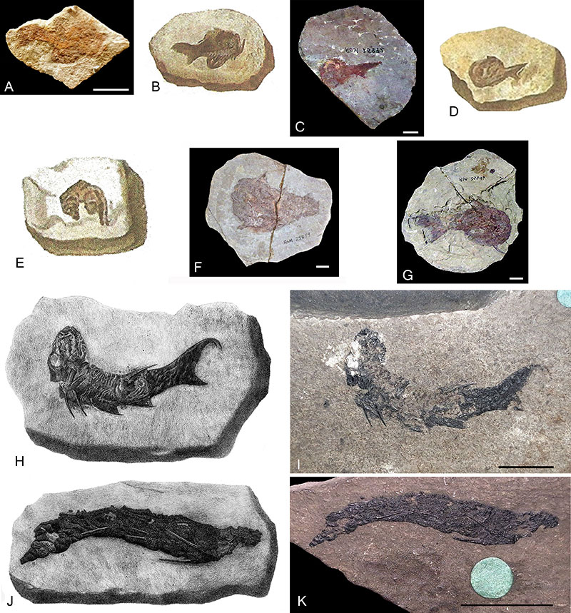
FIGURE 3. Orcadacanthus pusillus neotype NMHUK PV OR 35786 from Tynet Burn. A, photograph of the specimen. B, figure of specimen from Woodward 1891, plate 1, figure 5. C, close-up of the prepelvic spine. Abbreviations: a.fs, anal fin spine; a.fw, anal fin web; d.fs, dorsal fin spine; Mc, Meckel’s cartilage; pect.fs, pectoral fin spine; pelv.fs, pelvic spine; pelv.fw, pelvic fin web; prepelv.s, prepelvic spine; ot, otic capsule; sc, scapula; scl, sclerotic plate. Scale bar equals 10 mm in A, 5.0 mm in C.
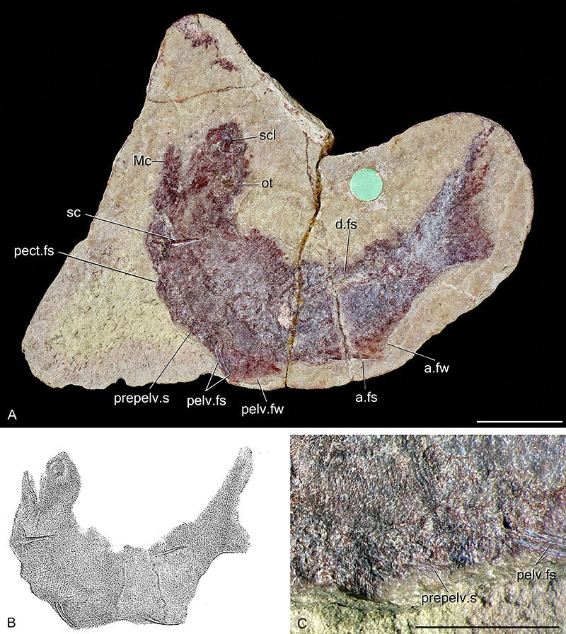
FIGURE 4. Orcadacanthus pusillus, complete specimens. A, NMS G.2021.7.27 from Achanarras Quarry. B, NMS G.2021.7.78 from Brims Ness. C, NMS G.1887.35 from Thurso (probably Pennyland). D, NMS G.2021.7.269 from Blacken Berry. E, NMS G.2022.10.8 from Button Geo, Stroma. Abbreviations: a.fs, anal fin spine; br, branchiostegal rays; d.fs, dorsal fin spine; d.fw, dorsal fin web; gr, gular rays; ot, otic capsule; pect.fs, pectoral fin spines; pelv.fs, pelvic fin spines; prepelv.s, prepelvic spines. Scale bars equal 10 mm.
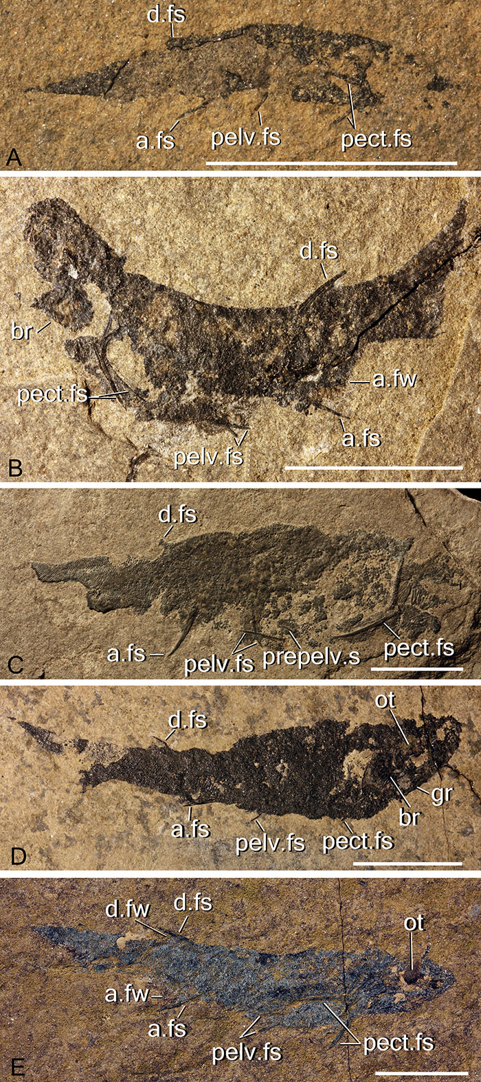
FIGURE 5. Orcadacanthus pusillus, details of the head. A, NMS G.2021.7.286 from Lythmore. B, NMS G.2022.10.686 from Blacken Berry. C, NMS G.2021.7.269 from Blacken Berry. D-E, NMS G.2021.7.125.5.3 from Cromarty, SEM image of a sclerotic plate. F, NMS G.2019.3.10 from Cromarty, detail of two sclerotic plates (modified from Burrow et al., 2020, figure 1g). G, GLAHM V3573 from Tynet Burn. H, NHMUK PV OR 35786 from Tynet Burn. I, NMS G.2022.10.2 from Lythmore, detail of main branchial cover. J, NMS G.2021.7.292 from Den of Findon (Gamrie), detail of main branchial cover. K-L, NMS G.2021.7.285 from Achanarras. K, palatoquadrate. L, Meckel’s cartilage. Abbreviations: br, branchial ray; cr.epq, extra-palatoquadrate ridge; ec, sclerotic plate eyeball contact area; en, external naris; l.Mc, left Meckel’s cartilage; nb, nasal bone; o, orbit; ot, otic capsule; pa.n, palatobasal articulation to the neurocranium; pop.c, preopercular canal; pq, palatoquadrate; r.Mc, right Meckel’s cartilage; sc, scapula, scl, sclerotic plate. Scale bars equal 2 mm in A-C, G-H; 100 µm in D-E; 1 mm in F, J-L; 500 µm in I.
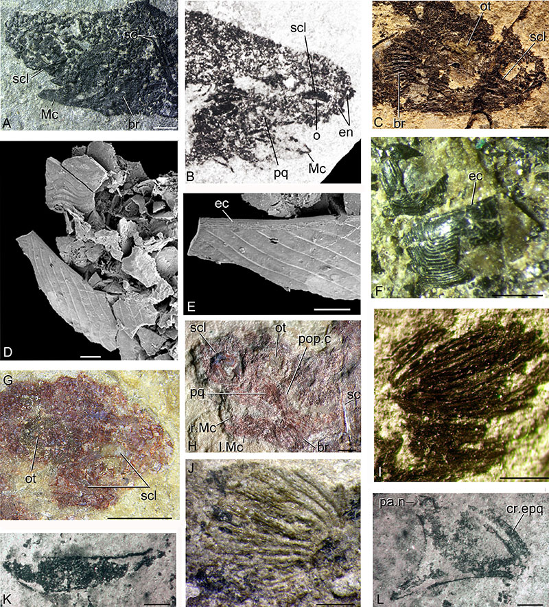
FIGURE 6. Orcadacanthus pusillus, details of the nasal area. A, NMS G.2022.10.686 from Blacken Berry showing right orbit and rostrum on the laterally flattened head. B, NMS G.2021.7.238 from Skinnet, orbit and one or two nasal openings. C, NMS G.2021.7.209 from Ness of Litter. D, NMS G.2021.7.93 from Cairnfield, dorsoventrally flattened rostrum. E, NMS G.2021.7.125.5s from Cromarty, ESEM of detached nasal plate, internal surface. F-G, NMS G. 2021.7.238 from Skinnet, displaced nasal plates. Abbreviations: db, denticle base; dpvnp, displaced posteroventral plate; en, external nare; ladnp, right anterodorsal nasal plate; np, nasal plate; o, orbit; radnp, right anterodorsal nasal plate; rpvnp, right posterovental nasal plate; scl, sclerotic plate; ts, tesserae. Scale bars equal 500 µm in B-D, F-G; 100 µm in E.
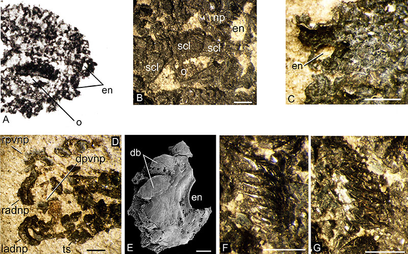
FIGURE 7. Orcadacanthus pusillus head and branchial elements, histology. A-B, NMS G.2021.7.136 from Cromarty, sclerotic plates, vertical section NMS G.2021.7.136.5, with close-up of one plate in (B). C, NMS G.2021.7.265 from Blacken Berry, NMS G.2021.7.265.3, pair of nasal plates. D-F, NMS G.2019.3.11 from Corbie Den: D, NMS G.2019.3.11.3, pair of nasal plates. E, NMS G.2021.7.265 from Blacken Berry, NMS G.2021.7.265.5, vertical section through head tesserae oblique section through scapulocoracoid. F, lower jaw transverse section NMS G.2019.3.11.4, ventral edge to left (the presumed transverse section through a posterior scale crown (s?) was mislabelled as a sclerotic plate in Burrow and den Blaauwen, 2021:figure 7E). G, NMS G.2021.7.126 from Cromarty, NMS G.2021.7.126.3, transverse section through jaw cartilages. H, NMS G.2021.7.162 from East Mey, NMS G.2021.7.162.5, transverse section through branchiostegal plates (ornament layer to bottom). Abbreviations: br, branchiostegal plate; cc, calcified cartilage; g, grooves; nc, nasal canals; np, nasal plates; scap, scapulocoracoid; scl, sclerotic plates; t, tesserae; tmt, tessellate mineralised tesserae; vp, vascular plexus. Scale bars equal 100 µm in A-F, 1.0 mm in G.
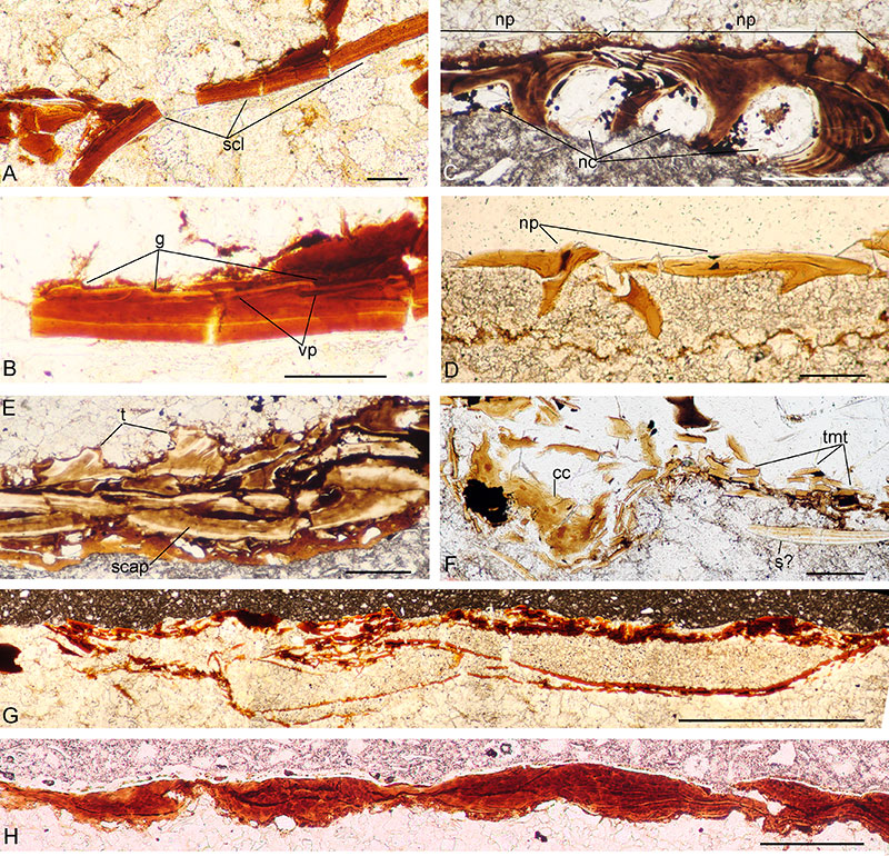
FIGURE 8. Orcadacanthus pusillus shoulder girdle, spines morphology. A, NMS G.2021.7.45 from Achanarras, articulated pectoral girdles and spines. B, NMS G.2019.3.10 from Cromarty, disarticulated pectoral spines and scales. C, NMS G.2021.7.135 from Cromarty, disarticulated pectoral spine, mid-spine ornament ridges. D, NMS G.2021.7.94 from Cromarty, articulated pectoral girdles and spines. E, NMS G.2021.7.290 from Cairnfield, pair of articulated pectoral girdles and spines, sliced by saw thus showing longitudinal sections through the elements. F, NMS G.2021.7.165 from East Murkle Bay, possibly the dorsal spine. G, NMS G.2021.7.202 from Blacken Berry, dorsal fin spine and fin rays. H, NMS G.2021.7.158 from East Murkle Bay, articulated pectoral girdles and spines. I, NMS G.2021.7.187 from Surrigarth, articulated fish, dorsal and anal fin spines. A-D, Achanarras Fish Bed Member horizon; E-F, lower part of the Mey Flagstone Formation horizon; G-I, upper part of the Mey Flagstone Formation horizon. Abbreviations: seg?, possible segmented rays. Scale bars equal 2.0 mm.
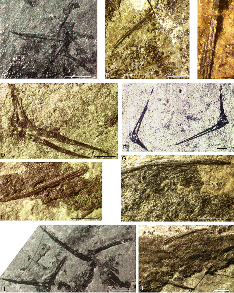
FIGURE 9. Orcadacanthus pusillus scapulocoracoid and paired spines histology, horizontal section through headless? articulated specimen NMS G.2022.10.1 from Blacken Berry. A, scapulocoracoids and pectoral fin spines, longitudinal sections of spines and oblique sections through scapulocoracoids (composite of three images); B, C, prepelvic spines; D, pelvic fin spines. Abbreviations: dt, dentine tubules/canals; fbs?, possible large scale near fin spine base; pect.fs, pectoral fin spine; pelv.fs, pelvic fin spine; prepelv.s, prepelvic spine; s, scales; sc, scapulocoracoid. Scale bars equal 10 mm in A, B, D, 1.0 mm in C.
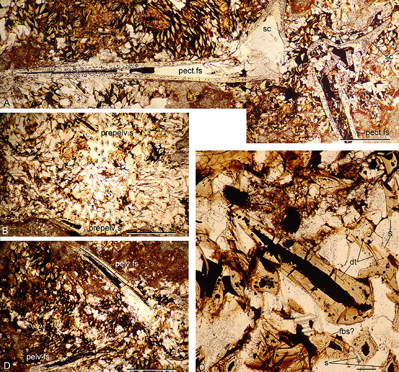
FIGURE 10. Orcadacanthus pusillus shoulder girdle, spines histology. A, NMS G.2022.10.13 from Castlehill, cross-section of scapulocoracoid shaft. B-C, transverse sections of spines from Achanarras: B, NMS G.2021.7.284.4, proximal end, possibly of pectoral spine; C, NMS G.2021.7.51.2, possible dorsal spine and small fin rays. D, NMS G.2019.3.11.7 from Corbie Den, NMS G.2019.3.11.2.7, transverse section, possible anal spine, proximal end. E, NMS G.2021.7.303.9 from Cromarty, NMS G.2021.7.303.9, transverse section, possible anal spine. F, NMS G.2021.7.136.9 from Cromarty, NMS G.2021.7.136.9, transverse section, possible anal spine. G-H, NMS G.2021.7.303 from Cromarty: G, NMS G.2021.7.303.6, transverse section, tip of possible dorsal spine and small fin rays; H, NMS G.2021.7.303.7, median spines and basal cartilage. I, NMS G.2022.10.15 from Scarfhall (Op beds horizon), transverse section, median spine and basal cartilage. J, NMS G.2022.10.17, bonebed sample from Cairn of Hattel, NMS G.2022.10.17.4, transverse section of distal end, possible dorsal spine. K, NMS G.2022.10.12 from Blacken Berry, proximal end of prepelvic spine. L-N, NMS G.2021.7.265 from Blacken Berry: L-M, NMS G.2021.7.265.14, dorsal fin and spine cross section: L, whole depth of fin plus dorsal spine; M, closeup of fin rays, large basal scale, and small outer fin ‘rays’; N, NMS G.2021.7.265.14, distal end of dorsal spine. A-H, Achanarras Fish Bed Member horizon; I-K, lower part of the Mey Flagstone Formation horizon; L-N, upper part of the Mey Flagstone Formation horizon. Abbreviations: bc, basal cartilage; d.fs, dorsal fin spine; fbs, scale from base of fin; fr, fin ray; frt, fin raylet; fw, fin web; lc, longitudinal canal; s, scale; sec.c, secondary longitudinal canal. Scale bars equal 100 µm in A-F, 1.0 mm in G, H.
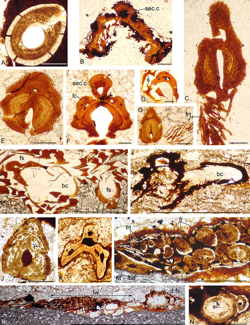
FIGURE 11. Orcadacanthus pusillus spines from articulated fish, Sollas serial section drawings. A, Achanarras Fish Bed Member horizon: pectoral spine (pect.fs) from Den of Findon, dorsal (d.fs) and anal (a.fs) spines from Tynet Burn. B, lower part of the Mey Flagstone Formation: all from west of Cairn of Hattel. C, upper part of the Mey Flagstone Formation horizon; pectoral spine from Blacken Berry, dorsal and anal spines from NMS G.2021.7.187, Surrigarth.
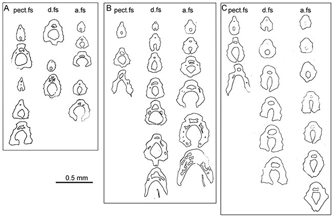
FIGURE 12. Orcadacanthus pusillus spines from bonebeds in the Mey Flagstone Formation, Sollas serial section drawings. A, pectoral, dorsal and three anal spines from Cairn of Hattel, at the lower part of the Mey Flagstone Formation horizon. B, dorsal and anal spines from Surrigarth (Westray), upper part of the Mey Flagstone Formation. C, prepelvic spines, a single section each of two from Clardon and one from Blacken Berry, upper part of the Mey Flagstone Formation.
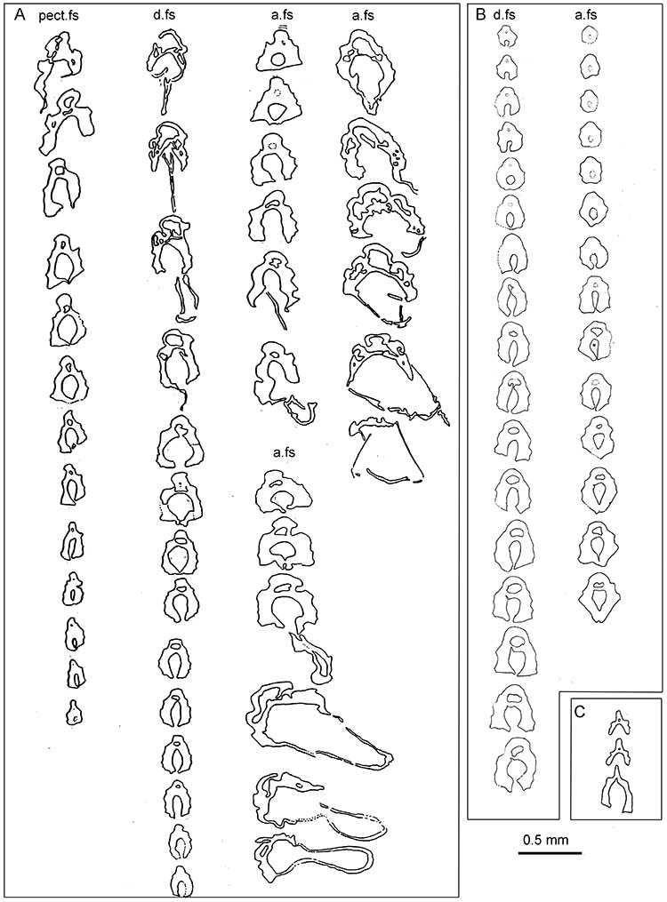
FIGURE 13. Achanarras Fish Bed Member horizon, Orcadacanthus pusillus scale morphology. Cromarty specimens, all ESEM images except F, light microscope image. A-E, NMS G.2021.7.125, disarticulated fish: A, NMS G.2021.7.125.5h, flank? scale, anterocrown view; B-C, NMS G.2021.7.125.5r, flank? Scale, anterocrown and anterior views; D, NMS G.2021.7.125.5d, clump of scales, showing posterior views; E, NMS G.2021.7.125.5l, clump of scales showing lateral and basal views. F-J, NMS G.2019.3.10, disarticulated fish: F, patch of scales; G, NMS G.2019.3.10.4d, clump of five scales; H, NMS G.2019.3.10.4e, dorsoventrally compressed scale with long posterior crown, lateral view; I, NMS G.2019.3.10.4c, dorsoventrally compressed scale with long posterior crown, basal view; J, NMS G.2019.3.10.4a, dorsoventrally compressed scale with long posterior crown, anterocrown view. K-L, NMS G.2021.7.284 from Achanarras: K, front half of specimen; L, disrupted mid-flank squamation showing scales in basal, crown and lateral views. Scale bars 1.0 mm in K, 0.5 mm in F, 0.1 mm in A-E, G-J, L. Arrows indicate anterior direction. br, branchiostegal plate impressions; pect.fs, pectoral fin spine. Scale bars equal 10 mm in K, 500 µm in F, 100 µm in A-E, G-J, L.
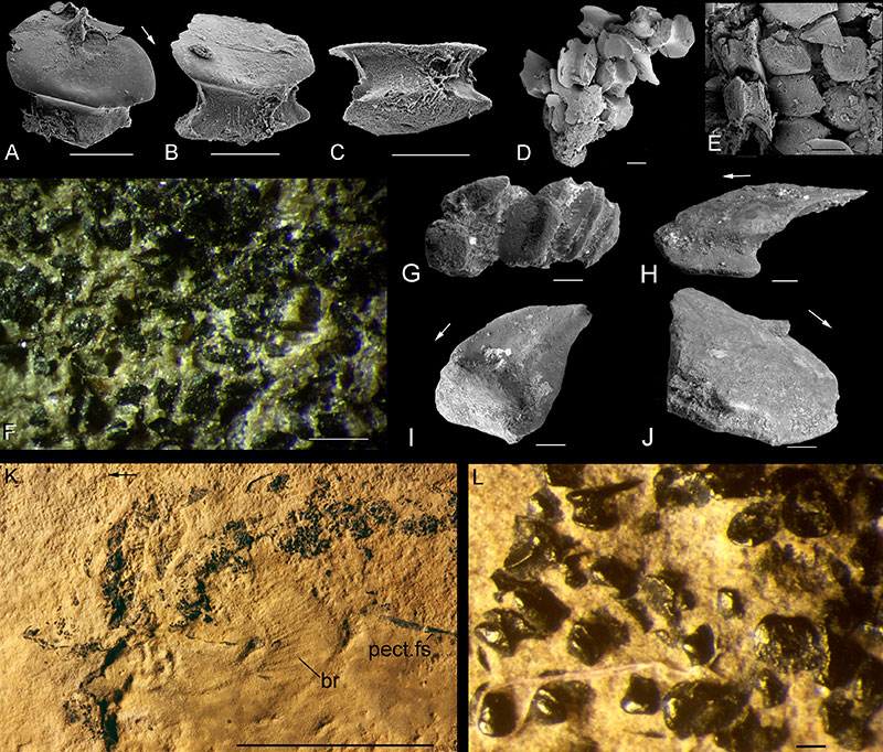
FIGURE 14. Achanarras Fish Bed Member horizon, Orcadacanthus pusillus scale histology. A-F, Cromarty specimens: A-E, NMS G.2021.7.136, disarticulated fish; A-B, NMS G.2021.7.136.2, scales and ?jaw cartilage tesserae; C, NMS G.2021.7.136.4, scales and cartilage; D-E, NMS G.2021.7.136.6, scales without and with a central pulp cavity, latter with radiating canals; F, NMS G.2021.7.303, dorsoventrally compressed articulated fish, NMS G.2021.7.303.6, transverse section posterior to ?pelvic spines through the body, tips of the spines and the fins. G, Achanarras specimen NMS G.2021.7.284, section NMS G.2021.7.284.3 through scales of two sides, centre of body trunk. H, Corbie Den specimen NMS G.2019.3.11, vertical longitudinal sections in NMS G.2019.3.11.4, centre of body trunk. ac, ascending canal; bs, body scales; calc, calcified cartilage blocks; cc, circular canal; en, enameloid; evc, embryonic crown zone vascular cavity; fins, finweb scales; fr, fin rays; n, node-like neck protuberance; pelv.fs, pelvic fin spine. Scale bars equal 100 µm.
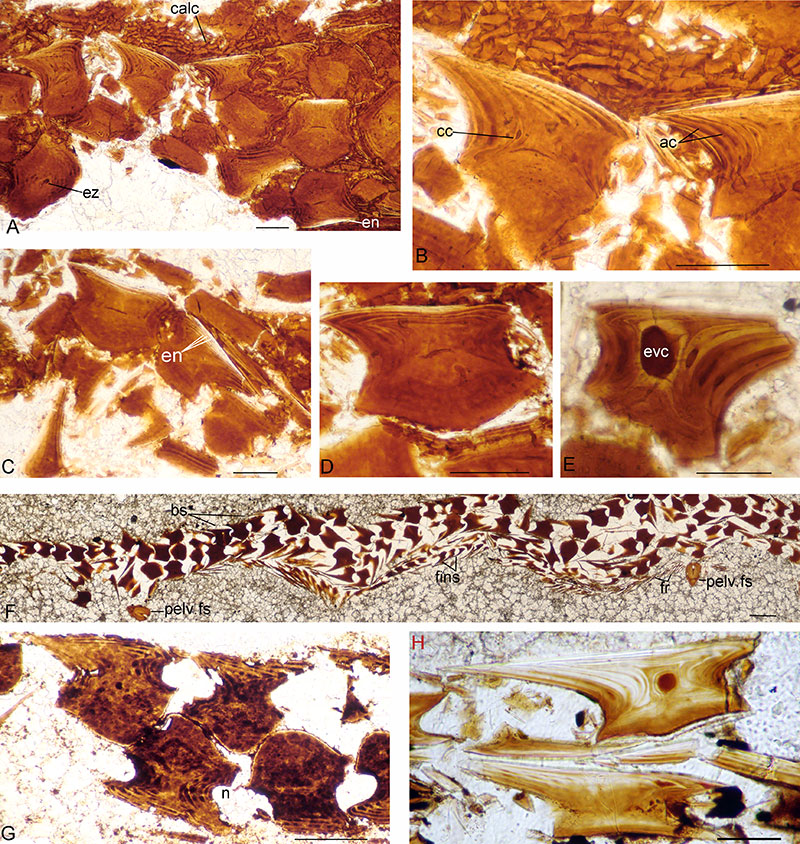
FIGURE 15. Lower part of the Mey Flagstone Formation, Orcadacanthus pusillus scale morphology. Light microscope images except D, ESEM. A-B, NMS G.2021.7.241 from Skinnet. A, whole specimen lacking head, left side squamation exposed. B, midflank squamation with lateral line. C, NMS G.2021.7.202 from Lythmore, eroded squamation on the left side below the dorsal fin. D, NMS G.2022.10.6 from Cairnfield squamation patch NMS G.2022.10.6.2b. Abbreviation: ll, lateral line. Scale bars equal 10 mm in A, 500 µm in B-D.
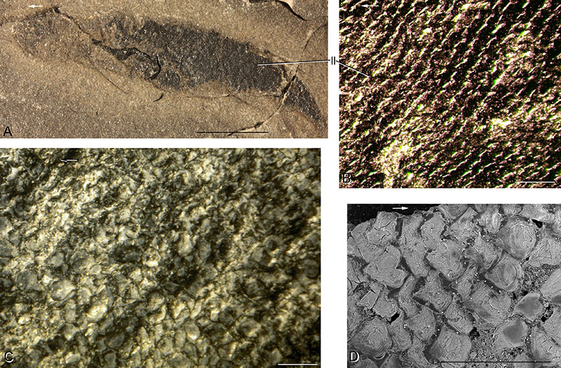
FIGURE 16. Lower part of the Mey Flagstone Formation (Osteolepis panderi horizon), Orcadacanthus pusillus scale histology. A, NMS G.2021.7.84 from Cairnfield, vertical section NMS G.2021.7.84.3 through squamation. B, NMS G.2021.7.251 from Skinnet, vertical section NMS G.2021.7.251.3. C-F, NMS G.2022.10.17 from Cairn of Hattel bonebed. C, NMS G.2022.10.17.3, oblique vertical section of convex based scale through large embryonic zone vascular cavity. D, NMS G.2022.10.17.4, vertical longitudinal section of small scale with vascular cavity opening out through the base. E, NMS G.2022.10.17.2, vertical longitudinal section of scale with long posterior crown and flat base. F, NMS G.2022.10.17.1, off-centre vertical transverse section of convex-based scale. G-I, bonebed section NMS G.2022.10.13 from Castlehill. G, vertical longitudinal section of scale with central vascular cavity. H, vertical transverse section of convex-based scale showing ascending canals. I, horizontal section through the crowns of two adjacent scales. Abbreviations: ac, ascending canal; cc, circular canal; cgz, crown growth zones; cW, canals of Williamson; dt, dentine canals/tubules; en, enameloid?; evc, embryonic crown zone vascular cavity; n, node-like protuberance; Sf, Sharpey's fibres. Arrows indicate anterior direction. Scale bars equal 500 µm in A, F, G, 100 µm in B-E, H-I.
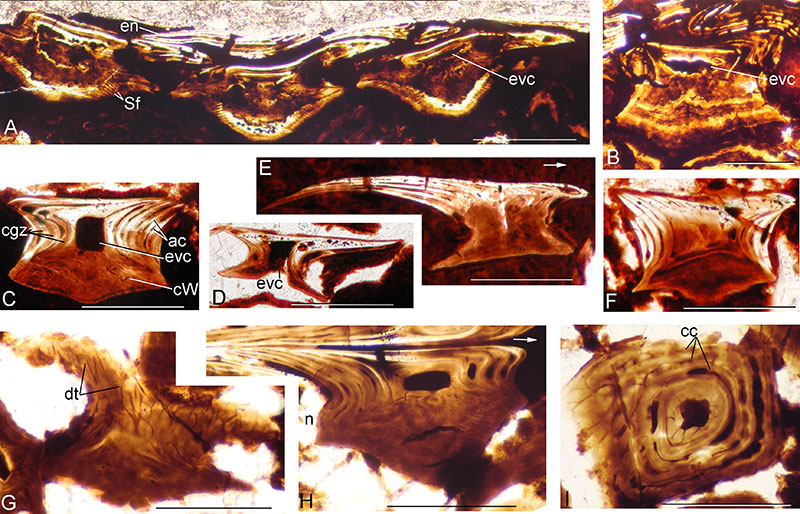
FIGURE 17. Upper part of the Mey Flagstone Formation Orcadacanthus pusillus scale morphology. A-C, Pennyland specimen NMS G.2022.10.5: A, NMS G.2022.10.5.2c, squamation patch, basal view; B, NMS G.2022.10.5.2b, encrusted articulated scales in crown view; C, NMS G.2022.10.5.2g, encrusted scale, crown view; D-J, Blacken Berry specimens. D-F, NMS G.2018.28.15: D, NMS G.2018.28.15.2a.9, crown view; E, NMS G.2018.28.15.2a.4, posterolateral view; F, NMS G.2018.28.15.2a.2, disarticulated scale clump, basal views; G-J, NMS G.2021.7.64: G, NMS G.2021.7.64.2e, squamation patch, basal view; H, scale NMS G.2021.7.64.2b, anterocrown view; I, NMS G.2021.7.64.2g, two scales from either side of the fish fused together, in lateral view; J, NMS G.2021.7.64.2a, scale with bifurcate posterior crown, in basal view showing open pulp cavity. Arrows indicate anterior direction. Abbreviation: pc, pulp canal. Scale bars equal 100 µm.
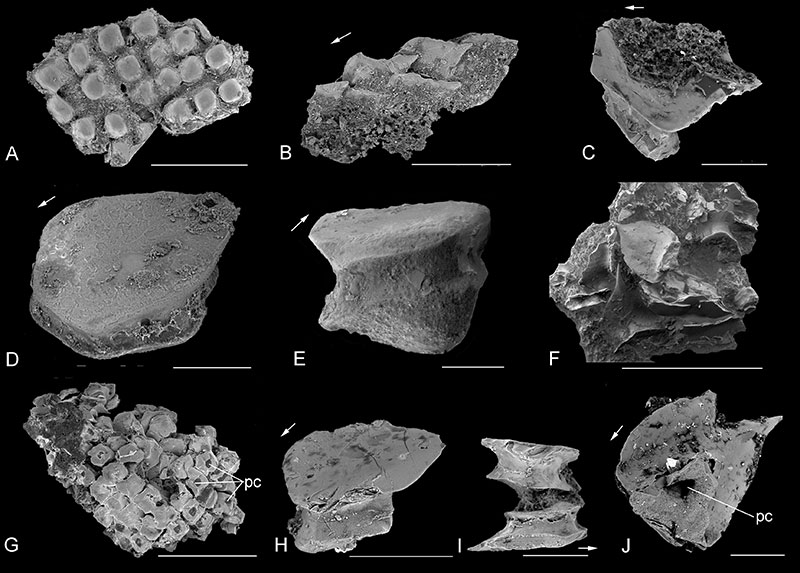
FIGURE 18. Upper part of the Mey Flagstone Formation Orcadacanthus pusillus scale histology. A-B, Pennyland specimen NMS G.2019.3.9, section NMS G.2019.3.9.2 through squamation patch. A, horizontal sections mid-scale; B, vertical longitudinal sections of scales from both sides of the fish, bases pressed together. C-H, Blacken Berry specimens. C-D, NMS G.2019.3.8, scales with two or three crown growth zones. C, NMS G.2019.3.8.9 showing wide central pulp cavities and canals extending down to base; D, NMS G.2019.3.8.11 showing ascending canals and dentine overlain by enameloid in the central levels of crown growth zones; E-G, NMS G.2018.28.15, juvenile fish, scales with only one growth zone; E-F, NMS G.2018.28.15.4, scales with open basal pulp canal, both sides of fish squashed together; G, NMS G.2018.28.15.5, one layer of scales with strongly overlapping posterior crowns, possibly from a fin. H, NMS G.2022.10.1, section NMS G.2022.10.1.2, horizontal section through scales with four crown growth zones. Abbreviations: cgz, crown growth zones; dt, dentine canals/tubules; en, enameloid; evc, embryonic crown zone vascular cavity; pc, pulp canal. Scale bars equal 100 µm.
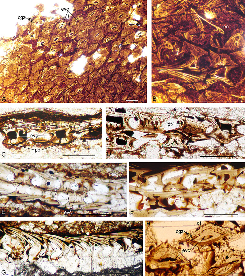
FIGURE 19. Mesacanthus mitchelli (upper) compared with Orcadacanthus pusillus (lower).
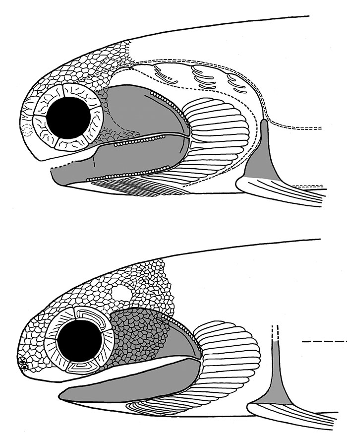
FIGURE 20. Orcadacanthus pusillus, preservation of specimens. A, GLAHM V3573 from Tynet Burn. B, NRM P1645 from Tynet Burn. C, NMS G.2021.7.29 from Achanarras. D, NMS G.2021.7.166 from East Murkle Bay. E, NMS G.1975.12.26 from Stroma (specific locality not recorded, but almost certainly Button Geo). F, NMS G.2021.7.27 from Achanarras. G, NMS G.2022.10.9 from Button Geo, Stroma. Scale bars equal 10 mm.