FIGURE 1. Anatomy of †Whiteia giganteus gen. et sp. nov. YPM VP 3928 in (A) left lateral, (B) right lateral, (C) dorsal, and (D) ventral views.
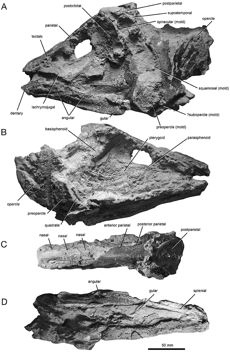
FIGURE 2. Anatomy of †Whiteia giganteus gen. et sp. nov. YPM VP 3928 in left lateral view, showing detail of rostrum.
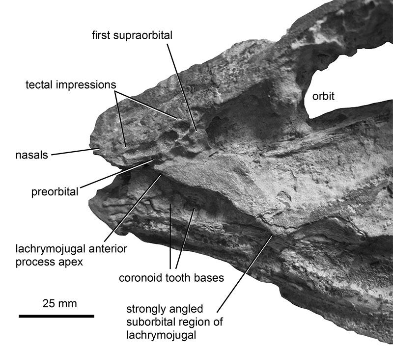
FIGURE 3. Anatomy of †Whiteia giganteus gen. et sp. nov. YPM VP 3928 in right lateral view, showing detail of exposed palate.
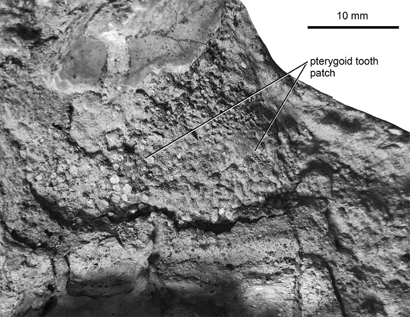
FIGURE 4. Anatomy of †Whiteia giganteus gen. et sp. nov. YPM VP 3928 in (a) left lateral view, showing detail of mandible.
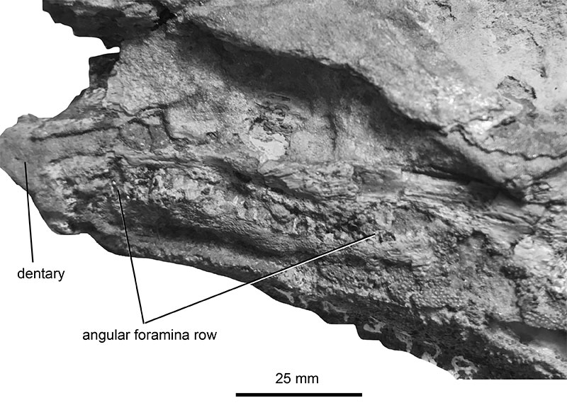
FIGURE 5. Comparative anatomy of †Whiteia giganteus gen. et sp. nov. Skull of †W. giganteus (A) compared to other species of coelacanths from the Late Paleozoic to Mesozoic of North America, including (B) †Whiteia spp. (after Schaeffer et al., 1976), (C) †Diplurus newarki (personal observation of YPM VPPU 14944), and (D) †Chinlea sorenseni (after Schaeffer, 1967). All drawings are traces over photographs. 1 = elongated preorbital region, 2 = lachrymojugal with straightened anterior process, 3 = opercle with rounded dorsal and posterior margins and pointed ventral apex. 1* = large angular foramina row, 2* = strongly angled lachrymojugal below orbit. Numbers indicate apomorphies of †Whiteia, numbers with asterisks indicate autapomorphies of the new species.
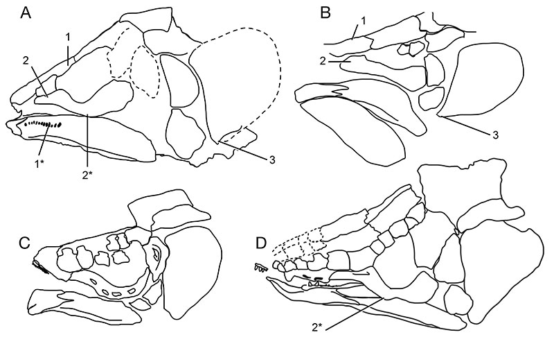
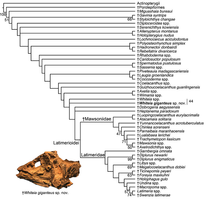
FIGURE 6. Phylogenetic position of †Whiteia giganteus gen. et sp. nov. Strict consensus topology derived from phylogenetic analysis of the 59 OTU, 110-character-dataset in the parsimony program TNT v. 1.5. Numbers indicate bootstrap support values.