FIGURE 1. The sterna of adult individuals of A) the non-pterodactyloid Rhamphorhynchus (based on Wellnhofer, 1975) and B) the pterodactyloid Pteranodon (based on Bennett, 2003). These are seen in i) dorsal, ii) lateral and iii) ventral views. Abbreviations here and throughout are: af, articulation facet; ca, coracoid articulation; cr, cristospine; pf, piercing foramen; pn, pneumatopore; rd ridge (or keel); sp, sternal plate; tb tubercle; xa, xiphoid articulation. Scale bars are A 10 mm and B 50 mm.
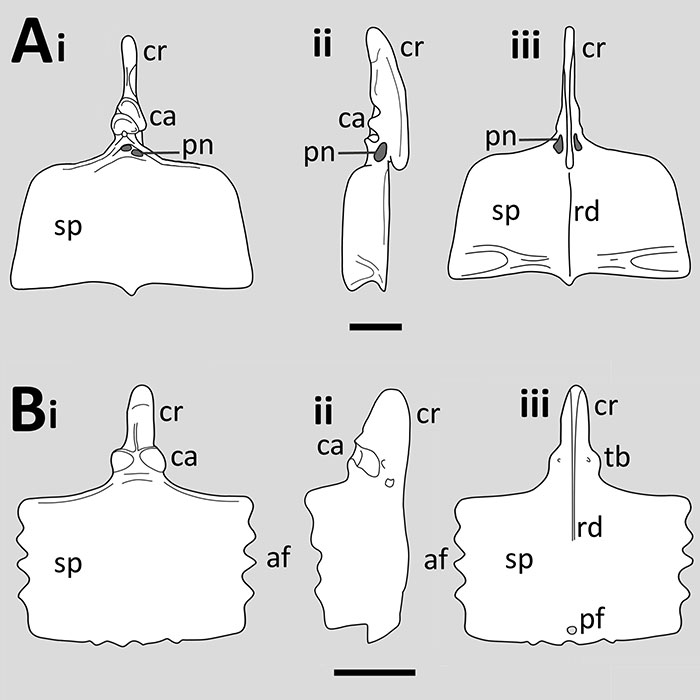
FIGURE 2. Photographs of a variety pterosaur sterna to show the variation in shape and preservation. A) The anurognathid Batrachognathus PIN 52-2, B) the scaphognathid Fenghuangopterus CYGB-0037, C) the rhamphorhynchine Rhamphorhynchus JME-SOS4009, D) the istiodactylid Nurhachius IVPP V 13288 and E) an indeterminate tapejarid MN 6599V. Images not shown to scale.
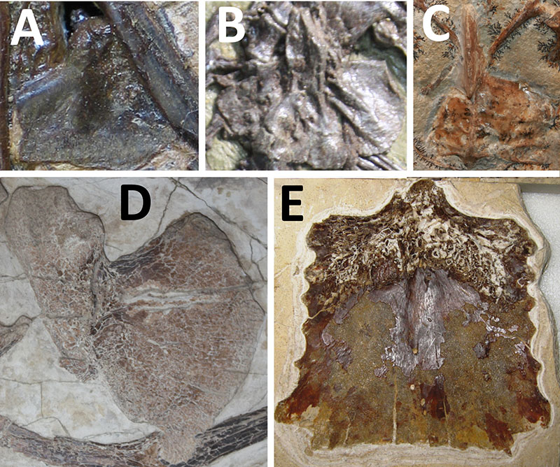
FIGURE 3. The sternum of A) an adult Eudimorphodon (modified from Renesto, 1993) and B) juvenile (traced from Wild 1993). These are seen in A, ventral and B dorsal views. Stippling in B indicates a differing bone texture. Scale bars are A 10 mm and B 5 mm.
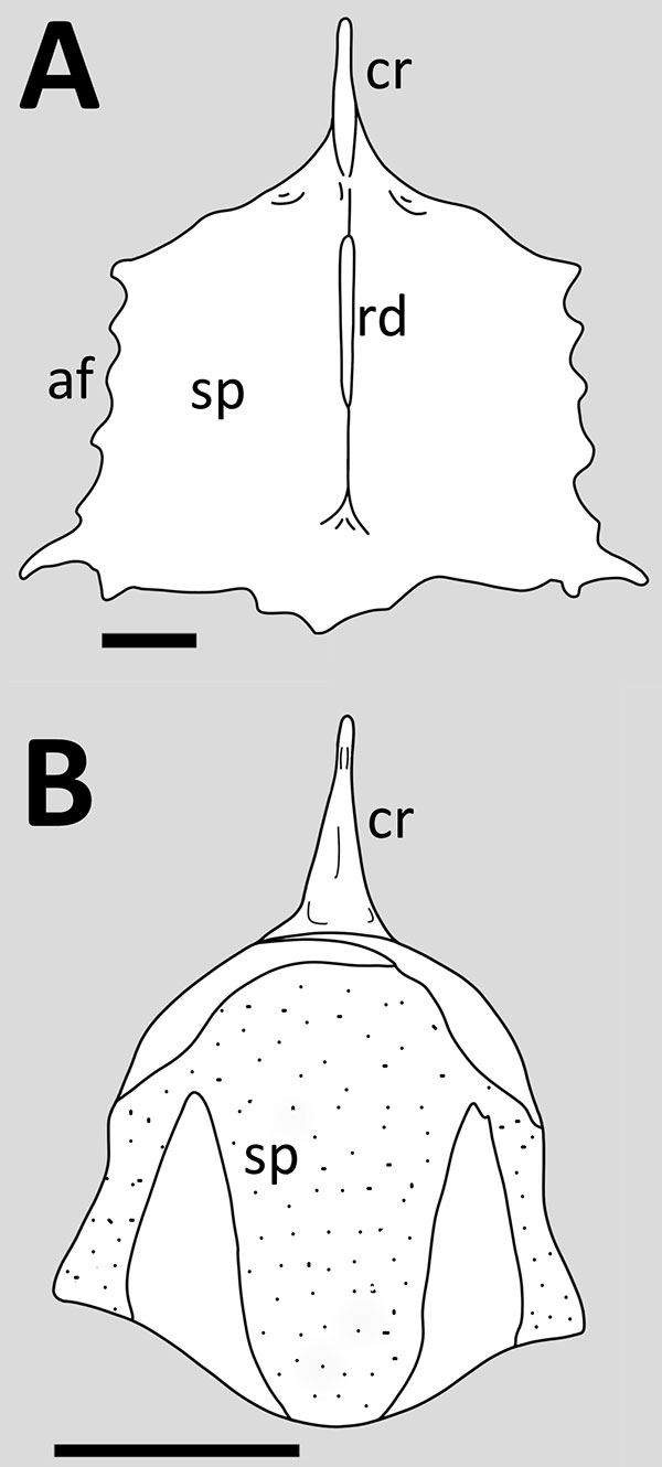
FIGURE 4. The sternum of Campylognathoides A) modified from Wellnhofer, 1978, and B) based on Padian, 2008b. Both shown in ventral view. Scale bar equals 10 mm and applies only to A, no scale was given for the specimen used in Padian’s figure.
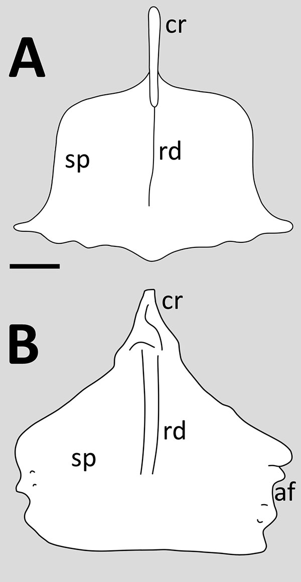
FIGURE 5. The sternum of the anurognathid Batrachognathus (restored based on a photo). This is seen in ?dorsal view. Scale bar equals 1 mm.
FIGURE 6. The sterna of the scaphognathids A) Fenghuangopterus (based on a photo), B) Scaphognathus (based on Wellnhofer, 1978) and C) Nesodactylus (based on Colbert et al., 1969). These are seen in, A ventral view, B dorsal view, Ci ventral and Cii left lateral view (mirrored from the origins right lateral view, dotted line indicates the restored part of the cristospine, following Colbert et al.). Scale bars are A 10 mm, B 10 mm, and C 20 mm.
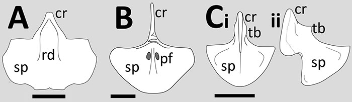
FIGURE 7. The sterna of rhamphorhynchid pterosaurs. A) an adult Rhamphorhynchus (based on Wellnhofer, 197), B) a ‘subadult’ Rhamphorhynchus (based on Wellnhofer, 1975), C) a juvenile Rhamphorhynchus (based on Wellnhofer, 1978), D) Bellubrunnus (drawn from Hone et al., 2012), and E) Dorygnathus (based on Padian, 2008a, scale bar determined from Wiman, 1925). These are seen in Ai ventral and ii lateral views, B dorsal view, Ci ventral and ii lateral views, and D and E ventral view. Scale bars equal 10 mm.
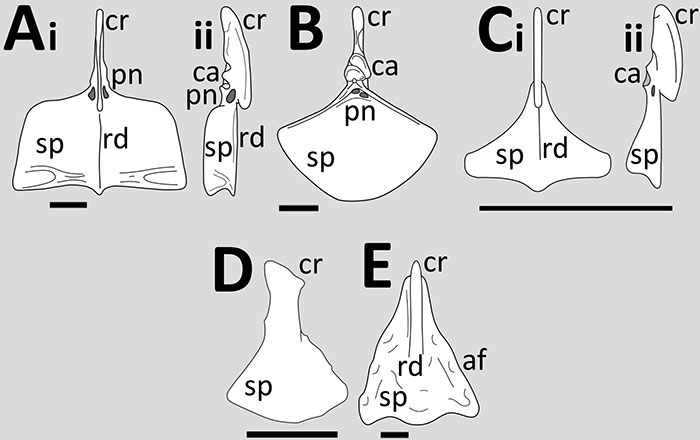
FIGURE 8. The sternum of the wukongopterid Kunpengopterus (drawn from Wang et al., 2010). Shown in dorsal view. Scale bar equals 10 mm.
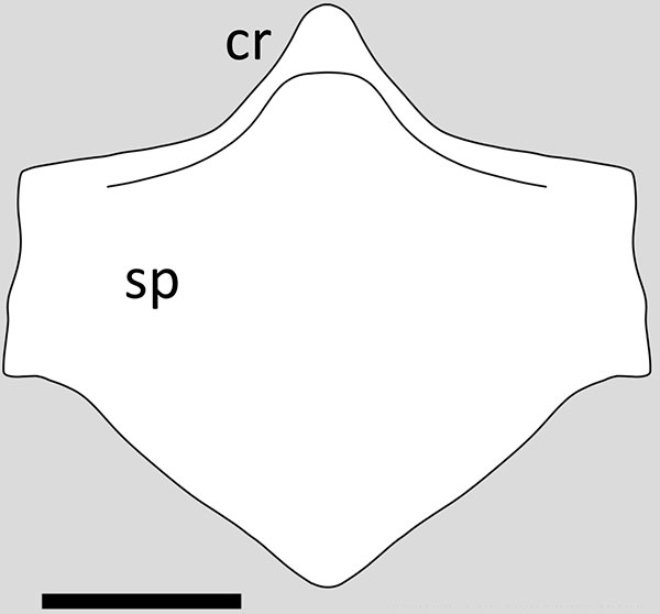
FIGURE 9. The sterna of the ctenochasmatoids A) Pterodactylus (traced from Wellnhofer, 1978), B) Cycnorhamphus (based on Wellnhofer, 1978), C) Ardeadactylus (based on Meyer, 1854) and D) Auroroazhdarcho (drawn from Frey et al., 2011). These are all seen in ventral view. Scale bars are A, B and G 10 mm, B 10 mm. No scalebar was given for C, but based on the published length of the wing phalanges in Wellnhofer, 1970 this would be c. 66 mm in diameter.
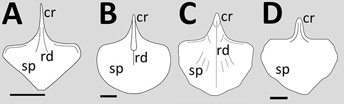
FIGURE 10. The sterna of the pteranodontoid pterosaurs A) Pteranodon (based on Bennett, 2001), B) Nyctosaurus (based on Jiang, 2016) and C) Musquizopteryx (based on Frey et al., 2006). These are all seen in ventral view. Scale bars are A, 50 mm and C, 50 mm, no scale bar was provided for B, but based on Willison, 1902, this would be c. 70 mm in diameter.
FIGURE 11. The sterna of istiodactyliform pterosaurs, A) Haopterus (based on Wang and Lü, 2001) and B) Luichibang (drawn from Hone et al., 2020) where the dashed line indicates the apparent absence of the cristospine. Both are shown in ventral view. Scale bars are A, 10 mm and B, 20 mm.
FIGURE 12. The sterna of the ornithocherids A) Santanadactylus (based on de Buisonje, 1981) and B) Anhanguera (based on Kellner and Tomida, 2000). These in seen in A dorsal view, Bi dorsal view, Bii ventral view, and Biii lateral view. Scale bars are both 50 mm.
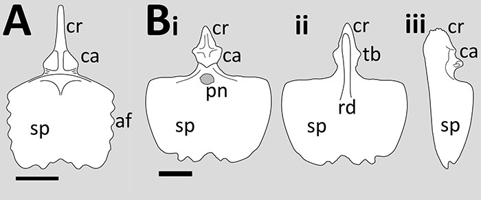
FIGURE 13. The sternum of the ornithocheirid Coloborhynchus (based on Veldmeijer, 2006). This in seen in A anterior, B posterior, C ventral and D lateral view. Scale bar equals 50 mm.
FIGURE 14. The sternum of the dsungaripterid pterosaur Dsungaripterus (based on Jiang et al., 2016) seen in ventral view. No scale bar was given in Jiang et al., 2016 and the original illustration in Young, 1973 also lacks a scale, but based on comparisons to the skull in Plate 3 appears to be c. 100 mm in diameter.
FIGURE 15. The sterna of tapejarid pterosaurs. A) Cauijara (drawn from Manzig et al., 2014), B) Keresdrakon (drawn from Kellner et al., 2019 with the cristospine restored to the sternal plate) and C) Tapejara (based on Eck et al., 2011). These in seen in A ventral view, B dorsal view and Ci anterior, Cii dorsal and Ciii ventral lateral views. Scale bars are A 10 mm, B 50 mm, and C 20 mm.
FIGURE 16. The sternum of Jidapterus (based on Wu et al., 2017). This is in dorsal or ventral view. Scale bar equals 10 mm.
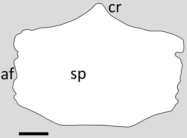
FIGURE 17. The incomplete sternum of the giant azhdarchid Quetzalcoatlus (based on Andres and Langston, 2021) which has most of the sternal plate missing. Shown in A) dorsal, B) ventral and C) anterior views. Scale bar equals 100 mm.
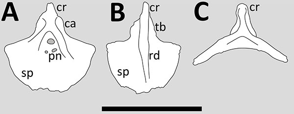
FIGURE 18. Generalised reptile sternum based on Cau et al., (2021) and reconstructed sternum of the dinosauromorph Tawa based on Bradley et al., (2019) both seen in ventral view. Abbreviations as follows: af, articulation facets; ca, clavicle; co, coradoic; ic interclavicle; sc, scapula; sp sternal plate.
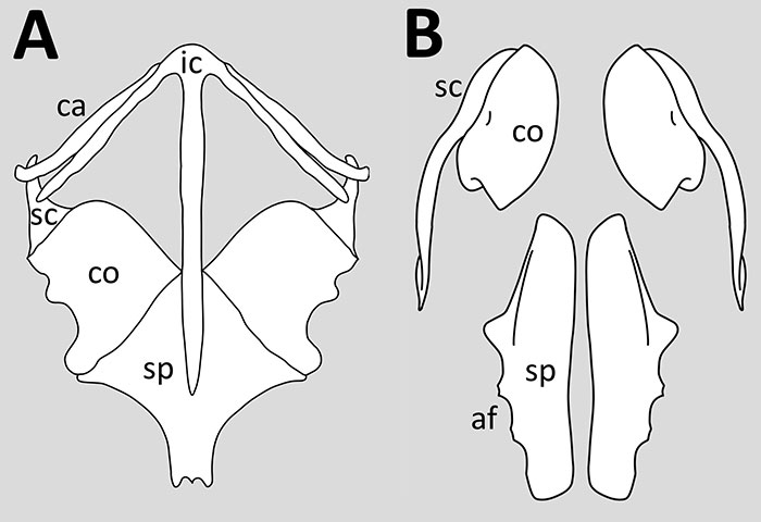
FIGURE 19. A) Identities of the elements that make up the pterosaur sternum according to Wild (1993) based on a juvenile Eudimorphodon and B) photograph of the sternum of a juvenile Altmulopterus in ?dorsal view (LF 2086P) showing a pair of dark areas either side of the midline of the sternal plate which are inferred to be centres of condensation.
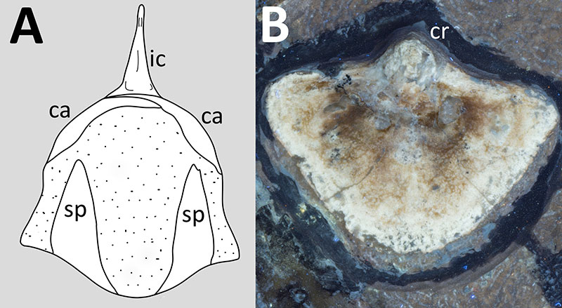
FIGURE 20. Hypothesised orientations and articulations of the pterosaurian sternum using Pteranodon as a model in A) anterior view and B) left lateral view based on Bennett, 2001 and C) based on Claessens et al., 2009. Note that A is shown at a larger size than B and C for clarity and shows only the sternum and coracoids, the coracoids are not shown in B or C as they would obscure much of the detail, but the upper part of the scapulocoracoids would articulate with the notarium. Abbreviations: co, coaracoids; dr, dorsal ribs; gs, gastralia; no, notarium; sa, scapula articulation; sr, sternal ribs.
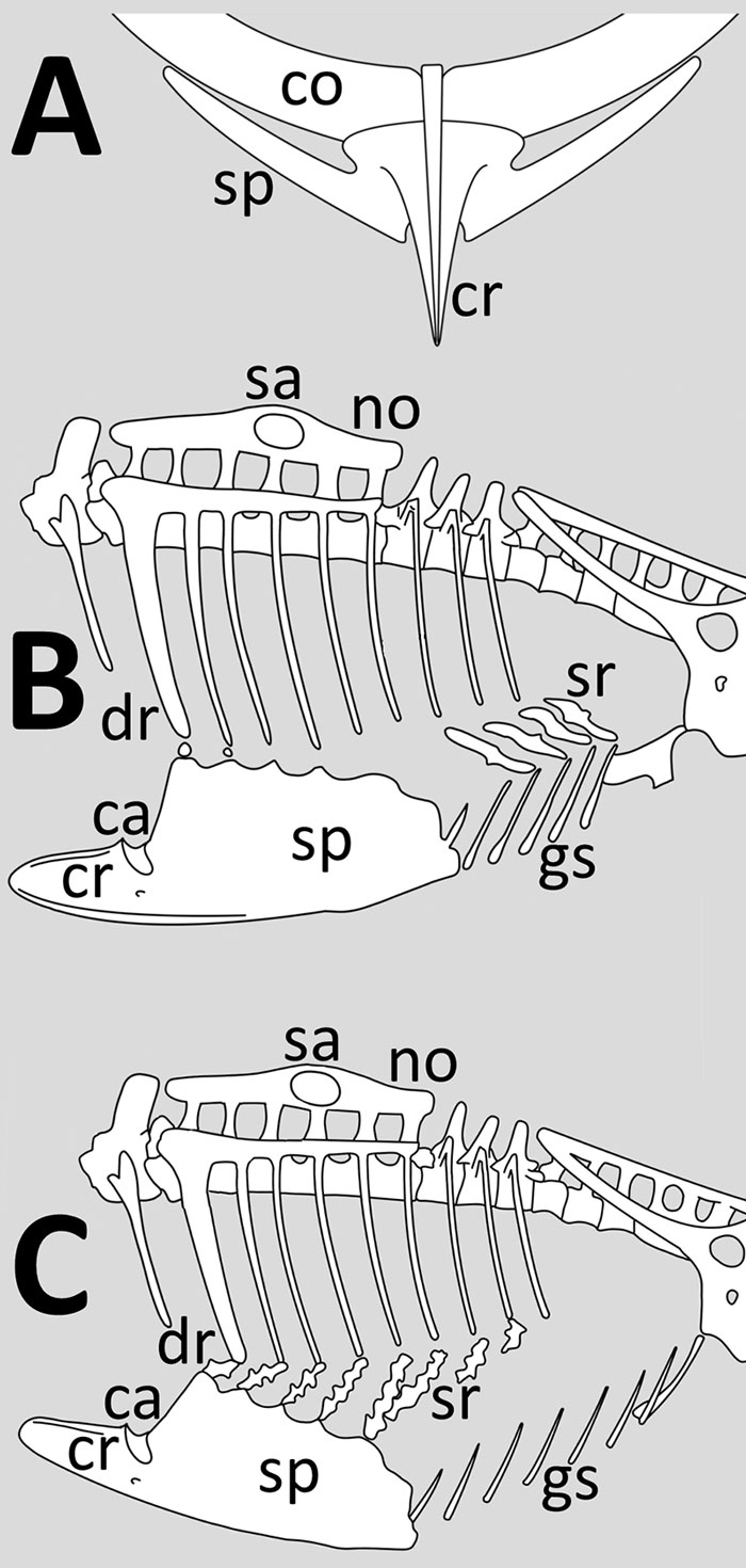
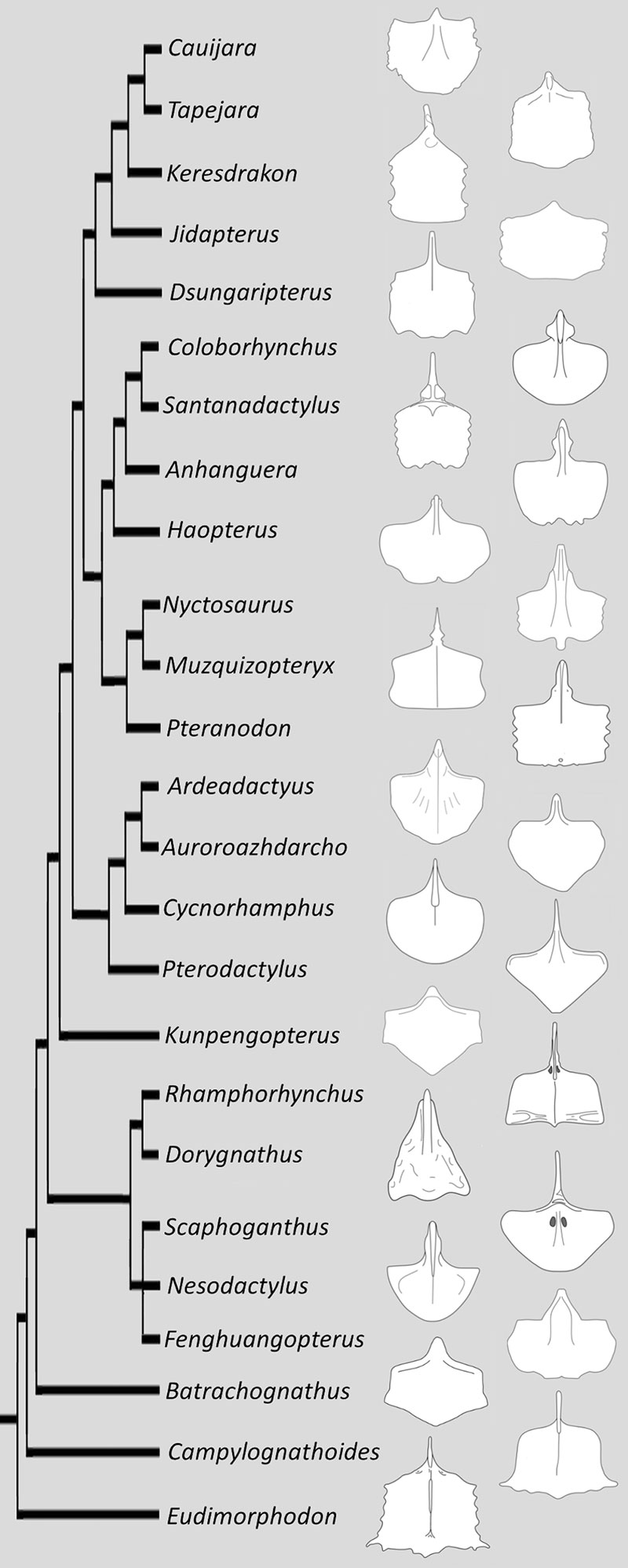
FIGURE 21. Figure of the diversity of shapes of pterosaur sterna plotted onto a dendrogram of pterosaurian relationships (images not to scale). This is based on Zhou et al., 2021 (non-pterodactyloids), Andres, 2021 (ctenochasmatids), Hone et al., 2020 (other pterodactyloids). Where there are multiple specimens, preference has been given to adult specimens and all sterna are shown in ventral view except Santanadactylus and Scaphognathus which are in dorsal view.