FIGURE 1. A. Simplified geographic map of Bohemian Cretaceous Basin indicating locations of the studied sites (in rectangle). B. Geographic position of nearshore deposits at Velim, Kaňk “Na Vrších” and Kamajka, where samples were taken (black pentangles).
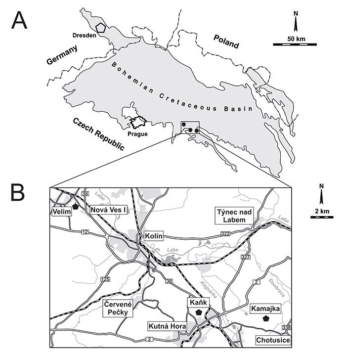
FIGURE 2. Stratigraphic provenance of serpulid tubes from Velim, Kamajka, and Kaňk. 1 - crystalline basement; 2 - basal Cenomanian conglomerate; 3 - redeposited Turonian conglomerate; 4 - bioclastic limestone with calcitic-clayey matrix; 5 - organodetritic clayey limestone; 6 - marly siltstone with intercalations of phosphatized horizon; 7 - sponge ‛meadows’; 8 - limestone layer with nodule-like bodies; 9 - calcareous claystone (modified from Košťák et al., 2010; Kočí, 2012). Full filled circles indicate position of serpulid fauna.
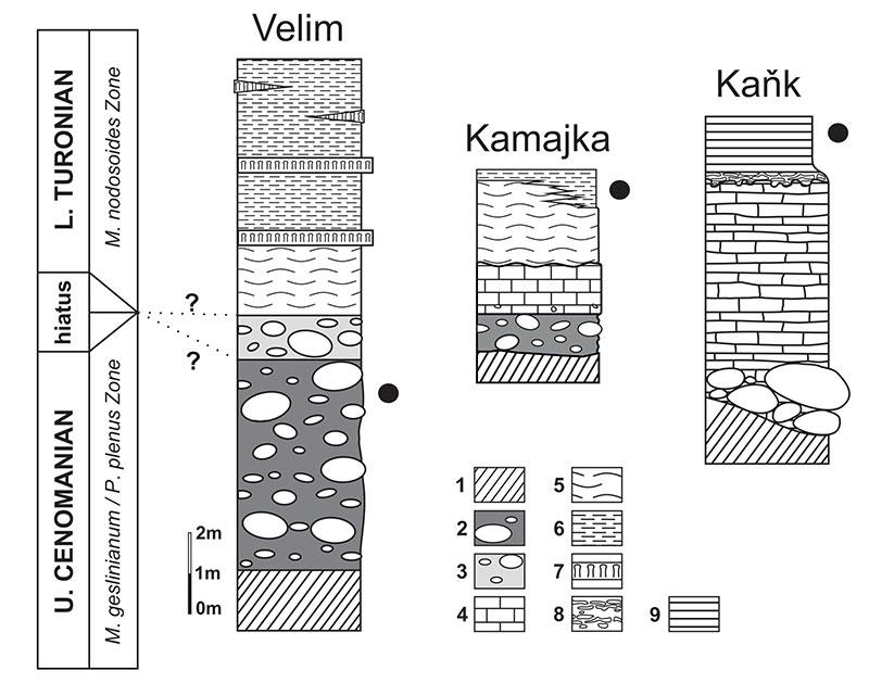
FIGURE 3. Cementula sp., a coiled tube, no. CZ2, from Velim. A-C. Scanning electron microscope images of resin cast, B-C insets in A showing microbioerosion beneath D-F. Micro-CT images. A, D and E. Identical views using different methods. B. SEM image of resin cast shows branching stolons of Iramena isp., below lower limit of micro-CT resolution. C. Detail showing microbioerosion beneath tube surface and shaft incompletely filled with epoxy resin. D. 2D section through both tubes. E. Semi-transparent rendering of 2D section. F. Volume reproduction image, 3D view to smooth inner surfaces of the tubes.
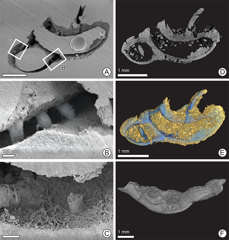
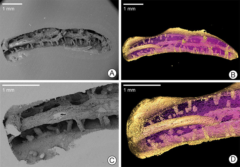
FIGURE 4. Placostegus zbyslavus (Ziegler, 1984), longitudinal section of a tube from Kamajka near Chotusice, no. NM O8727. A. SEM image of the resin cast showing a high degree of silicification that led to incomplete dissolution of the tube wall in HCl; image shows only indeterminate non-branching shafts. B. The same view of the specimen using micro-CT clearly shows relatively frequent Maeandropolydora isp. and shallow shafts of Trypanites isp.
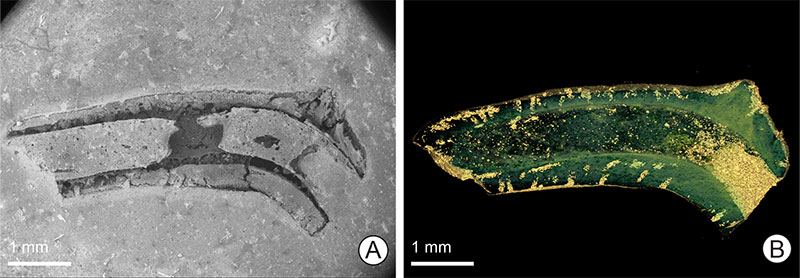
FIGURE 5. Pyrgopolon (Septenaria) cf. tricostata (Goldfuss, 1841), longitudinal section of a tube from Kaňk “Na Vrších”, no. NM O8728. A. SEM image of the resulting cast providing a three-dimensional view of three bi-camerate specimens of Entobia isp. and numerous shafts of Trypanites isp. cut by the longitudinal boring Maeandropolydora isp.; galleries are duplicated, more or less parallel, partially touching each other. B. The same view to the specimen by using micro-CT. C. detail of the resin cast showing a pair of bi-camerate Entobia isp. D. micro-CT scan from the same view, details of Entobia chambers are below the lower limit of micro-CT resolution.