FIGURE 1. Map of study area. Black dots denote cities/towns. Red squares denote primary field localities. A: Swift Current Creek locality. B-C: Lac Pelletier main primate-bearing exposures.

FIGURE 2. New upper molar of Trogolemur from the Swift Current Creek locality, with comparisons. A, E: New specimen of Trogolemur, a left M1, RSM P3450.1466. A: Photograph of RSM P3450.1466 on pin. E: Surface model of RSM P3450.1466 from µCT scan. This specimen was scanned at the Duke University Shared Materials Instrumentation Facility using a Nikon XTH 225 ST µCT scanner, with a voxel size of 0.018 mm, a voltage of 135 kV, an amperage of 107µa, and 2500 projections. A surface model was reconstructed in Avizo 8.0 (2013; FEI Visualization Sciences Group). B-D: Trogolemur leonardi from Lac Pelletier. B: RM2 (RSM P1899.1007, reversed image, originally labeled M1). C: RM3 (RSM P1899.1016, reversed image, originally labeled as M2). D: LM3 (RSM P1899.1014). F: Stereo pair of cast of Trogolemur myodes RM2-M3 (UM 31201, reversed image). Compare with USNM 417396, M1 of Trogolemur (Emry, 1990, fig. 6H). Scale bar is 1 mm long.
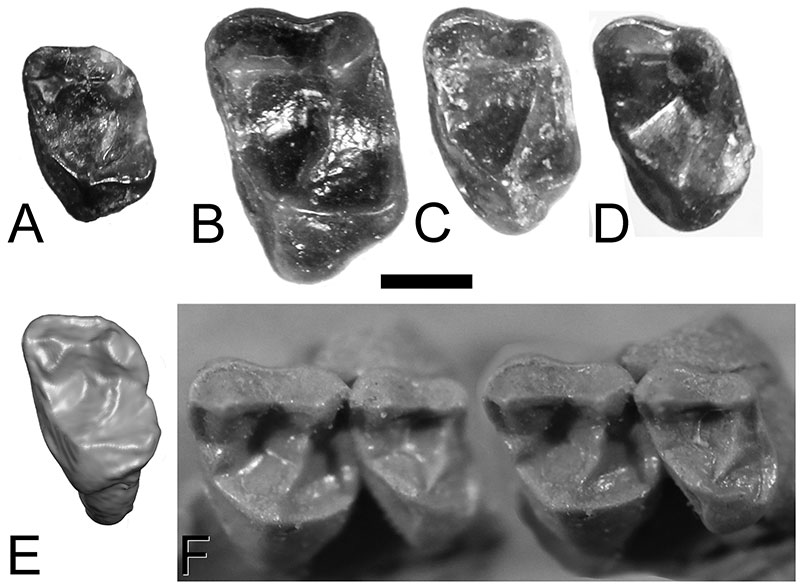
FIGURE 3. Lower teeth of Trogolemur from Lac Pelletier, previously described by Storer (1990). A: Lp4 (RSM P1899.1001). B: Lm1 or m2 (RSM P1899.1002). C: Rm1 or m2 (RSM P1899.1003). Scale bar is 1 mm long.
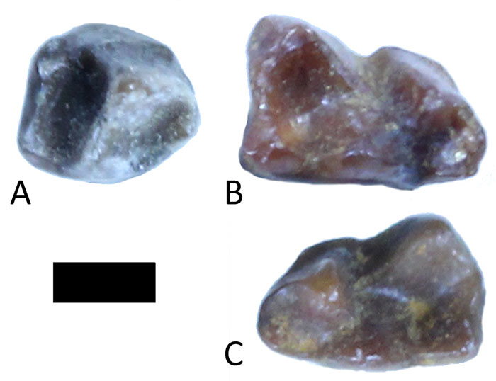
FIGURE 4. New lower third molars of Trogolemur from Swift Current Creek. A: Lm3, RSM P3450.1467. B: Rm3, RSM P3450.1468. Compare with m3s of Trogolemur myodes AMNH 12599 (Matthew, 1909, fig. 5), USNM 417355 (Emry, 1990, fig. 6D-F), and USNM 417356 (Emry, 1990, fig. 6A-C). Scale bar is 1 mm long.
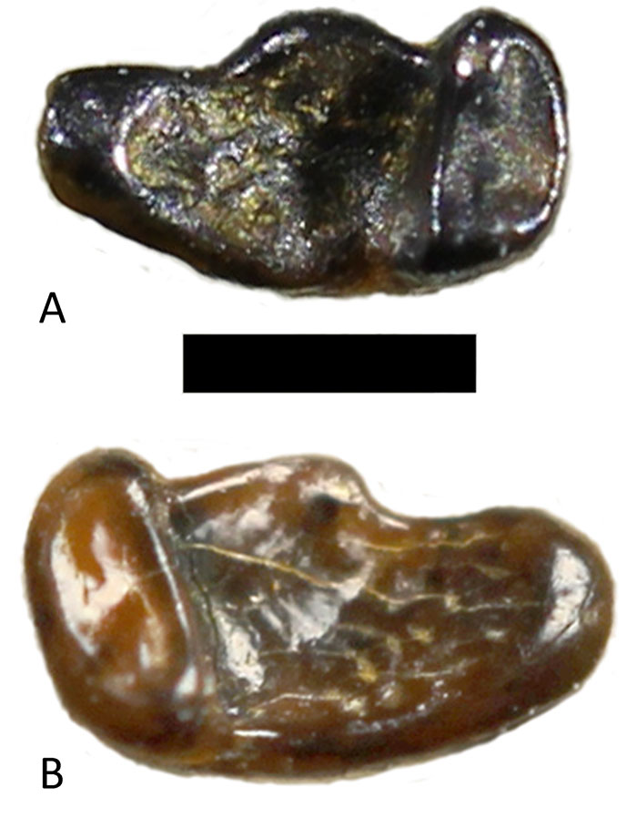
FIGURE 5. Stereopair of RM1 (RSM P3450.1469) from Saskatchewan, assigned to Walshina mcgrewi. Maximum buccolingual dimension is 3.03 mm. Maximum mesiodistal dimension is 2.14 mm. Scale bar is 1 mm long.
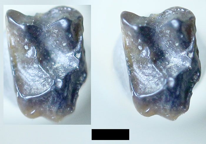
FIGURE 6. Macrotarsius cf. M. montanus from Lac Pelletier. A: Rm1 (RSM P1899.1009). B: Rp2? (RSM P1899.1008). Scale bar is 1 mm long.
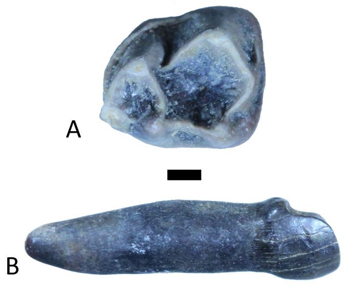
FIGURE 7. Upper teeth of Saskomomys lindsayorum from Lac Pelletier, described by Storer (1990) as “Omomys sp.”. A: LM1 (RSM P1899.1011, originally identified as M2). B: LM2 (RSM P1899.1010, originally identified as M1). C: LM3 (RSM P1899.1013). D: RM1 or M2 (RSM P1899.1015). E: LM2 (RSM P1899.1012). Scale bar is 1 mm long.
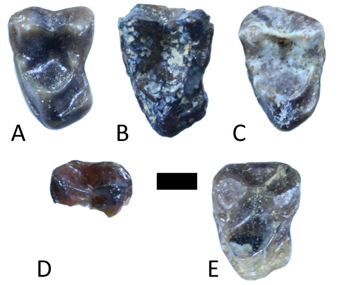
FIGURE 8. Lower teeth of Saskomomys lindsayorum from Lac Pelletier, described by Storer (1990) as “ Omomys sp.”. A: Lp3 (RSM P1899.1017) in occluso-buccal and lingual views. B: Lm1 (RSM P1899.1019). C: Lm1 (RSM P1899.1021, originally identified as m2). D: Lm2 (RSM P1899.1023). E: Rm2 (RSM P1899.1025). Scale bar is 1 mm long.
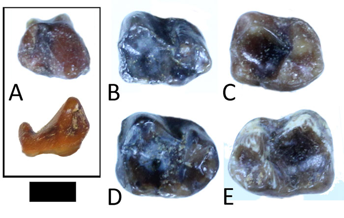
FIGURE 9. Teeth from the Swift Current Creek locality described by Storer (1984) as Omomyidae. A-B: Teeth described as “Omomyidae sp. 1”. A: Rm3 of Saskomomys lindsayorum (RSM P1654.343), holotype. B: Rm3 of Saskomomys lindsayorum (RSM P1654.344). C: Molar of artiodactyl originally described as RM3 of “Omomyidae sp. 2”. Scale bar is 1 mm long.
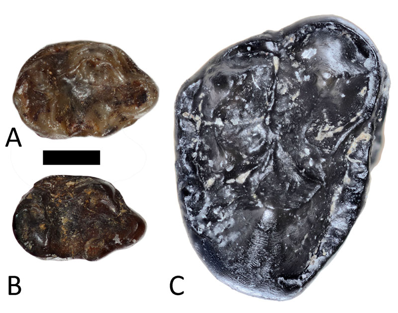
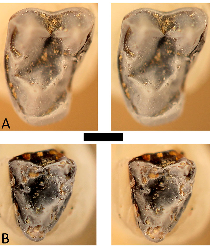
FIGURE 10. Stereo pairs of upper molars of Saskomomys lindsayorum from the Swift Current Creek locality. A: RSM P3450.1470, a left M1. B: RSM P3450.1471, a right M3. Scale bar is 1 mm long.