FIGURE 1. Overview of the main bones preserved in the specimens of Danielsavis nazensis Houde et al., 2023 from the London Clay of Walton-on-the-Naze (Essex, UK). A, holotype, NMS.Z.2021.40.1 (not shown are the radii, which were figured by Houde et al., 2023: figure 5Q, R). B, referred specimen NMS.Z.2021.40.2 (the two separate blocks of matrix containing the beak and the neurocranium, respectively, were assembled for the photo; not shown is the cranial portion of the sternum, which was figured by Houde et al., 2023: figure 7E, F). C, referred specimen NMS.Z.2021.40.3. D, referred specimen NMS.Z.2021.40.6. Abbreviations: for, foramen piercing skull roof; wdn, widening of scapus claviculae. The scale bars equal 10 mm.
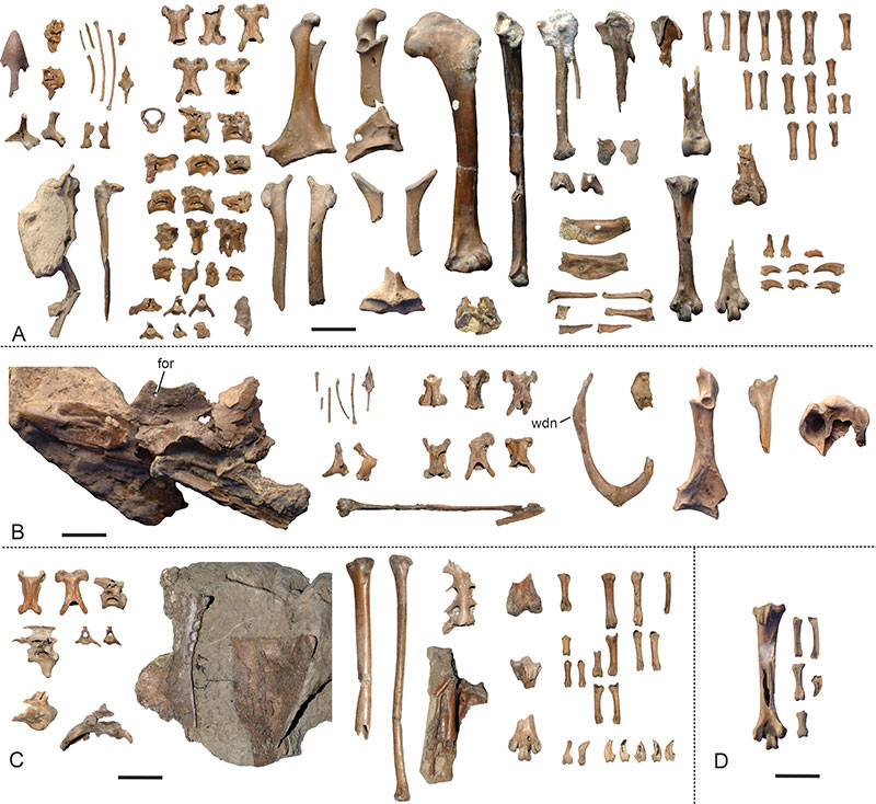
FIGURE 2. Danielsavis nazensis Houde et al., 2023, upper beak, palate, and mandible. A‒C, D. nazensis, upper beak of NMS.Z.2021.40.2 in right lateral (A), left lateral (B), and dorsal (C) view. D, D. nazensis, tip of the upper beak of the holotype (NMS.Z.2021.40.1) in dorsal view. E, D. nazensis, neurocranium of NMS.Z.2021.40.2 in ventral view. F, G, D. nazensis, palatine bones of NMS.Z.2021.40.2 in ventral view, in F, surrounding bones and matrix were digitally brightened. H‒J, palatines (ventral view) of H, Alectura lathami (Megapodiidae, Galliformes; SMF 7243); I, Chauna torquata (Anhimidae, Anseriformes; SMF 19920); J, Anseranas semipalmata (Anseranatidae, Anseriformes; SMF 11276). K, L, D. nazensis, left ramus mandibulae of the holotype (NMS.Z.2021.40.1) in dorsal (K) and lateral (L) view; the arrows denote details of the caudal end. M, N, D. nazensis, right ramus mandibulae of the holotype (NMS.Z.2021.40.1) in medial (M) and dorsal (N) view. O, P, D. nazensis, partial mandible of NMS.Z.2021.40.2 in dorsal (O) and dorsolateral (P) view; the arrow denotes a detail of the caudal end. Q, mirrored right ramus mandibulae of Anachronornis anhimops from the late Paleocene of Wyoming (holotype, coated with ammonium chloride; from Houde et al., 2023: figure 1, published under a CC BY 4.0 license). R, mandible of C. torquata (SMF 19920) in lateral view. S, mandible of A. semipalmata (SMF 11276) in lateral view. Abbreviations: acl, angulus caudolateralis; ame, flange-like process that served for the attachment of musculus adductor mandibulae externus; crv, crista ventralis; ctl, cotyla lateralis; ctm, cotyla medialis; for, foramen piercing skull roof; ldc, lamella dorsalis, pars choanalis palatini; pbp, processus basipterygoideus; pla, pars lateralis; pra, processus retroarticularis; rps, rostrum parasphenoidale. The scale bars equal 10 mm.
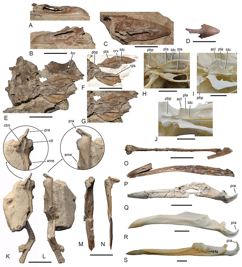
FIGURE 3. Cranial elements and hyoid bone of Danielsavis nazensis Houde et al., 2023 in comparison with those of other galloanserine birds. A‒F, D. nazensis (holotype, NMS.Z.2021.40.1), right (A‒D) and left (E, F) quadrate in lateral (A, E), caudal (B), medial (C, F), and ventral (D) view. G, H, D. nazensis (NMS.Z.2021.40.2), left (G) and right (H) quadrate in lateral view. I, J, right quadrate of Pipile jacutinga (Cracidae, Galliformes; SMF 4139) in lateral (I) and medial (J) view. K, L, mirrored left quadrate of Anseranas semipalmata (Anseranatidae, Anseriformes; SMF 11276) in lateral (K) and medial (L) view. M, N, Anachronornis anhimops from the late Paleocene of Wyoming (holotype, coated with ammonium chloride; from Houde et al., 2023: figure 1, published under a CC BY 4.0 license), left quadrate in lateral (M) and medial (N) view. O‒R, D. nazensis (holotype, NMS.Z.2021.40.1), right (O, P) and left (Q, R) pterygoid in dorsal (O, Q) and medial (P, R) view. S, D. nazensis (NMS.Z.2021.40.2), basiurohyal in dorsal view. T, U, D. nazensis (holotype, NMS.Z.2021.40.1), basiurohyal and putative paraglossum in lateral (T) and dorsal (U) view. V, Alectura lathami (Megapodiidae, Galliformes; SMF 19785), basiurohyal and paraglossum in dorsal view. W, Chauna torquata (Anhimidae, Anseriformes; SMF 19920), basiurohyal and paraglossum in dorsal view; the arrow denotes an enlarged detail of the basihyal. X, A. semipalmata (SMF 19902), basiurohyal and paraglossum in dorsal view; the arrow denotes an enlarged detail of the basihyal. Abbreviations: arf, articular facet for pterygoid; bsh, os basihyale; cbr, os ceratobranchiale; cdl, condylus lateralis; cdm, condylus medialis; cdp, condylus pterygoideus; cpo, capitulum oticum; cps, capitulum squamosum; cqj, cotyla quadratojugalis; fab, facies articularis basipterygoidea; fpb, foramen pneumaticum basiorbitale; fpc, foramen pneumaticum caudomediale; fqv, facies quadratojugalis ventralis; orb, processus orbitalis; pgl, os paraglossum; tsc, tuberculum subcapitulare; urh, os urohyale. The scale bars equal 5 mm.
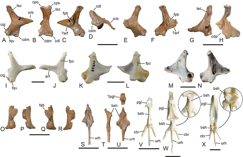
FIGURE 4. Danielsavis nazensis Houde et al., 2023, comparison of the vertebrae with those of other galloanserine birds. A, D. nazensis, cranialmost five cervical vertebrae (atlas: NMS.Z.2021.40.1; axis, third, and fourth vertebra: NMS.Z.2021.40.2; fifth vertebra: NMS.Z.2021.40.3). B‒D, cranialmost five cervical vertebrae of B, Chauna torquata (Anhimidae, Anseriformes; SMF 11885); C, Dendrocygna arborea (Anatidae, Anseriformes; SMF 6591); D, Alectura lathami (Megapodiidae, Galliformes; SMF 7243). E, D. nazensis, selected cervical and thoracic vertebrae of the holotype (NMS.Z.2021.40.1); the arrows denote enlarged details of the bones to show irregular lamellate projections. F‒I, axis in lateral view of F, D. nazensis (NMS.Z.2021.40.2); G, C. torquata (SMF 11885); H, D. arborea (SMF 6591); I, A. lathami (SMF 7243). J, D. nazensis, fourth cervical vertebra of the holotype (NMS.Z.2021.40.1) in dorsal and lateral view. K, D. nazensis, fourth cervical vertebra of the referred specimen NMS.Z.2021.40.2 in dorsal and lateral view. L, D. nazensis, thoracic vertebra in lateral view (NMS.Z.2021.40.3). Abbreviations: dns, dens; for, foramen; ftv, foramen transversarium; ntc, notch; ote, ossified tendons; pno, pneumatic opening; ptr, processus transversus; pvt, processus ventralis; zcd, zygapophysis caudalis; zcr, zygapophysis cranialis. The scale bars equal 5 mm.
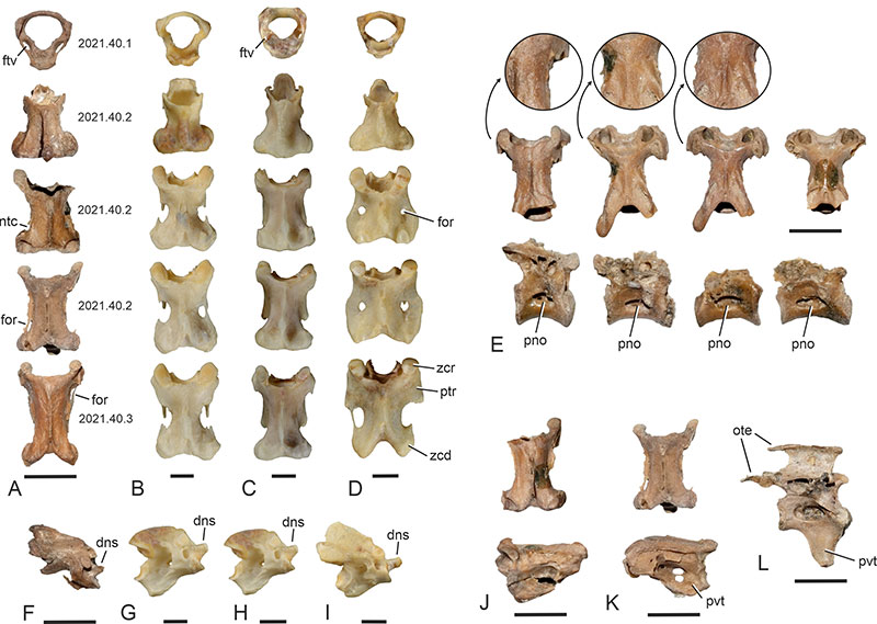
FIGURE 5. Danielsavis nazensis Houde et al., 2023, comparison of pectoral girdle and wing bones with those of other galloanserine birds. A, B, D. nazensis (holotype, NMS.Z.2021.40.1), right coracoid in ventral (A) and dorsal (B) view. C, right coracoid of an undescribed stem group galliform from Walton-on-the-Naze in dorsal view. D, mirrored left coracoid of Anachronornis anhimops from the late Paleocene of Wyoming (holotype, coated with ammonium chloride; from Houde et al., 2023: figure 2, published under a CC BY 4.0 license) in dorsal view. E, right coracoid of Nettapterornis oxfordi from Walton-on-the-Naze (holotype, NHMUK A 5922) in dorsal view. F, mirrored left coracoid of the presbyornithid Telmabates antiquus from the early Eocene of Argentina (AMNH 3181). G, partial furcula of D. nazensis (NMS.Z.2021.40.2) in craniomedial view. H, furcula of Anseranas semipalmata (Anseranatidae, Anseriformes; SMF 11276) in caudomedial view. I, furcula of Alectura lathami (Megapodiidae, Galliformes; SMF 7243) in caudomedial view. J, K, cranial extremity of right scapula of D. nazensis (J: holotype, NMS.Z.2021.40.1; K: NMS.Z.2021.40.2) in lateral view. L, M, left humerus of D. nazensis (holotype, NMS.Z.2021.40.1) in caudal (L) and cranial (M) view. N, left humerus of A. anhimops (holotype, coated with ammonium chloride; from Houde et al., 2023: figure 2, published under a CC BY 4.0 license) in caudal view. O, left humerus of N. oxfordi (holotype, NHMUK A 5922) in cranial view. P, mirrored proximal end of a right humerus from the early Eocene of Egem in Belgium (IRSNB Av 163); coated with ammonium chloride. Q, left humerus of Gallinuloides wyomingensis from the early Eocene Green River Formation (Wyoming, USA; WDC CGR−012) in cranial view; surrounding matrix was digitally removed, the dotted line indicates the reconstructed outline of the ventral portion of the proximal end. R‒T, proximal end of left ulna in cranial view of R, D. nazensis (holotype, NMS.Z.2021.40.1); S, an unnamed galliform from the early Eocene of Egem in Belgium (IRSNB Av 164); T, Chauna torquata (Anhimidae, Anseriformes; SMF 11885); the arrows point to the distal end of the cotyla dorsalis. U, V, D. nazensis (holotype, NMS.Z.2021.40.1), right (U) and proximal left (V) carpometacarpus in ventral view. W, N. oxfordi (holotype, NHMUK A 5922), left carpometacarpus in ventral view. X, A. anhimops (holotype, coated with ammonium chloride; from Houde et al., 2023: figure 2, published under a CC BY 4.0 license), left carpometacarpus in ventral view. Y, Z, D. nazensis (holotype, NMS.Z.2021.40.1), left (Y) and right (Z) os carpi radiale in cranial view. AA, Dendrocygna viduata (Anatidae, Anseriformes; SMF 2418), left os carpi radiale in cranial view. BB, Pternistis squamatus (Phasianidae, Galliformes; SMF 10151), left os carpi radiale in cranial view. The dotted lines in B‒F indicate the angle between the medio-omal and latero-omal portions of the processus acrocoracoideus. Abbreviations: acr, acromion; cpc, crista procoracoidei; csc, cotyla scapularis; ctd, cotyla dorsalis; ctv, cotyla ventralis; fah, facies articularis humeralis; flx, processus flexorius; fns, foramen nervi supracoracoidei; ila, impressio ligamenti acrocoracohumeralis; mpr, medial projection; olc, olecranon; pac, processus acrocoracoideus; pla, processus lateralis; ppc, processus procoracoideus; pra, processus acromialis; tbd, tuberculum dorsale; trc, trochlea carpalis. The scale bars equal 10 mm.
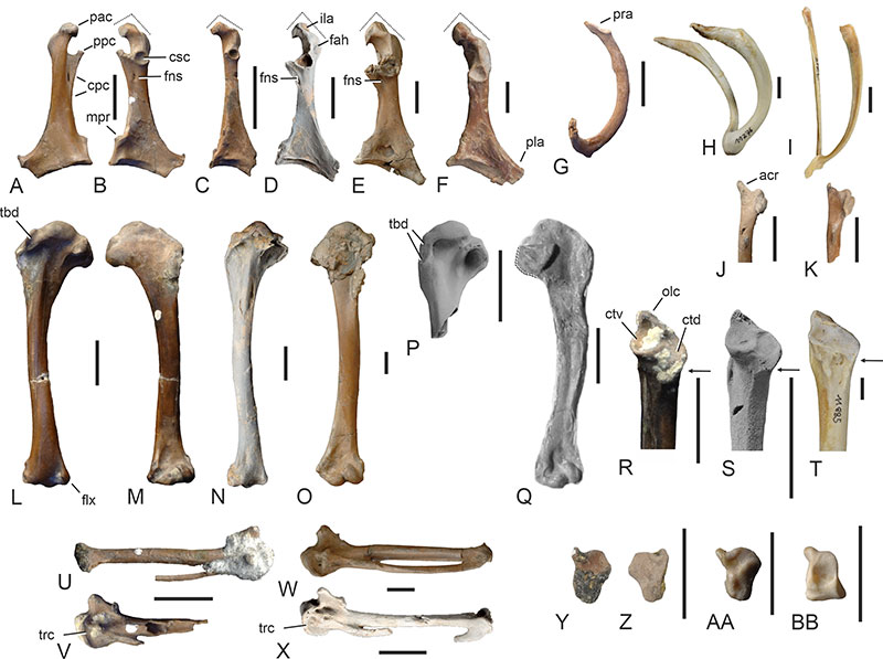
FIGURE 6. Danielsavis nazensis Houde et al., 2023, comparison of tibiotarsus, tarsometatarsus, pedal phalanges with other galloanserine birds. A, D. nazensis (holotype, NMS.Z.2021.40.1), distal end of right tibiotarsus; the arrow denotes an enlarged detail of the bone. B, distal end of right tibiotarsus of the anseriform Saintandrea chenoides from the late Oligocene of France (holotype, NMB Mar. 874e). C, mirrored distal end of left tibiotarsus of an unnamed galliform from the early Eocene of Egem in Belgium (IRSNB Av 168); coated with ammonium chloride. D‒G, D. nazensis (holotype, NMS.Z.2021.40.1), right tarsometatarsus in distal (D), proximal (E), dorsal (F), and plantar (G) view; the arrow denotes an enlarged detail of the proximal end of the bone. H, I, D. nazensis (NMS.Z.2021.40.3), proximal end of right tarsometatarsus in proximal (H) and plantar (I) view. J, D. nazensis (NMS.Z.2021.40.3), distal end of right tarsometatarsus in plantar view; the arrow denotes an enlarged detail of the bone. K, D. nazensis (holotype, NMS.Z.2021.40.1), non-ungual pedal phalanges; the asterisk indicates a mirrored phalanx. L, mirrored left foot of Chauna torquata (Anhimidae, Anseriformes; SMF 19920). M, right foot of Pavo cristatus (Phasianidae, Galliformes; SMF 20342). N, mirrored distal end of left tarsometatarsus of Anachronornis anhimops (holotype, coated with ammonium chloride; from Houde et al., 2023: figure 2, published under a CC BY 4.0 license) in plantar view. The dotted lines in A‒C indicate the midline of the distal tibiotarsus. The arrows in K‒M denote the length of the third phalanx of the fourth toe. Abbreviations: cdl, condylus lateralis; cdm, condylus medialis; cmh, crista medialis hypotarsi; ext, sulcus extensorius; fdl, sulcus for musculus flexor digitorum longus; fhl, sulcus for musculus flexor hallucis longus; ltr, lateral rim of plantar articular surface of trochlea metatarsi III; mdr, medial rim of plantar articular surface of trochlea metatarsi III; pst, pons supratendineus. The scale bars equal 10 mm.
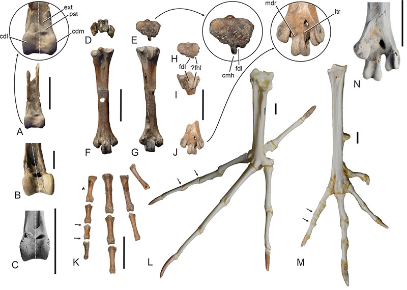
FIGURE 7. Specimens of Perplexicervix paucituberculata, sp. nov. and Perplexicervix sp. from the early Eocene London Clay of Walton-on-the-Naze (Essex, UK). A, P. paucituberculata, sp. nov. (holotype, NMS.Z.2021.40.7). B, cf. P. paucituberculata, sp. nov. (NMS.Z.2021.40.91). C, cf. P. paucituberculata, sp. nov. (NMS.Z.2021.40.92). D, Perplexicervix sp. (NMS.Z.2021.40.9). E, Perplexicervix sp. (NMS.Z.2021.40.10). Abbreviation: for, foramen perforating caudoventral portion of corpus pygostyli. The scale bars equal 10 mm.
FIGURE 8. Selected skeletal elements of Perplexicervix paucituberculata, sp. nov. from the early Eocene London Clay of Walton-on-the-Naze (Essex, UK) and P. microcephalon from the latest early or earliest middle Eocene of Messel (Germany). A, B, atlas of P. paucituberculata, sp. nov. (holotype, NMS.Z.2021.40.7) in dorsal (A) and cranial (B) view; the arrow denotes a detail of the incomplete arcus atlantis. C‒E, axis of P. paucituberculata, sp. nov. (holotype, NMS.Z.2021.40.7) in left lateral (C), dorsal (D), and ventral (E) view; the dotted line indicates the reconstructed shape of the broken processus ventralis. F, axis (lateral view) of P. microcephalon (SMF-ME 3548); coated with ammonium chloride. G, H, third cervical vertebra of P. paucituberculata, sp. nov. (holotype, NMS.Z.2021.40.7) in dorsal (G) and ventral (H) view; the arrows denote details of the tuberculate surface. I, cervical vertebra of P. paucituberculata, sp. nov. (holotype, NMS.Z.2021.40.7) in ventral view; the arrow denotes a detail of the tuberculate surface. J, fourth and fifth cervical vertebrae of P. microcephalon (holotype, SMF-ME 11211a); coated with ammonium chloride. K, thoracic vertebra of P. paucituberculata, sp. nov. (holotype, NMS.Z.2021.40.7). L, thoracic vertebra of P. microcephalon (SMF-ME 2559a); coated with ammonium chloride. M, os basiurohyale of P. paucituberculata, sp. nov. (holotype, NMS.Z.2021.40.7). N, caudal end of right mandibular ramus of Perplexicervix sp. (NMS.Z.2021.40.9) in dorsal view. O, cervical vertebra of Perplexicervix sp. (holotype, NMS.Z.2021.40.7); the arrow denotes a detail of the tuberculate surface. P, Perplexicervix sp., third cervical vertebra in lateral view (NMS.Z.2021.40.10); the arrow denotes a detail of the tuberculate surface. Q, R, left coracoid of P. paucituberculata, sp. nov. (NMS.Z.2021.40.91) in dorsal (Q) and ventral (R) view. S, T, right coracoid of P. paucituberculata, sp. nov. (NMS.Z.2021.40.92) in dorsal (S) and ventral (T) view. U, extremitas omalis of right coracoid of P. microcephalon (HLMD-Me 14996a) in ventral view; coated with ammonium chloride. V, extremitas cranialis of left scapula of P. paucituberculata, sp. nov. (NMS.Z.2021.40.92) in lateral view. W, extremitas cranialis of left scapula of P. microcephalon (HLMD-Me 14996b) in lateral view; coated with ammonium chloride. X, Y, cranial portion of sternum of P. paucituberculata, sp. nov. (NMS.Z.2021.40.92) in lateral (X) and cranial (Y) view. Z, proximal portion of right humerus of P. paucituberculata, sp. nov. (NMS.Z.2021.40.91) in cranial view. AA, left wing of P. microcephalon (HLMD-Me 14996a); coated with ammonium chloride. BB, CC, distal end of right humerus of P. paucituberculata, sp. nov. (NMS.Z.2021.40.92) in caudal (BB) and cranial (CC) view. DD, distal end of right humerus of P. microcephalon (HLMD-Me 14996b) in cranial view; coated with ammonium chloride. EE, left carpometacarpus of P. paucituberculata, sp. nov. (NMS.Z.2021.40.92) in ventral view. FF, right carpometacarpus of P. microcephalon (SMF-ME 11211b) in dorsal view; coated with ammonium chloride. Abbreviations: acr, acromion; ctc, cotyla caudalis; ctl, cotyla lateralis; ctm, cotyla medialis; dns, dens; fah, facies articularis humeralis; fns, foramen nervi supracoracoidei; icf, incisura fossae; pno, pneumatic opening; pvc, processus ventralis corporis; tbc, tuberculum coracoideum. The scale bars equal 5 mm.
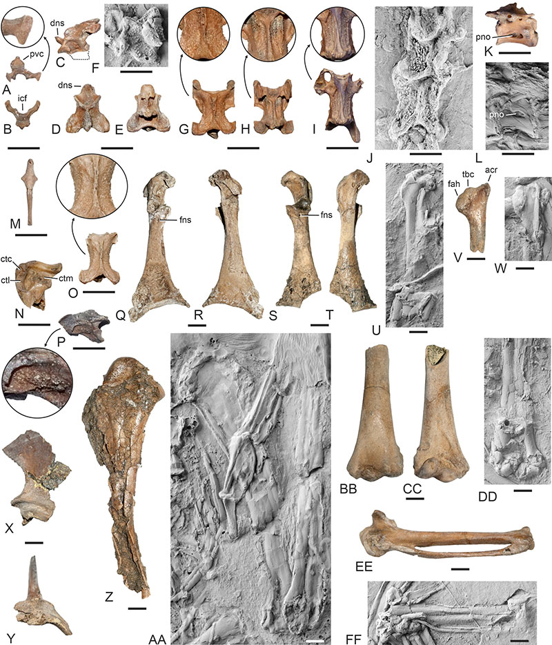
FIGURE 9. Comparison of selected skeletal elements of Perplexicervix paucituberculata, sp. nov. and Eupodotis senegalensis (Otidiformes) to illustrate similar proportions and morphologies. A‒C, E. senegalensis (SMF 21520), first three cervical vertebrae (A), left coracoid (B), and right humerus (C). D‒G, P. paucituberculata, sp. nov., first three cervical vertebrae (D), left coracoid (E), and proximal and distal ends of right humerus (F, G) (D: holotype, NMS.Z.2021.40.7; E, F: tentatively referred specimen NMS.Z.2021.40.91; G: tentatively referred specimen NMS.Z.2021.40.92). Abbreviations: cbp, crista bicipitalis; cdd, condylus dorsalis; cdv, condylus ventralis; cph, caput humeri; ppc, processus procoracoideus. The scale bars equal 10 mm.
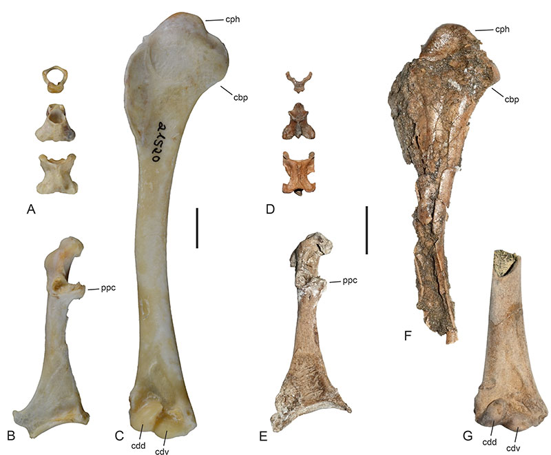
FIGURE 10. Undetermined birds that were likened to Danielsavis by Houde et al. (2023). A, partial skeleton of Aves indet. A (NMS.Z.2021.40.5). B, C, left tarsometatarsus of Aves indet. A (NMS.Z.2021.40.8) in plantar (B) and dorsal (C) view. D, partial skeleton of Aves indet. B (NMS.Z.2021.40.4). E‒H, right quadrate of Aves indet. A (NMS.Z.2021.40.5) in lateral (E), caudal (F), medial (G), and ventral (H) view. I, J, right pterygoid of Aves indet. B (NMS.Z.2021.40.4) in lateral (I) and dorsal (J) view. K, distal end of right tarsometatarsus of Aves indet. B (NMS.Z.2021.40.4) in distal view. L, distal end of left tarsometatarsus of Aves indet. A (NMS.Z.2021.40.8) in distal view. M, proximal end of left tarsometatarsus of Aves indet. A (NMS.Z.2021.40.8) in proximal view. Abbreviations: cdc, condylus caudalis; cdm, condylus medialis; cdp, condylus pterygoideus; cpo, capitulum oticum; cps, capitulum squamosum; for, foramen; orb, processus orbitalis; tsc, tuberculum subcapitulare. The scale bars equal 10 mm.
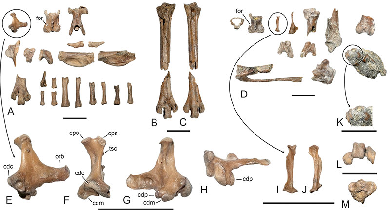
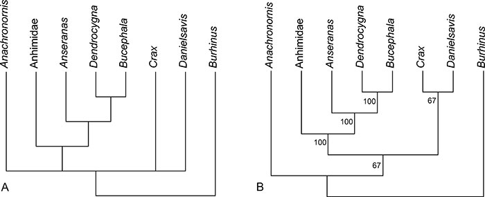
FIGURE 11. Results of the analysis of the emended dataset 1 of Houde et al. (2023). A, strict consensus tree of three most parsimonious trees (length = 317, consistency index = 0.69, retention index = 0.50). B, majority rule consensus tree with values indicating the percentage of trees in which the respective node is retained.