FIGURE 1. Geological map of the Seymour Island showing the fossiliferous sites where MLP-PV 13-XI-28-546 and MLP-PV 14-I-10-199 were found (A) and the corresponding stratigraphy of the locality IAA 2/13 (B), in the James Ross Basin (C), West Antarctica (D). Modified from Montes et al. (2019).
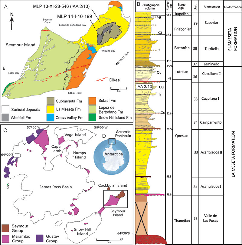
FIGURE 2. Anatomical terms used for descriptions and comparisons (after Baumel et al., 1993) in A, dorsal; B, lateral; and C, proximal views. Abbreviations: ap. phal., apex phalanx; corp. phal., corpus phalangis; cot. art., cotyla articularis (articular or proximal face); cot. art. lat., cotyla articularis lateralis; cot. art. med., cotyla articularis medialis; sulc. neur., sulcus neurovascularis (neurovascular sulcus); tuberc. ext., tuberculum extensorium (extensor tubercle); tuberc. flex., tuberculum flexorium (flexor tubercle).
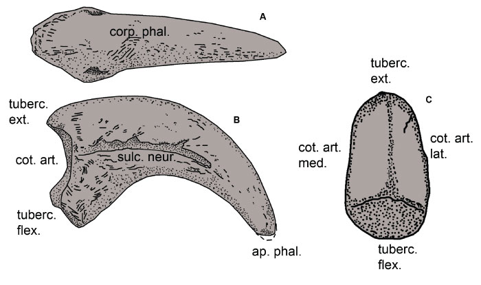
FIGURE 3. Linear and angular measurements taken on the schematic ungual phalanges in A-B lateral, and C, proximal views. Abbreviations: BH, basal height; HAF, maximum height of the articular facet; ICA, inner curvature angle; LFT, flexor tubercle length; OCA, outer curvature angle; TL, total length; and WAF, maximum width of articular facet.
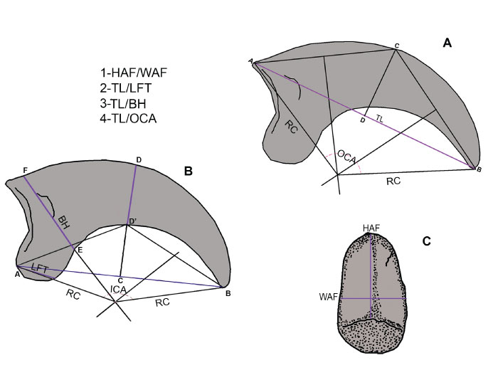
FIGURE 4. Fossil cariamiforms examined here. Ungual phalanx of the second right digit MLP-PV 13-XI-28-546 (A, C, E, G) in lateral (A), dorsal (C), medial (E), and proximal (G) views, and ungual phalanx of second digit MLP-PV 14-I-10-199 (B, D, F) in lateral or medial (B, F) and dorsal (D) views. Scale bar: 10 mm.
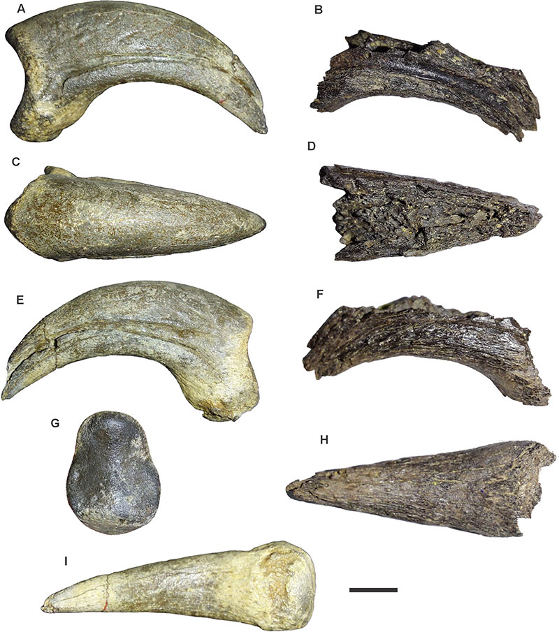
FIGURE 5. Morphological differences between the ungual phalanges of each digit (I, II, II, and IV) of Cariama cristata (A-D) and Psilopterus colzecus (E) in A, lateral; B, dorsal; C, plantar; D and E, proximal (articular) views (not scaled).
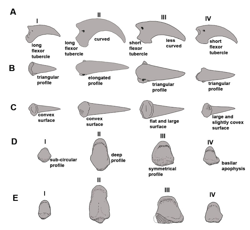
FIGURE 6. Biplot of the Principal Component Analyses (PCA). A, Analysis without normalization of data; B, Analysis with variables converted into indexes. Abbreviations: BH, basal height; HAF, maximum height of the articular facet; ICA, inner curvature angle; LFT, flexor tubercle length; OCA, outer curvature angle; TL, total length; and WAF, maximum width of articular facet.
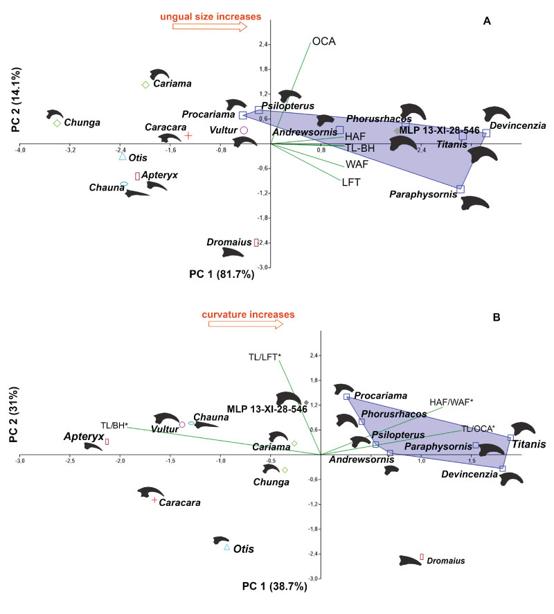
FIGURE 7. Ungual phalanx of representatives of the most relevant groups compared with the Antarctic specimens described in this study. A, Antarctic fossil MLP-PV 13-XI-28-546; B, Chunga incerta (Cariamiformes); C, Vultur gryphus (Cathartiformes); D, Caracara plancus (Falconiformes); E, Geranoaetus melanoleucus (Accipitriformes); F, Ninox novaeseelandiae (Strigiformes); G, Casuarius casuarius (Casuariformes); H, Dromaius novaehollandiae (Struthioniformes); I, Rhea americana (Rheiformes); J, Tinamus solitarius (Tinamiformes); K, Penelope obscura and L, Crax fasciolata (Galliformes); M, Otis tarda (Otidiformes); N, Chauna torquata (Anseriformes); O, Macronectes giganteus (Procellariiformes); P, Anthropornis grandis (giant Antartic Sphenisciformes); and Q, Pygoscelis antarctica (modern Sphenisciformes). Scale bar: 10 mm.
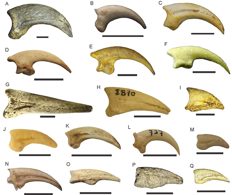
FIGURE 8. Antarctic ungual phalanx compared with different Phorusrhacidae species in lateral view. A, MLP-PV 13-XI-28-546; B, Phorusrhacos longissimus (AMNH 9497 taken from Sinclair and Farr 1932 and mirrored); C, Procariama simplex MACN 8275; D, Phorusrhacus (MLP-PV 20-572); E, Paraphysornis brasiliensis; F, Patagornis marshi MLP-PV 20-184; G, Procariama simplex MACN 8225; H, Devincenzia pozzi MACN Pv 6681; I, Titanis walleri (calcotype UF 10417); J, Psilopterinae indet. MLP-PV 90-III-5-56; K, Mesembriornis milneedwardsi MACN Pv 5944; L, Patagornis marshi MLP-PV 20-164; M, Psilopterus colzecus MLP-PV 76-VI-12-2; N, Psilopterinae indet. MPEF-PV 12256; O, MMP s/n Phorusrhacidae (re-drawn from Cenizo et al., 2012). Scales bar: 10 mm (except for C, G, M, and N where the scale represents 20 mm).
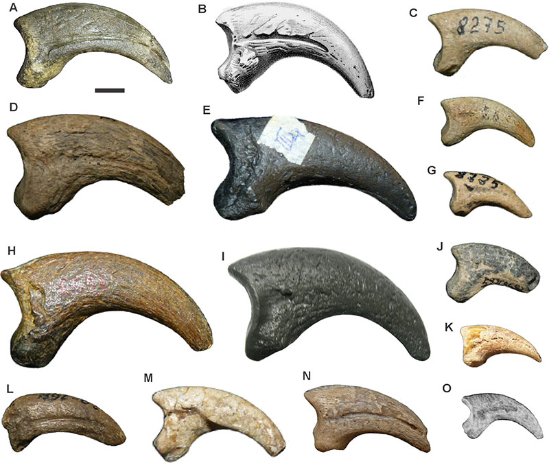
FIGURE 9. Three-dimensional reconstructions (not scaled) of different ungual phalanges. The dotted lines indicate the transversal cut areas shown below each phalanx. The arrowheads mark the path of the neurovascular sulcus and/or neurovascular canal along the phalanx. A, Patagornis marshi (MLP-PV 20-85, digit III); B, Patagornis marshi (MLP-PV 20-86, digit III); C, Psilopterus colzecus (MLP-PV 76-VI-12-2, digit II); D, Brontornis burmeisteri (MLP-PV 20-570, digit III); E, Phorusrhacos longissimus (MLP-PV 67-VIII-28-1, digit II); F, Andrewsornis abbotti MLP-PV 59-II-26-83 (digit II).
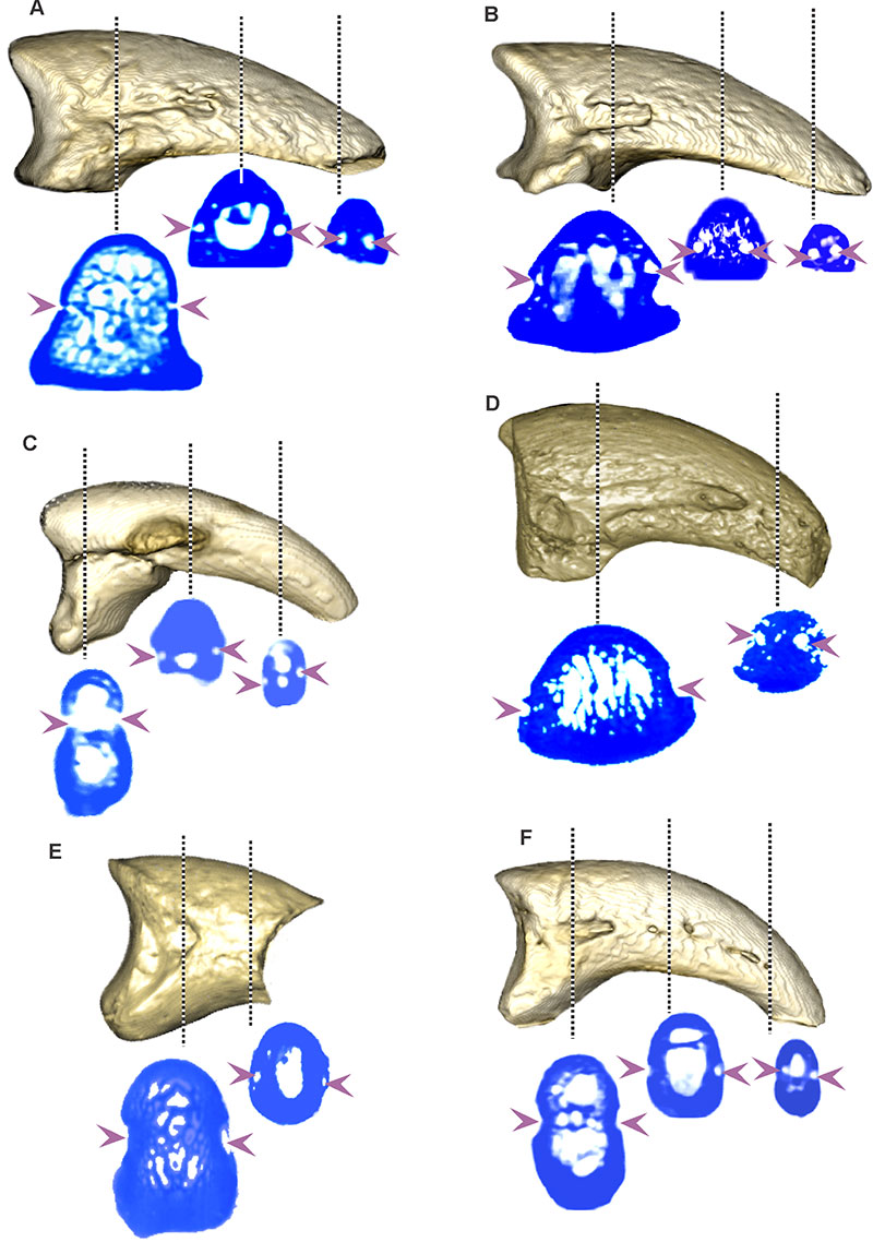
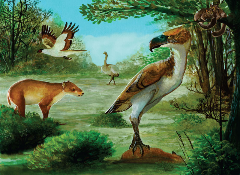
FIGURE 10. Paleoenvironmental reconstruction of the Ypresian continental communities of Seymour Island. A large Cariamiform hunting a medium-sized ungulate and staring at Notiolofos regueroi (Mammalia: Sparnotheriodontidae), a couple of marsupials on a tree, Antarctoboenus carlinii (Aves, Falconiformes) flying on the sky, and a flightless Ratites in the back.