FIGURE 1. Simplified geographical map of Slovenia showing (marked with star) the fossiliferous locality “Neža” near Trbovlje.
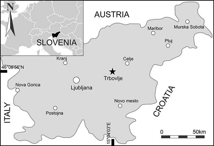
FIGURE 2. Lithostratigraphic section showing exposed strata at the “Neža” locality. Depths on the right indicate the fish fauna mass mortality layers (marked with a fish icon) and layers with fossil hymenosomatid crabs, which are marked with a crab icon.
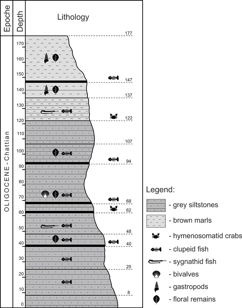
FIGURE 3. Halicarcinus popeius sp. nov., schematic reconstruction. A. Carapace in dorsal view; B. Detailed view of second type of rostrum and robust chelipeds (male); C. Third pereopod (P3) with detailed view of dactylus.
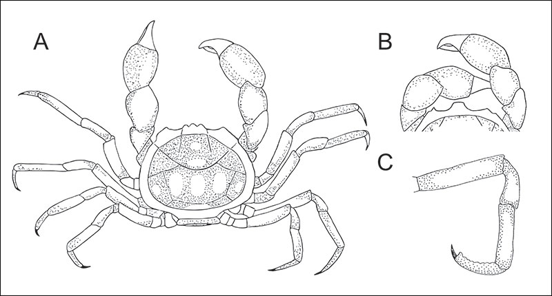
FIGURE 4. Halicarcinus popeius sp. nov. (A-D) and Lucascinus trifailensis sp. nov. (E, F): A. RGA/SMNH 6377, dorsal, (holotype), visible hymenosomian rim (hr) and coloration patterns; B. RGA/SMNH 6373, dorsal, (paratype), male with well-preserved chelipeds and pereopods (P2-P5); C. RGA/SMNH 6385, dorsal, (paratype), male with bulbous chelipeds; D. RGA/SMNH 2041, ventral, (paratype), female with missing sternum; E. RGA/SMNH 6384, dorsal, (holotype), indicated rostrum and hymenosomian rim (hr); F. RGA/SMNH 6391, dorsal, (paratype), elongated chelipeds and well preserved pereopods (P2-P5). All scale bars equal 5 mm.
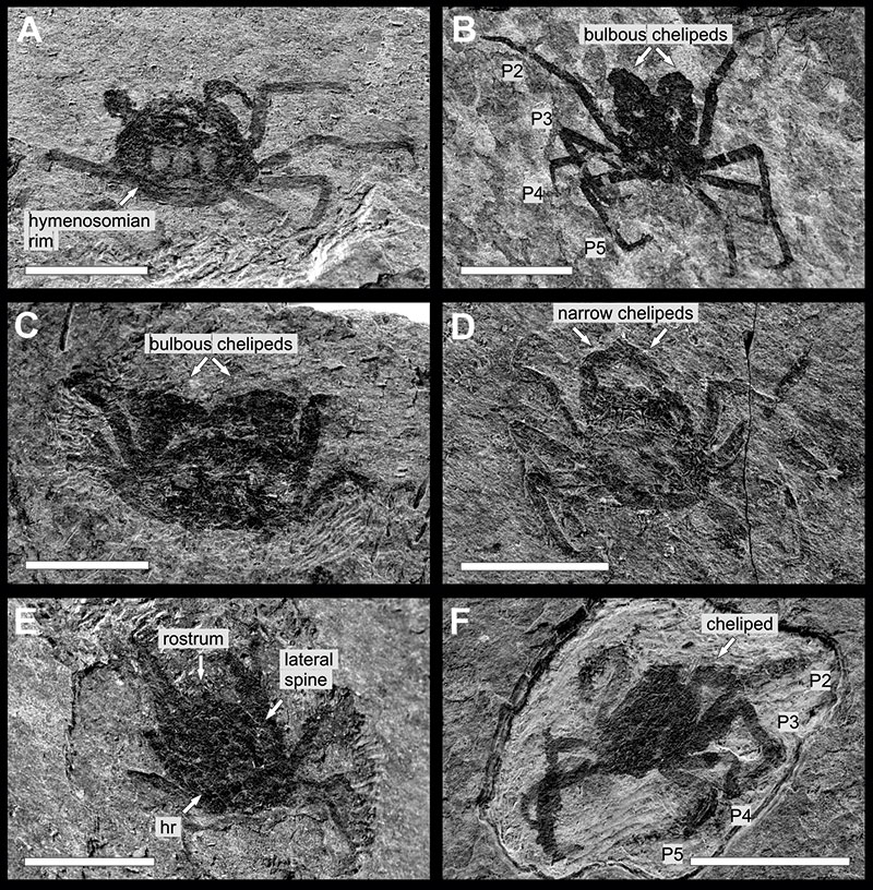
FIGURE 5. Scanning electron photomicrographs of Halicarcinus popeius sp. nov. (A-C) and Lucascinus trifailensis sp. nov. (D): A. RGA/SMNH 6385, details of anterior and left lateral border in dorsal view, pereopods (P2-P5), (composite of several photomicrographs); B. RGA/SMNH 2041, ventral side with missing sternum, details of anterior margin and buccal cavity, coxa of pereopods (cx2-cx5), pereopods (P2-P5), (composite of several photomicrographs); C. RGA/SMNH 6373, detailed view of pereopodal (P3) dactylus; D. RGA/SMNH 6387, ventral side with male pleon, pereopods (P2-P5), abdominal somites (s1-s5), pleotelson (pt), sternites (st4-st8), sternal shield (s), third maxilliped (mxp3). Scale bars indicated on the photomicrographs.
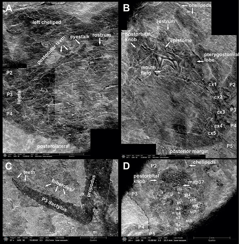
FIGURE 6. Lucascinus trifailensis sp. nov., schematic reconstruction. A. Carapace in dorsal view; B. Detailed view of male pleon.
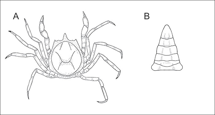
FIGURE 7. Carapace length vs. maximum carapace width of hymenosomatid specimens from the “Neža” locality. Halicarcinus popeius sp. nov. are marked in blue triangles (circles when trapezoid rostrum), Lucascinus trifailensis sp. nov. specimens are marked with orange squares. All measurements in mm.
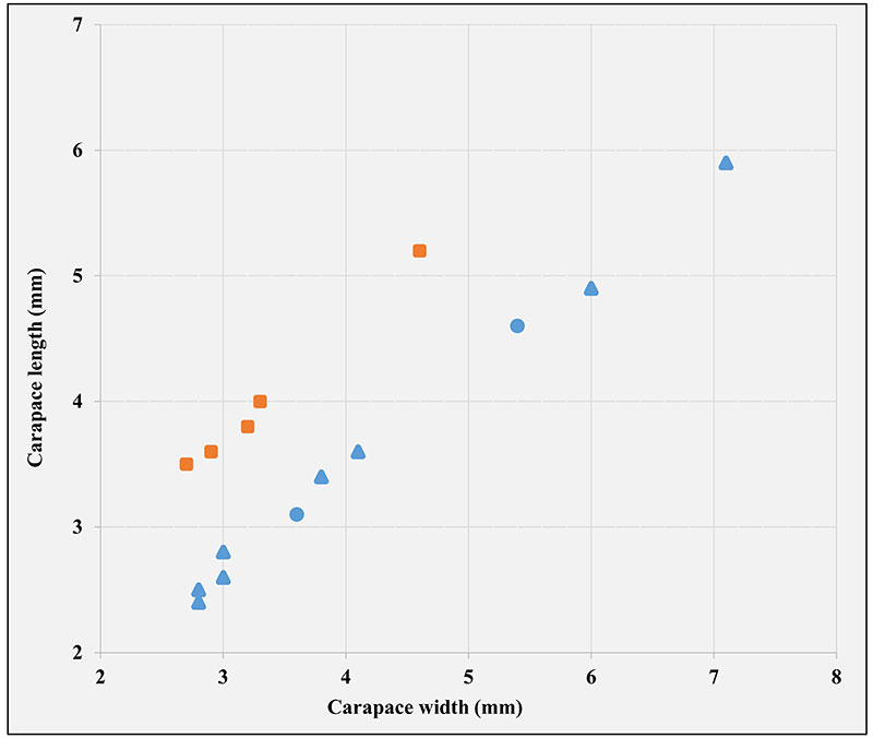
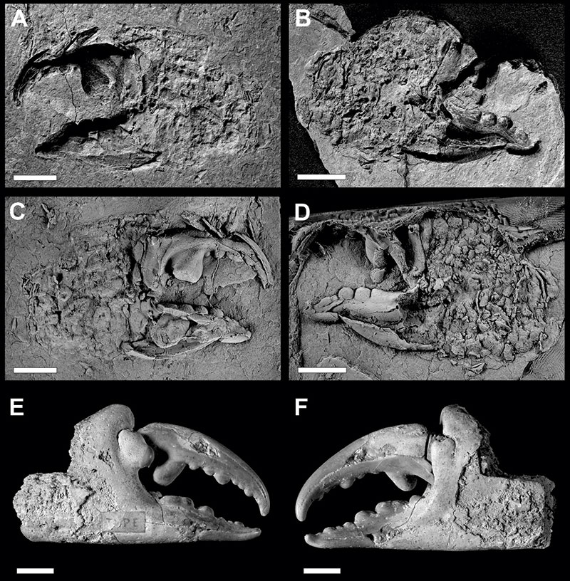
FIGURE 8. Necronectes cf. michelini Milne-Edwards, 1860 (A-D) and Necronectes michelini Milne-Edwards, 1860 (E, F): A. RGA/SMNH 5009, imprint of the outer side of chela; B. RGA/SMNH 5010, imprint of the inner side of chela; C. RGA/SMNH 5009, whitened latex cast; D. RGA/SMNH 5010, whitened latex cast; E. MNHN.F.R03775, (paralectotype), outer side of chela; F. MNHN.F.R03775, (paralectotype), inner side of chela. All scale bars equal 10 mm. Photos A and D: RECOLNAT (ANR-11-INBS-0004) - Jocelyn FALCONNET - 2017.