FIGURE 1. Location of the Hambach opencast lignite mine in northwestern Germany (from Mörs and Stefen, 2010).

FIGURE 2. Cryptobranchidae indet. from Hambach: trunk vertebra (IPB-HaH 2009) in anterior (A), dorsal (B), ventral (C) and left lateral (D) views; fragment of trunk vertebra (IPB-HaH 2191) in dorsal view (E); left humerus (IPB-HaH 2395) in posterior (F) and anterior (G) view. Scale bars equal 2 mm. Abbreviations: np, notochordal pit; ns, neural spine; pz, prezygapophysis.

FIGURE 3. Skull elements of Palaeoproteus cf. miocenicus from Hambach: left dentary (IPB-HaR 2043) in medial (A) and lateral (B) views. Scale bar equals 1 mm. Abbreviations: pd, pars dentalis; s, shelf; vg, ventral groove.

FIGURE 4. Vertebrae of Palaeoproteus cf. miocenicus from Hambach: atlas (IPB-HaH 2117) in dorsal (A), ventral (B), anterior (C), posterior (D) and right lateral (E) views; atlas (IPB-HaH 2175) in dorsal (F), ventral (G), anterior (H) and posterior (I) views; anterior trunk vertebra (IPB-HaR 2183) in dorsal (J), ventral (K), anterior (L), posterior (M) and right lateral (N) views; trunk vertebra (IPB-HaR 2008) in dorsal (O), ventral (P), anterior (Q), posterior (R), right lateral (S) and left lateral (T) views; trunk vertebra (IPB-HaR 2408) in dorsal (U), ventral (V), anterior (W), posterior (X), right lateral (Y) and left lateral (Z) views. Scale bars equal 1 mm. Abbreviations: avc, anterior ventral crest; b, basapophyses; c, cotyle; k, keel; n, neurapophysis; np, notochordal pit; ns, neural spine; oj, occipital joint; pl, processus lateralis; po, processus odontoideus; pvc, posterior ventral crest; tp, transverse process.

FIGURE 5. Limb bones of possible Palaeoproteus cf. miocenicus from Hambach: humerus (IPB-HaH 2390) in anterior (A) and posterior (B) views. Scale bar equals 1 mm.

FIGURE 6. Mioproteus cf. wezei from Hambach: trunk vertebra (IPB-HaR 2000) in dorsal (A), ventral (B) and right lateral (C) views; trunk vertebra (IPB-HaR 2006) in dorsal (D), ventral (E) and right lateral (F) views; trunk vertebra (IPB-HaR 2007) in dorsal (G), ventral (H) and left lateral (I) views; trunk vertebra (IPB-HaR 2403) in dorsal (J) and ventral (K) views. Scale bars equal 1 mm. Abbreviations: b, basapophysis; k, keel; n, neurapophysis; ps, posterior spine; tp, transverse process; vl, ventral lamina.

FIGURE 7. Skull elements of Chelotriton sp. from Hambach: left premaxilla (IPB-HaH 2005) in anterior (A), posterior (B), dorsal (C), ventral (D) and lateral (E) views; right maxilla (IPB-HaH 2007) in lateral (F) and medial (G) views; left frontal (IPB-HaH 2000) in ventral (H) and dorsal (I) views; right frontal (IPB-HaH 2001) in ventral (J) and dorsal (K) views; left frontal (IPB-HaH 2337) in ventral (L) and dorsal (M) views; right dentary (IPB-HaH 2004) in lateral (N) and medial (O) views; left dentary (IPB-HaH 2039) in lateral (P) and medial (Q) views. Scale bars equal 1 mm. Abbreviations: pa, pars palatina; pf, pars faciale; po, processus postorbitalis.

FIGURE 8. Vertebrae of Chelotriton sp. from Hambach: trunk vertebra (IPB-HaH 2010) in dorsal (A), left lateral (B), anterior (C) and ventral (D) views; trunk vertebra (IPB-HaH 2038) in left lateral (E), dorsal (F), ventral (G), right lateral (H), posterior (I) and anterior (J) views; trunk vertebra (IPB-HaH 2177) in dorsal (K) and ventral (L) views; trunk vertebra (IPB-HaH 2114) in dorsal (M), anterior (N), left lateral (O) and ventral (P) views. Scale bars equal 1 mm. Abbreviations: n, neurapophysis; pzc, posterior zygapophyseal crest; tp, transverse process.

FIGURE 9. Limb bones of possible Chelotriton sp. from Hambach: left humerus (IPB-HaH 2389) in anterior (A) and posterior (B) views; right humerus (IPB-HaH 2393) in ventral (C) and dorsal (D) views. Scale bars equal 1 mm. Abbreviations: cvf, cubital ventral fossa; dc, dorsal crest of the humerus; h, humeral head; of, olecranon fossa; vc, ventral crest of the humerus.

FIGURE 10. Lissotriton sp. from Hambach 6C: atlas (IPB-HaH 2118) in anterior (A), dorsal (B), left lateral (C), ventral (D) and posterior (E) views; trunk vertebra (IPB-HaH 2110) in dorsal (F), anterior (G), left lateral (H) and ventral (I) views; trunk vertebra (IPB-HaH 2115) in dorsal (J), anterior (K), right lateral (L) and ventral (M) views; trunk vertebra (IPB-HaH 2116) in dorsal (N), anterior (O), left lateral (P) and ventral (Q) views. Scale bars equal 1 mm. Abbreviations: lr, longitudinal ridge; n, neck; oj, occipital joint; po, processus odontoideus.

FIGURE 11. Lissotriton sp. from Hambach 11: trunk vertebra (IPB-HaR 2009) in dorsal (A), left lateral (B), anterior (C), right lateral (D) and ventral (E) views; trunk vertebra (IPB-HaR 2011) in dorsal (F), right lateral (G), anterior (H) and ventral (I) views; caudal vertebra (IPB-HaR 2010) in dorsal (J), left lateral (K), anterior (L), right lateral (M) and ventral (N) views; caudal vertebra (IPB-HaR 2012) in dorsal (O), right lateral (P) and ventral (Q) views. Scale bars equal 1 mm.

FIGURE 12. Triturus sp. from Hambach: atlas (IPB-HaH 2196) in dorsal (A), anterior (B), left lateral (C), posterior (D) and ventral (E) views; trunk vertebra (IPB-HaH 2101) in dorsal (F) and left lateral (G) views; trunk vertebra (IPB-HaH 2103) in dorsal (H), ventral (I) and left lateral (J) views; trunk vertebra (IPB-HaH 2224) in dorsal (K), anterior (L), right lateral (M), posterior (N) and ventral (O) views. Scale bars equal 1 mm. Abbreviations: avc, anterior ventral crest; azc, anterior zygapophyseal crest; n, neurapophysis; oj, occipital joint; op, odontoid process; sc, secondary dorsal crest.

FIGURE 13. Skull elements of Latonia sp. from Hambach: premaxilla (IPB-HaH 2050) in anterior (A) and posterior (B) views; right maxilla (IPB-HaH 2053) in lateral (C) and medial (D) views; right maxilla (IPB-HaH 2059) in lateral (E) and medial (F) views; left maxilla (IPB-HaR 2016) in lateral (G) and medial (H) views; frontoparietal (IPB-HaH 2002) in dorsal (I) and ventral (J) views; left angular (IPB-HaH 2051) in lateral (K), medial (L) and dorsal (M) views; right angular (IPB-HaR 2117) in dorsal (N) and medial (O) views; left angular (IPB-HaR 2142) in lateral (P), medial (Q) and dorsal (R) views; right angular (IPB-HaR 2416) in dorsal view (S). Scale bars equal 1 mm. Abbreviations: cme, crista mandibulae externa; if, incrassatio frontoparietalis; lh, lamina horizontalis; pc, processus coronoideus; pd, posterior depression; pp, processus palatinus; pr, processus paracoronoideus.

FIGURE 14. Axial elements of Latonia sp. from Hambach: atlas (IPB-HaH 2071) in anterior (A), dorsal (B), left lateral (C), ventral (D) and posterior (E) views; trunk vertebra (IPB-HaH 2073) in anterior (F), dorsal (G) and posterior (H) views; trunk vertebra (IPB-HaR 2032) in anterior (I), dorsal (J) and left lateral (K) views; sacral vertebra (IPB-HaH 2219) in dorsal view (L); sacral vertebra (IPB-HaR 2014) in dorsal (M), anterior (N) and ventral (O) views; urostyle (IPB-2199) in dorsal view (P); urostyle (IPB-HaH 2200) in dorsal view (Q); urostyle (IPB-HaR 2034) in dorsal view (R). Scale bars equal 1 mm. Abbreviations: f, fissure; k, keel.

FIGURE 15. Appendicular elements of Latonia sp. from Hambach: left scapula (IPB-HaH 2324) in dorsal (A) and ventral (B) views; left scapula (IPB-HaH 2325) in dorsal (C) and ventral (D) views; left scapula (IPB-HaH 2329) in dorsal (E) and ventral (F) views; right humerus (IPB-HaH 2061) in ventral (G) and dorsal (H) views; left ilium (IPB-HaR 2015) in lateral (I) and medial (J) views; right ilium (IPB-HaR 2083) in lateral (K) and medial (L) views; right ilium (IPB-HaR 2417) in lateral (M) and medial (N) views. Scale bars equal 1 mm. Abbreviations: ca, crista anterior; dt, dorsal tubercle; ig, interiliac groove; pa, pars acromialis.

FIGURE 16. Palaeobatrachus eurydices from Hambach: right maxilla (IPB-HaR 2429) in dorsal (A), medial (B) and lateral (C) views; sphenethmoid (IPB-HaR 2021) in dorsal (D), ventral (E), anterior (F) and right lateral (G) views; left humerus (IPB-HaR 2148) in ventral (H) and dorsal (I) views; left humerus (IPB-HaR 2152) in ventral (J) and dorsal (K) views; left humerus (IPB-HaR 2420) in ventral (L) and dorsal (M) views, right ilium (IPB-HaR 2099) in lateral view (N); right ilium (IPB-HaR 2100) in lateral view (O); left ilium (IPB-HaR 2101) in lateral view (P); right ilium (IPB-HaR 2147) in lateral view (Q). Scale bars equal 1 mm. Abbreviations: dt, dorsal tubercle; pp, processus palatinus; ps, contact surface for the parasphenoid.

FIGURE 17. Individual variation in the angulars of Palaeobatrachus eurydices from Hambach: left angular (IPB-HaR 2111) in dorsal view (A); left angular (IPB-HaR 2132) in dorsal view (B); left angular (IPB-HaR 2144) in dorsal view (C); right angular (IPB-HaR 2110), detail of the processus coronoideus in dorsal view (D); left angular (IPB-HaR 2112), detail of the processus coronoideus in dorsal view (E); left angular (IPB-HaR 2130), detail of the processus coronoideus in dorsal view (F); right angular (IPB-HaR 2131), detail of the processus coronoideus in dorsal view (G); right angular (IPB-HaR 2145), detail of the processus coronoideus in dorsal view (H); right angular (IPB-HaR 2146), detail of the processus coronoideus in dorsal view (I). Scale bars equal 1 mm. Abbreviations: pc, processus coronoideus; t, tubercle.

FIGURE 18. Possible Palaeobatrachidae indet. from Hambach: fragment of maxilla (IPB-HaH 2279) in medial (A), lateral (B), dorsal (C) and ventral (D) views. Scale bar equals 1 mm.

FIGURE 19. Pelobatids from Hambach: sacral vertebra (IPB-HaR 2179) of cf. Eopelobates sp. in dorsal (A), anterior (B), posterior (C) and ventral (D) views; left humerus (IPB-HaR 2154) of cf. Eopelobates sp. in ventral (E) and dorsal (F) views; left ilium (IPB-HaR 2084) of cf. Eopelobates sp. in lateral (G) and medial (H) views; right ilium (IPB-HaR 2103) of cf. Eopelobates sp. in lateral (I) and medial (J) views; right humerus (IPB-HaR 2414) of ?Eopelobates sp. in ventral (K) and dorsal (L) views; left humerus (IPB-HaR 2415) of ?Eopelobates sp. in ventral view (M); right ilium (IPB-HaR 2421) of ? Eopelobates sp. in lateral (N) and medial (O) views; trunk vertebra (IPB-HaH 2220) of Pelobatidae indet. in right lateral (P), anterior (Q), dorsal (R) and posterior (S) views; left humerus (IPB-HaH 2399) of Pelobatidae indet. in ventral (T) and dorsal (U) views. Scale bars equal 1 mm. Abbreviations: dae, dorsal acetabular expansion; fcv, fossa cubitalis ventralis.

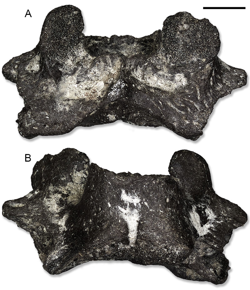
FIGURE 20. Hyla sp. from Hambach: sacral vertebra (IPB-HaH 2195) in dorsal (A), ventral (B), anterior (C) and posterior (D) views; left ilium (IPB-HaR 2088) in lateral view (E). Scale bars equal 1 mm. Abbreviations: dt, dorsal tubercle; pz, preacetabular zone.

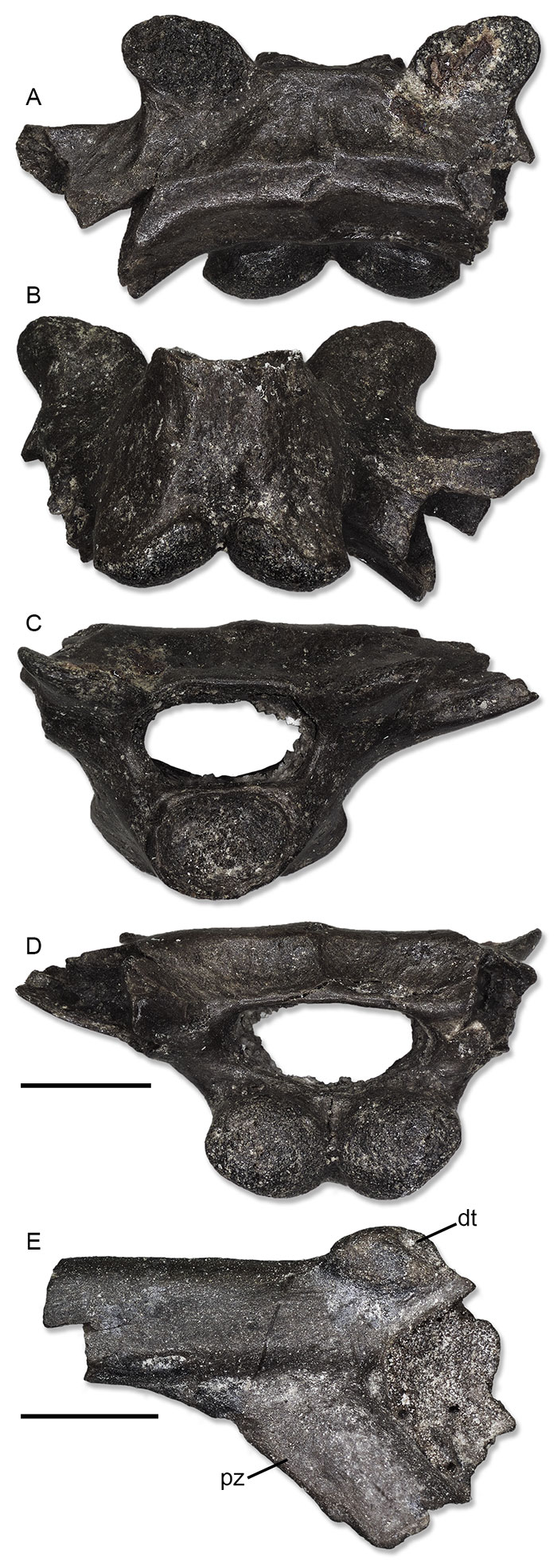
FIGURE 21. Bufo gr. bufo from Hambach: sacral vertebra (IPB-HaR 2020) in dorsal (A) and ventral (B) views. Scale bar equals 1 mm.

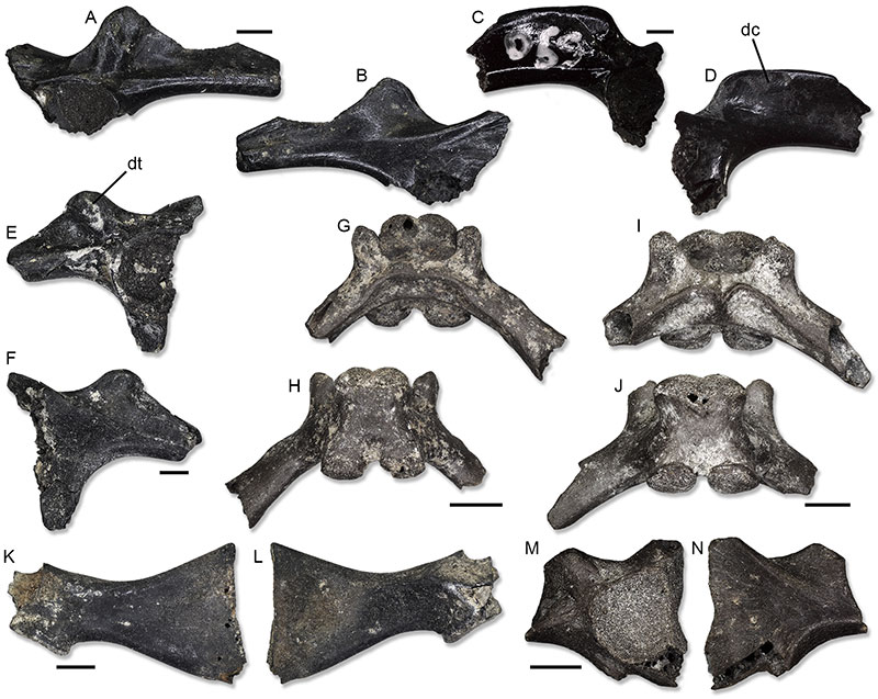
FIGURE 22. Ranids from Hambach: right ilium (IPB-HaH 2307) of Pelophylax sp. in lateral (A) and medial (B) views; left ilium (IPB-HaH 2387) of Pelophylax sp. in lateral (C) and medial (D) views; left ilium (IPB-HaH 2321) of Rana sp. in lateral (E) and medial (F) views; sacral vertebra (IPB-HaR 2018) of Ranidae indet. in dorsal (G) and ventral (H) views; sacral vertebra (IPB-HaR 2019) of Ranidae indet. in dorsal (I) and ventral (J) views; left scapula (IPB-HaH 2323) of Ranidae indet. in ventral (K) and dorsal (L) views; left ilium (IPB-HaR 2087) of Ranidae indet. in lateral (M) and medial (N) views. Scale bars equal 1 mm. Abbreviations: dc, dorsal crest; dt, dorsal tubercle.

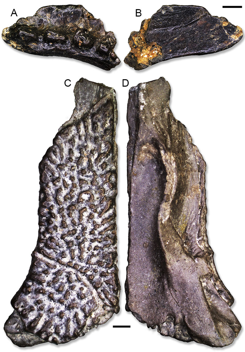
FIGURE 23. Reptiles from Hambach: right squamosal (IPB-HaH 2340) of Chamaeleo aff. andrusovi in lateral (A) and medial (B) views; left frontal (IPB-HaR 2171) of Pseudopus cf. pannonicus in dorsal (C) and ventral (D) views. Scale bars equal 1 mm.


