FIGURE 1. Map of the location of the study areas. A - Map of the Mexican Republic. B - Map of the state of Oaxaca showing the location of the Jurassic units. C - Geological map of the Cualac Formation; Zepeda-Martinez (2021).
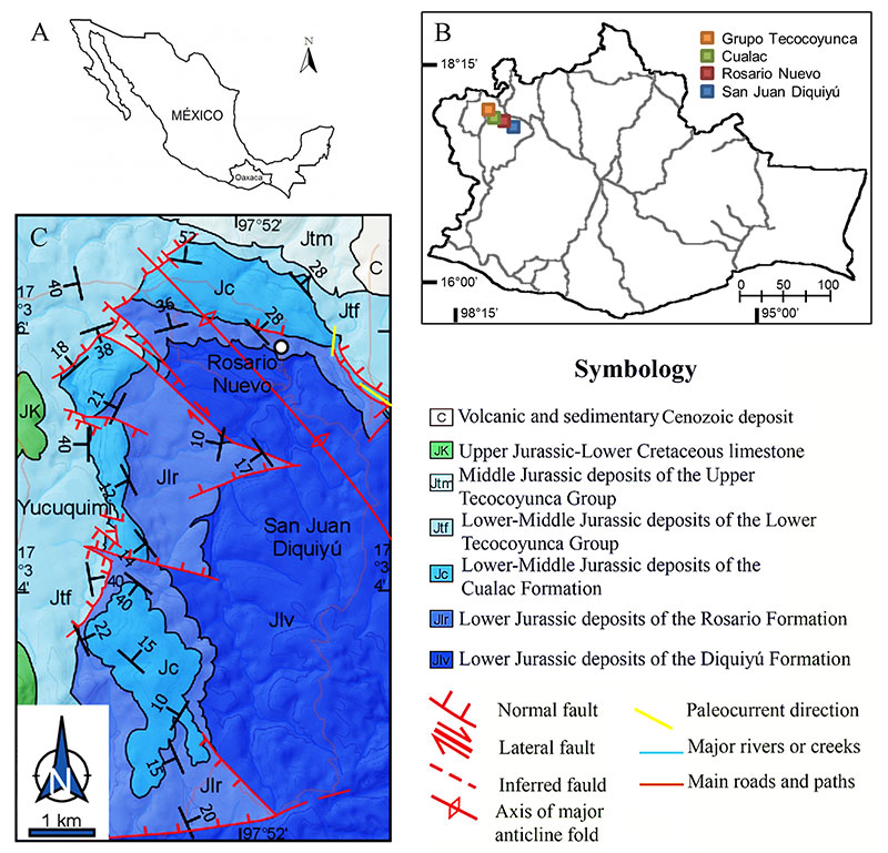
FIGURE 2. Stratigraphic section of the collection zone. A - Sequence of rocks that outcrop in the Rosario locality. B - Image of the outcrop. C - image of the stratigraphic section where the shale strata are observed. Symbols: 1) Clay, 2) Silt, 3) Very Fine, 4) Fine, 5) Medium, 6) Thick, 7) Very Thick, and 8) Conglomerate.
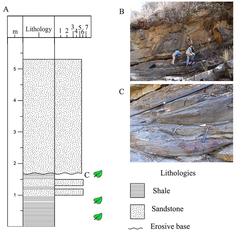
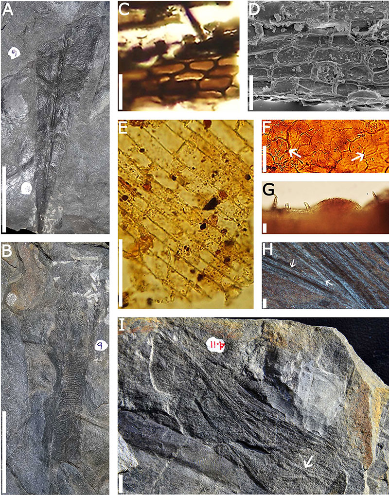
FIGURE 3. Fossil flora of the Rosario locality, Cualac Formation. A - incomplete leaf of Anomozamites, the diagnostic characters of the genus are observed; B - close-up of a leaflet of the same specimen; C - Cycadolepis cf. C. mexicana, towards the upper right the hairs are observed, D - and E - Zamites diquiyui with subopposite leaflets, in the approach of the leaflets strong and robust veins are observed, some of these veins bifurcate near the base. F-H - Cuticle of Zamites diquiyui . F - epidermal cells with slightly sinuous thick walls; G - butterfly-shaped stoma typical for Bennettitales; H - hooked trichome. Scale in A 2 cm; B 1 cm; C 1 cm; D 5 cm; E 2 cm; F 20 μm; G 10 μm; H 10 μm.
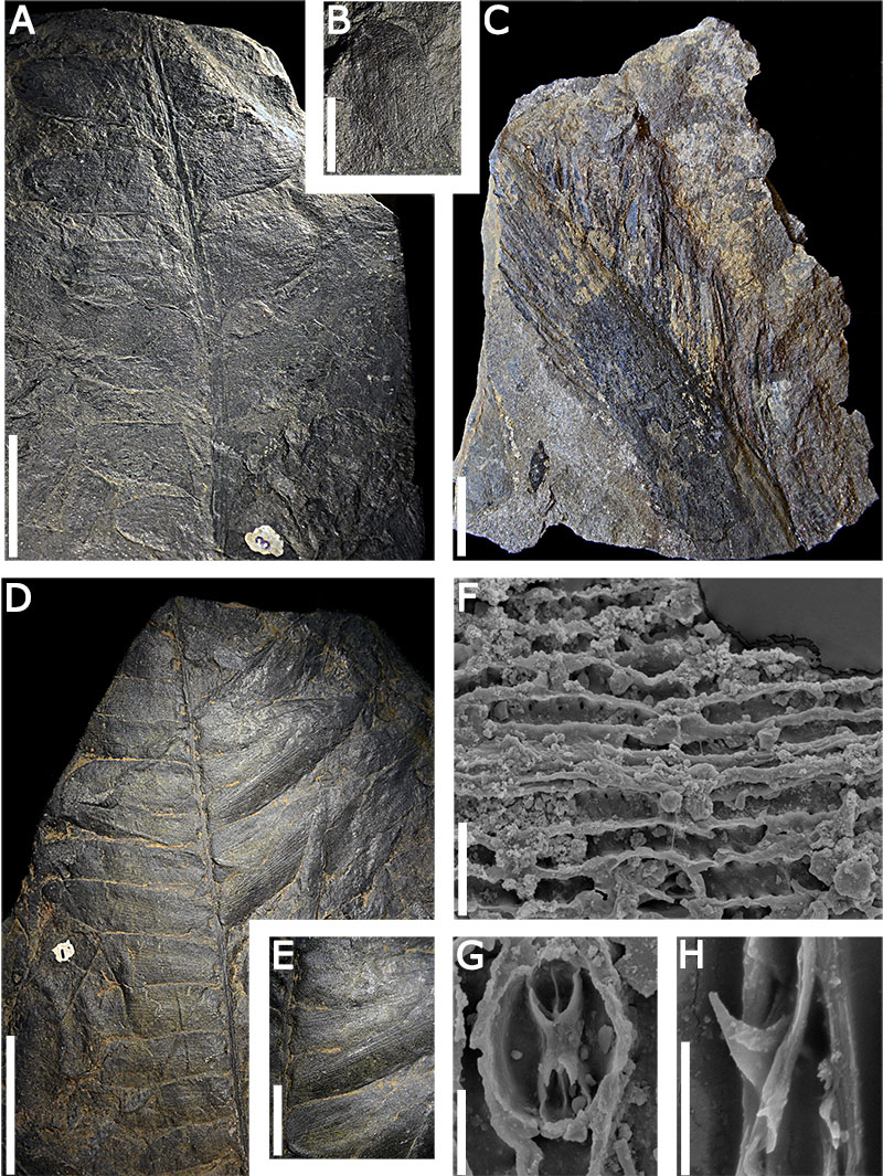
FIGURE 4. Williamsoniella rosarensis sp. nov. A - type specimen CFZr115, hermaphroditic structure where the peduncle is preserved attached to the rest of the structure and is characteristic of the material studied, the arrows point to the microsporophylls (side arrow) surrounding the gynoecium (central arrow). B - paratype CFZCr68, the arrows indicate the opening of the sporophylls (right side) and another flower in abaxial view (left side). C - upper part of specimen CFZCr115 the arrow pointing to the crown sensu Thomas (1915). D - upper part of gynoecium of specimen CFZCr 68. E - Specimen CFZCr82, the arrow upper part points to hair on the margin. F - Peel of peduncle with sting-shaped trichome. G - Peel where it is observed. Oval syndetocheilic stomata in peduncle. Scale in A 2 cm; B 1 cm; C 1 mm; D 1 mm; E 5 mm; F 15 μm; G 10 μm.
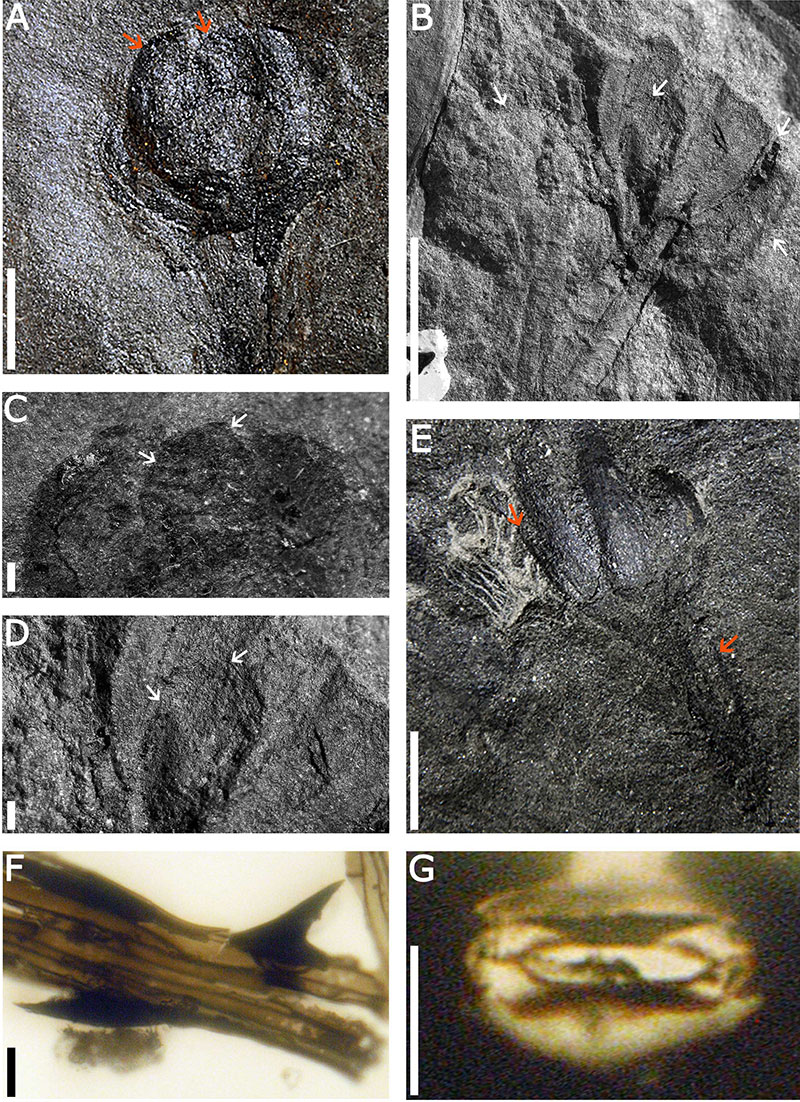
FIGURE 5. Leaves of Mexiglossa and Czekanowskia . A-B - whole leaves of Mexiglossa varia, note that the midvein cells; C-E- F-G light microscopic images, rectangular epidermal cells of Mexiglossa with straight walls; D - electron microscopy image, thick walls of epidermal cells; F - arrows indicate two stomata; G - trichomes present on leaves of Mexigossa varia ; H - linear leaves of the genus Czekanowskia, the arrows indicate the middle vein and the fork of the leaf; I - specimen CFZcr105 shows base of bundle of laciniae, the arrow indicates a bifurcation. Scale in A 3 cm; B 3 cm; C 10 μm; D 50 μm; E 15 μm; F 15 μm ; G 2.5 μm; H 1 mm; I 5 mm.