APPENDIX 1.
Supplementary data relative to the proceedings performed for the quantitative analyses of the Spanish Lutetian podocnemidid turtle Neochelys zamorensis and its comparison with extant podocnemidid representatives.
TABLE S1. Definition of landmarks (blue dots) and semilandmarks (red dots) location for the nuchal of Neochelys zamorensis (Pleurodira, Podocnemididae) from the Lutetian (middle Eocene) of the Zamora Province (Spain).
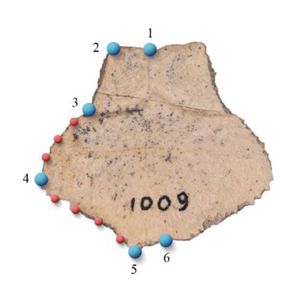
| View | Landmark | Anatomical definition |
| Dorsal | 1 | Medium point of anterior margin |
| Dorsal | 2 | Anterior left contact between the nuchal and first peripheral |
| Dorsal | 3 | Contact of the first marginal sulcus with the lateral margin of the nuchal |
| Dorsal | 4 | Posterior left contact between the nuchal and first peripheral |
| Dorsal | 5 | Posterior left contact between the nuchal and the first element of the neural row |
| Dorsal | 6 | Medium point of posterior margin |
| Dorsal | 7-13 | Semilandmarks: plate outline between LM3-LM4 and LM4-LM5 |
TABLE S2. Definition of landmarks (blue dots) and semilandmarks (red dots) location for the epiplastron of Neochelys zamorensis (Pleurodira, Podocnemididae) from the Lutetian (middle Eocene) of the Zamora Province (Spain).
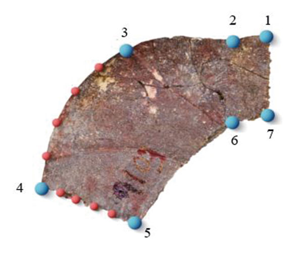
| View | Landmark | Anatomical definition |
| Ventral | 1 | Anterior right contact between the epiplastron and entoplastron |
| Ventral | 2 | Anterior right contact between the gular and the extragular |
| Ventral | 3 | Maximum curvature of the anterior margin |
| Ventral | 4 | Posterior lateral contact between the epiplastron and hyoplastron |
| Ventral | 5 | Posterior medial contact between the epiplastron, entoplastron and hyoplastron |
| Ventral | 6 | Posterior right contact between the epiplastron and entoplastron |
| Ventral | 7 | Posterior right contact between the epiplastron and entoplastron |
| Ventral | 8-15 | Semilandmarks: plate outline between LM3-LM4 and LM4-LM5 |
TABLE S3. Definition of landmarks (blue dots) and semilandmarks (red dots) location for the entoplastron of Neochelys zamorensis (Pleurodira, Podocnemididae) from the Lutetian (middle Eocene) of the Zamora Province (Spain).
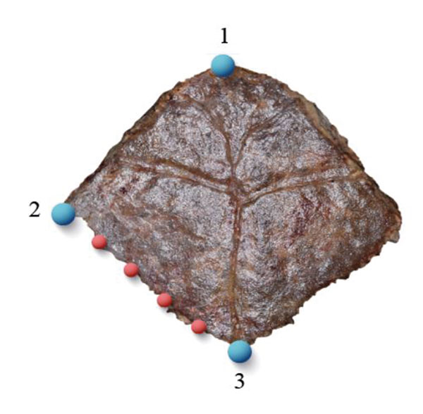
| View | Landmark | Anatomical definition |
| Ventral | 1 | Anterior-most point of entoplastron |
| Ventral | 2 | Posterior contact between the entoplastron and epiplastron |
| Ventral | 3 | Posterior-most point of entoplastron |
| Ventral | 4-7 | Semilandmarks: plate outline between LM2-LM3 |
TABLE S4. Definition of landmarks (blue dots) and semilandmarks (red dots) location for the anal notch of xiphiplastra of Neochelys zamorensis (Pleurodira, Podocnemididae) from the Lutetian (middle Eocene) of the Zamora Province (Spain).
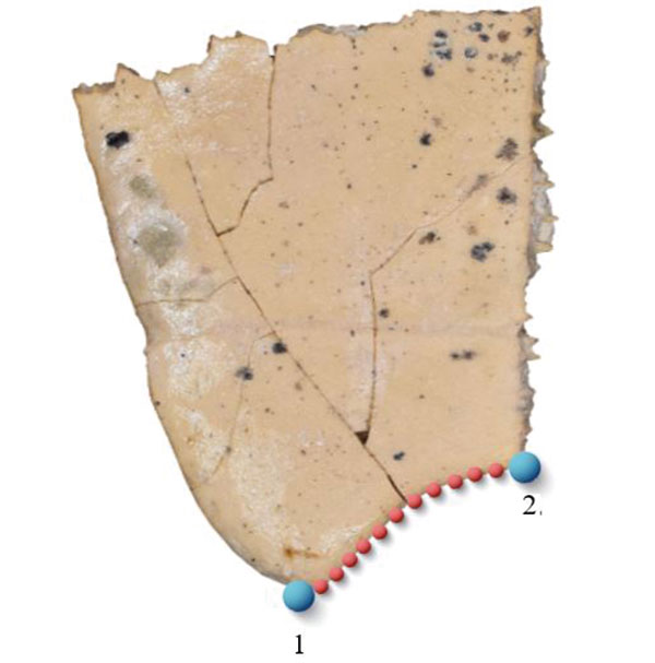
| View | Landmark | Anatomical definition |
| Ventral | 1 | Posterior-most point of the right xiphiplastral process |
| Ventral | 2 | Posterior contact between both xiphiplastra |
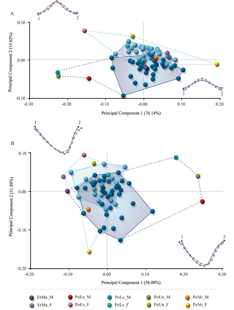
| Ventral | 3-13 | Semilandmarks: plate outline between LM1-LM2 |
TABLE S5. Definition of landmarks (blue dots) and semilandmarks (red dots) location for the for the xiphiplastral process of of Neochelys zamorensis (Pleurodira, Podocnemididae) from the Lutetian (middle Eocene) of the Zamora Province (Spain).
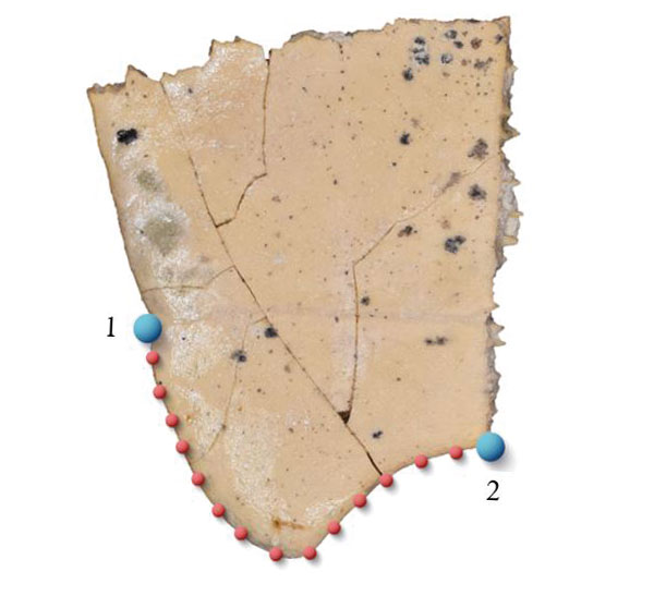
| View | Landmark | Anatomical definition |
| Ventral | 1 | Lateral contact between the xiphiplastron and the anal scute |
| Ventral | 2 | Posterior contact between both xiphiplastra |
| Ventral | 3-14 | Semilandmarks: plate outline between LM1-LM2 |
FIGURE S1. Shape differences of the nuchal plate of Neochelys zamorensis (Pleurodira, Podocnemididae) from the Lutetian (middle Eocene) of the Zamora Province (Spain). A, Principal component 1 (x) and 2 (y). B, Principal component 1 (x) and 3 (y).
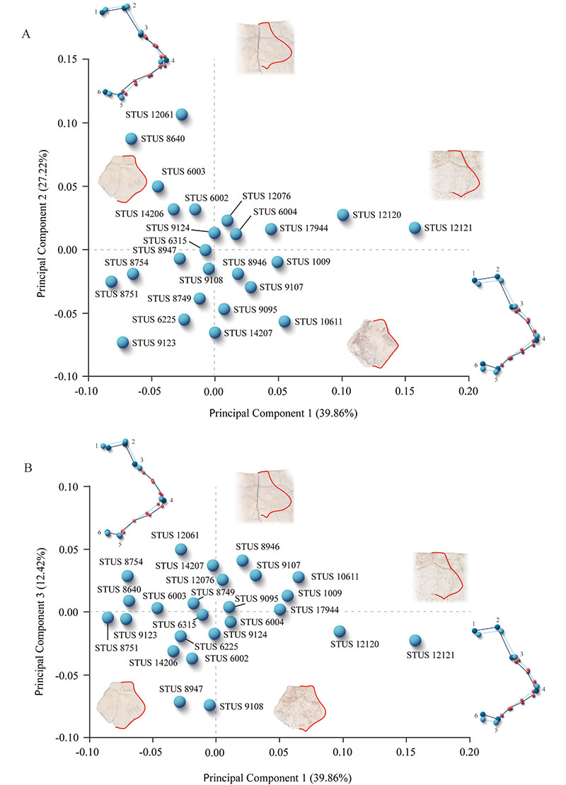
FIGURE S2. Shape differences of the epiplastron of Neochelys zamorensis (Pleurodira, Podocnemididae) from the Lutetian (middle Eocene) of the Zamora Province (Spain). A, Principal component 1 (x) and 2 (y). B, Principal component 1 (x) and 3 (y).
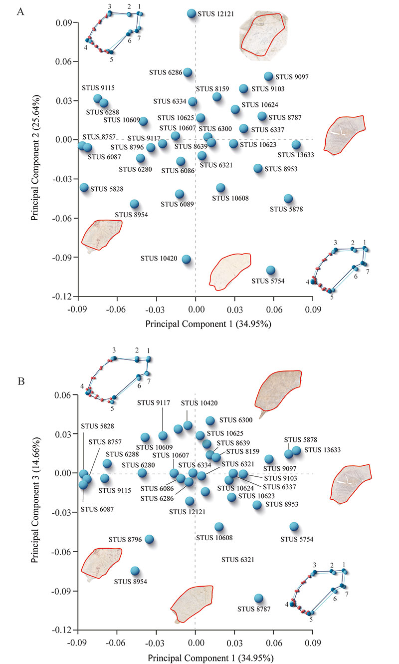
FIGURE S3. Shape differences of the entoplastron of Neochelys zamorensis (Pleurodira, Podocnemididae) from the Lutetian (middle Eocene) of the Zamora Province (Spain). A, Principal component 1 (x) and 2 (y). B, Principal component 1 (x) and 3 (y).
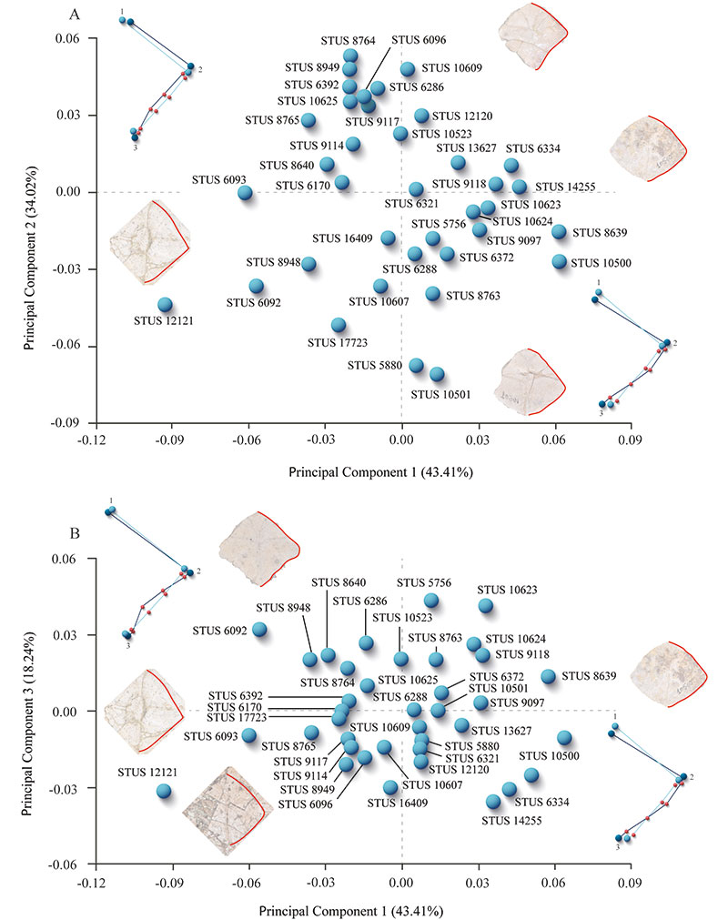
FIGURE S4. Linear regression between Procrustes coordinates (dependent variable) and logged-centroid size values (independent variable) of the nuchal (A), epiplastron (B), and entoplastron (C) of Neochelys zamorensis (Pleurodira, Podocnemididae) from the Lutetian (middle Eocene) of the Zamora Province (Spain).
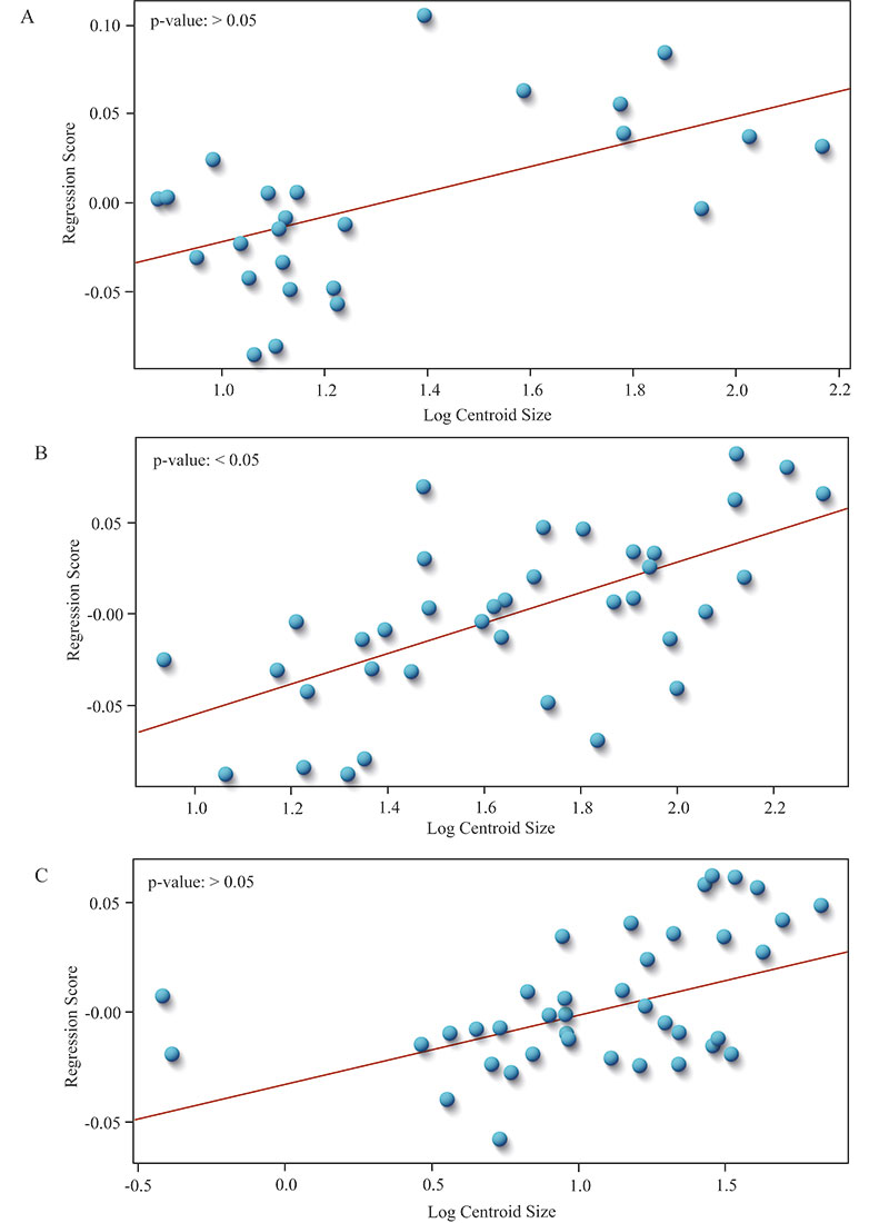
FIGURE S5. Reference shape of female and male xiphiplastral region of extant podocnemidids. A, female of Erymnochelys madagascariensis (MNHN.RA.932.578). B, male of Erymnochelys madagascariensis (MNHN.RA.609.6). C, female of Podocnemis expansa (MNHN.RA.1876.200). D, male of Podocnemis expansa (specimen without number of the MNHN.RA). E, female of Podocnemis lewyana (MNHN.RA.1944.285). F, male of Podocnemis lewyana (MNHN.RA.607.1). G, female of Podocnemis unifilis (MNHN.RA.1974.59). H, male of Podocnemis unifilis (MNHN.RA.608.16). I, female of Podocnemis vogli (MNHN.RA.0612.4). J, male of Podocnemis vogli (MNHN.RA.0612.1). Scale bars equal 2cm.
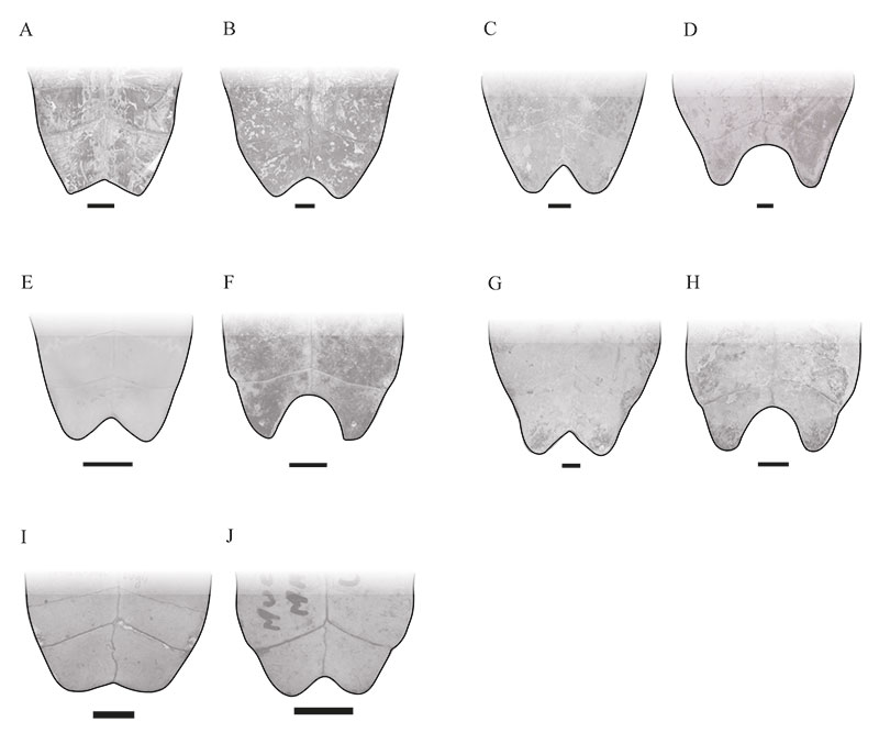
FIGURE S6. Principal Component Analysis (PCA) performed on the xiphiplastral region of extant podocnemidids and the specimens of Neochelys zamorensis (Pleurodira, Podocnemididae) from the Lutetian (middle Eocene) of the Zamora Province (Spain). A, anal notch. B, xiphiplastral process. Abbreviations: ErMa_M, male of Erymnochelys madagascariensis (MNHN.RA.609.6); ErMa_M, female of Erymnochelys madagascariensis (MNHN.RA.932.578); PoEx_M, male of Podocnemis expansa (specimen without number of the MNHN.RA); PoEx_F, female of Podocnemis expansa (MNHN.RA.1876.200); PoLe_M, male of Podocnemis lewyana (MNHN.RA.607.1); PoLeF, female of Podocnemis lewyana (MNHN.RA.1944.285); P oUn_M, male of Podocnemis unifilis (MNHN.RA.608.16); PoUn_F, female of Podocnemis unifilis (MNHN.RA.1974.59); PoVo_M, male of Podocnemis vogli (MNHN.RA.0612.1); PoVo_F, female of Podocnemis vogli (MNHN.RA.0612.4). Dark blue concerns the first sexual morphotype and light blue represents the second sexual morphotype.
