FIGURE 1. Photographs of the cranium of Axestemys infernalis MAB13742 embedded in matrix. A anterior view, B right lateral view, C oblique overview, D dorsal view. Both scale bars equal 1 cm; scale bar A applies to the anterior view; scale bar B applies to both right lateral and dorsal view.
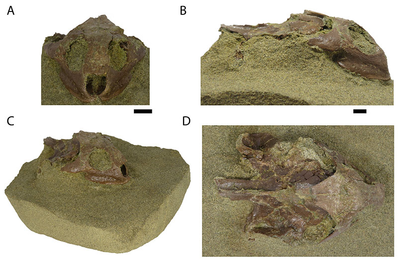
FIGURE 2. Reconstruction of the cranium of Axestemys infernalis MAB13742 in anterior view. A shows the digital rendering of the CT-scan and B the interpretative line drawing. Dashed lines are observed sutures, dotted lines indicate damage. Scale bar equals 1 cm. Abbreviations: ane. apertura narium externum, orb. orbit, fr. frontal, jug. jugal, max. maxilla, par. parietal, po. postorbital, prf. prefrontal, quad. quadrate.
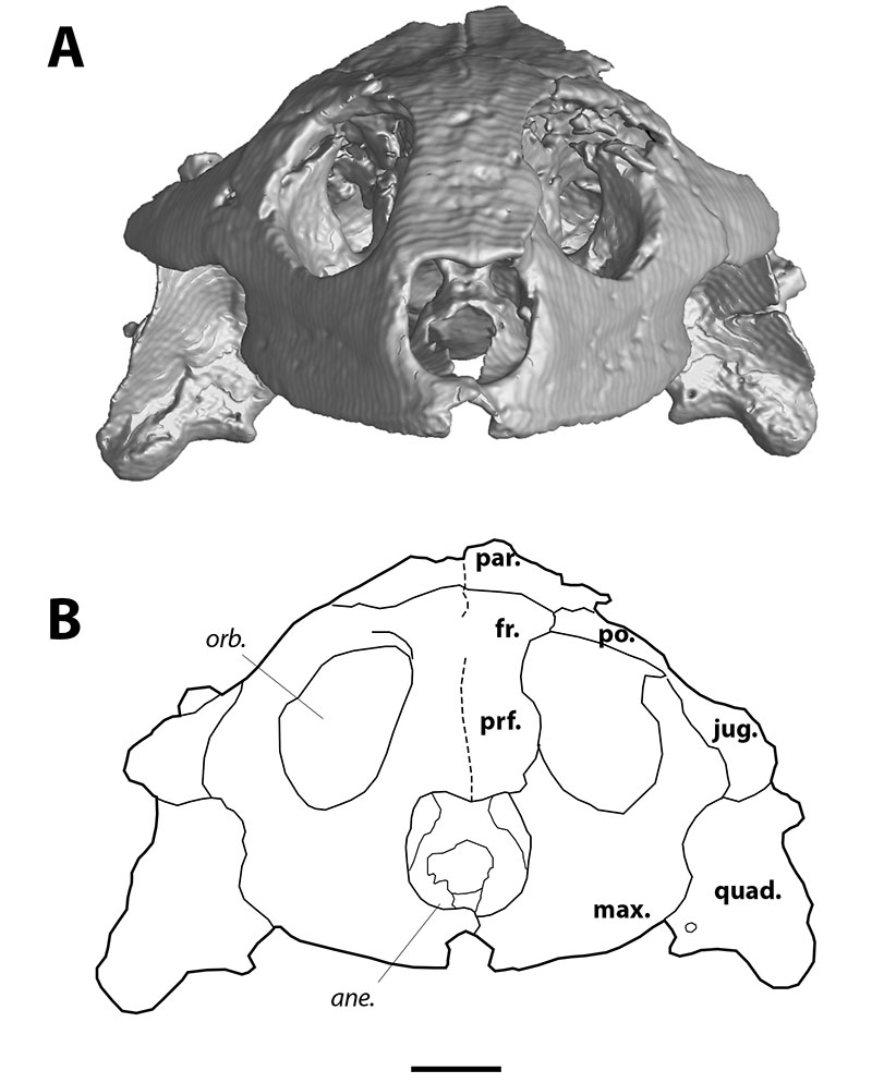
FIGURE 3. Reconstruction of the cranium of Axestemys infernalis MAB13742 in dorsal view. A shows the digital rendering of the CT-scan and B the interpretative line drawing. Dashed lines are observed sutures, dotted lines indicate damage. Scale bar equals 1 cm. Abbreviations: cso. crista supraoccipitalis, fst. foramen stapedio-temporale, pt. processus trochlearis, fr. frontal, jug. jugal, par. parietal, po. postorbital, prf. prefrontal, pro. prootic, quad. quadrate, so. supraoccipital.
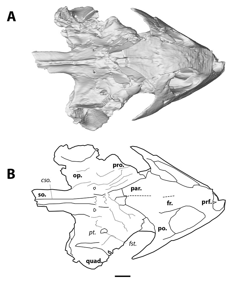
FIGURE 4. Reconstruction of the cranium of Axestemys infernalis MAB13742 in right lateral view. A shows the digital rendering of the CT-scan and B the interpretative line drawing. Dashed lines are observed sutures, dotted lines indicate damage. Scale bar equals 1 cm. Abbreviations: cso. crista supraoccipitalis, ica. incisura columellae auris, jug. jugal, max. maxilla, par. parietal, po. postorbital, quad. quadrate, so. supraoccipital.
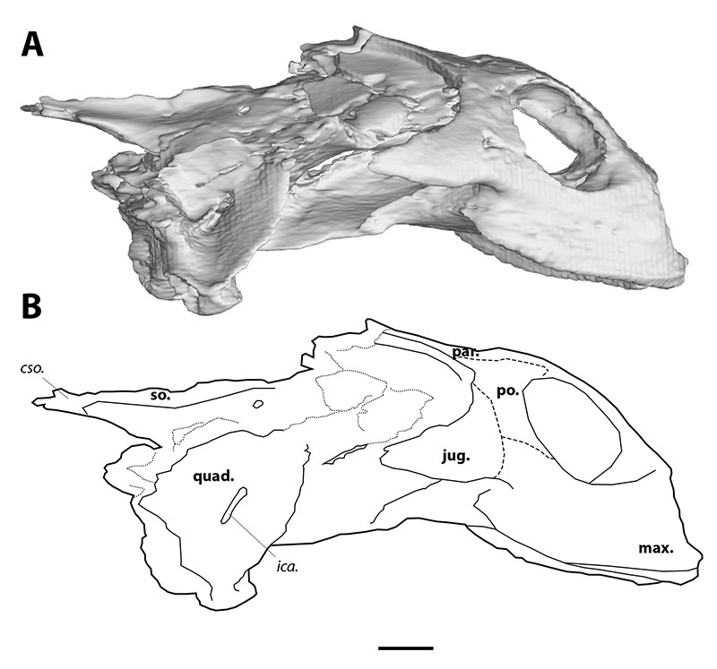
FIGURE 5. Reconstruction of the cranium of Axestemys infernalis MAB13742, digital rendering of the CT-scan showing the internal view of right lateral side and the supraalveolar foramen of the maxilla. Scale bar equals 1 cm. Abbreviations: saf. supraalveolar foramen, max. maxilla. Mirrored.
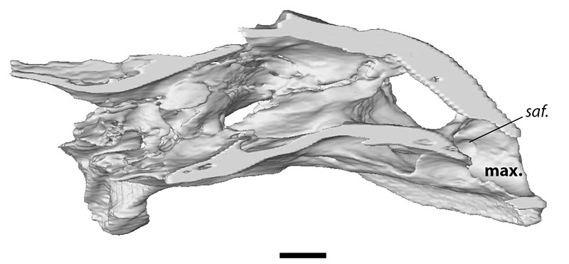
FIGURE 6. Reconstruction of the cranium of Axestemys infernalis MAB13742 in ventral view. A shows the digital rendering of the CT-scan and B the interpretative line drawing. Scale bar equals 1 cm. Abbreviations: fpp. foramen palatinum posterius, bs. basisphenoid, max. maxilla, pal. palatine, pter. pterygoid, vom. vomer.
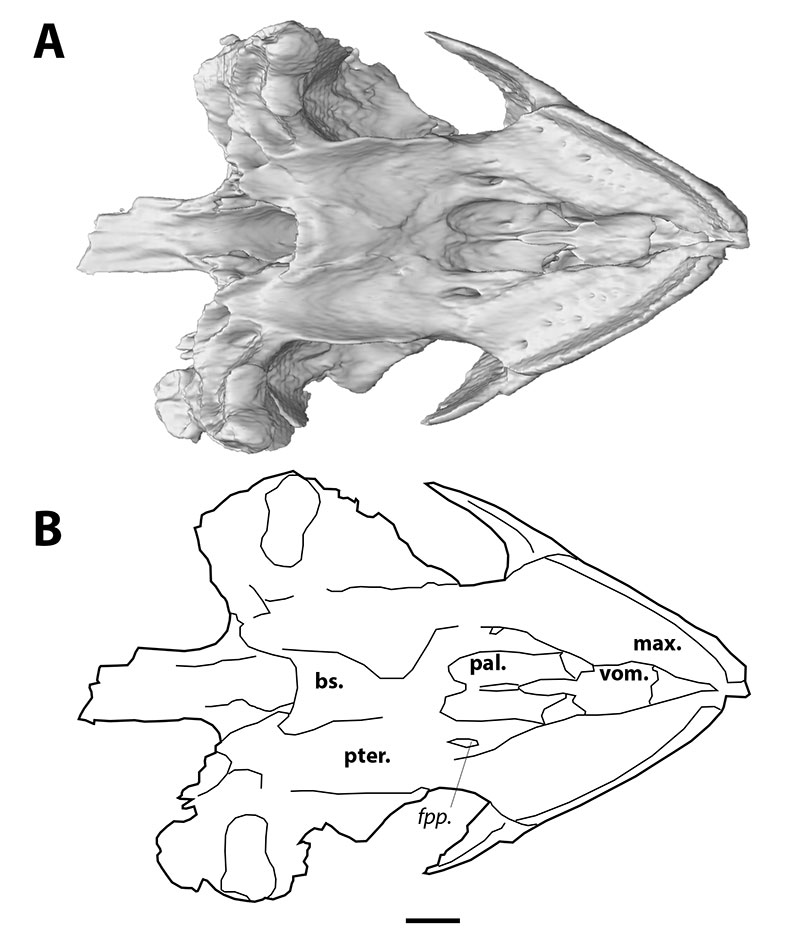
FIGURE 7. Reconstruction of the cranium of Axestemys infernalis MAB13742 in posterior view. A shows the digital rendering of the CT-scan and B the interpretative line drawing. Scale bar equals 1 cm. Abbreviations: fjp. foramen jugulare posterius, fm. foramen magnum, fpcci. foramen posterius canalis carotici interni, ex. exoccipital, op. opisthotic, pter. pterygoid, quad. quadrate, so. supraoccipital.
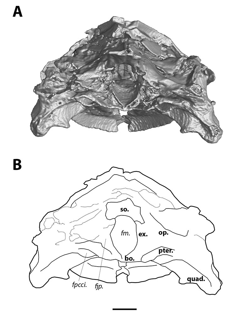
FIGURE 8. Digital reconstruction of the braincase endocast of Axestemys infernalis MAB13742 in A dorsal, B right lateral and C ventral views. Scale bar equals 1 cm. Abbreviations: acs. anterior semicircular canal, cc. common crus, cr. cartilaginous rider, el. endosseous labyrinth, fo. foramen ovalis, nc. nasal cavity, ocr. optic and cerebellar region, pf. pituitary foramen, pcs. posterior semicircular canal, suo. sulcus olfactorius.
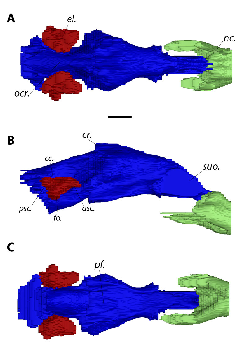
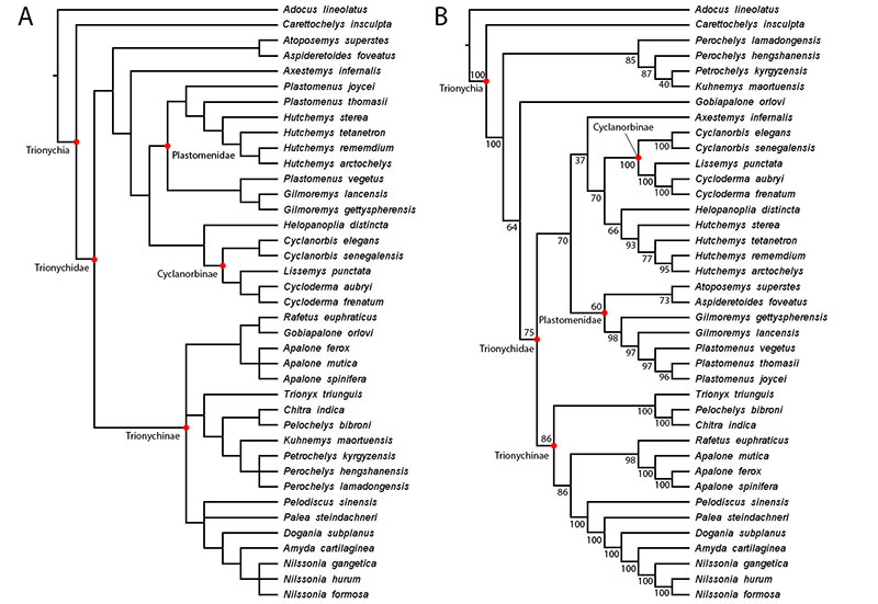
FIGURE 9. Cladograms showing A the results of the parsimony analysis in TNT, using a concavity constant of K = 12. B shows the results of the Bayesian analysis. Support values are indicated for each node.