FIGURE 1. Map of the SW Baltic Sea area displaying the finding locations of the individual vertebrae. Note the indicated cliff outcrops of Lower Cretaceous deposits at Arnager and Madsegrav on Bornholm (Denmark). Blue line indicates maximal Weichselian ice extent.
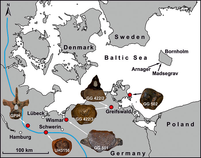
FIGURE 2. GG 501, photographs A, B of (?anterior pectoral) vertebral centrum in A anterodorsolateral and B posteroventrolateral view. CT renderings C-F of GG 501 in C, E anterodorsolateral view and D, F posterolateral view. Abbreviations: aaf, anterior articular facet; f, foramen; in, internal network; p, process; paf, posterior articular facet.
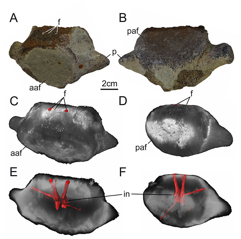
FIGURE 3. Overview scan of thin section GG 501-TS A with microscopical close-up photos of various domains of the extracted and processed cuttings. Representative microscopic views of the sediment composition, under B plane-polarized light (ppl) and under C crossed polarized light (xpl), showing the high amounts of subrounded quartz (Qz), glauconite (Glt) and the calcitic cement (Cal). Note the dispersed phosphate nodules (PN) at the bottom left. D Unspecified calcitic spheroidal microfossil (black arrow) next to a phosphate nodule (ppl). E Peripheral view of a chambered calcareous foraminifera (black arrow), possibly resembling a Muricohedbergella portsdownensis (Williams-Mitchell, 1948) from the Cenomanian surrounded by quartz and glauconite grains (ppl). F Unspecified fragment of a chambered calcareous foraminifera fossil (black arrow). Note the glauconite (Glt) sand-sized grains in typical greenish and greenish-brown color at plane polarized light.
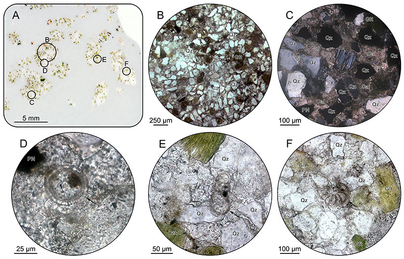
FIGURE 4. Overview scan of thin section MV400005-TS A displaying a domination of quartz and glauconite grains within calcitic cement. Note several lithoclasts indicated, which exhibit a reddish and brownish matrix, quartz grains, and dispersed glauconite. The evenly distributed glauconite (Glt) appear in typical green and brownish color. B Subrounded sand-sized quartz grains (Qz) and smaller subangular quartz grains dominate the thin section, whereas glauconite (Glt) and phosphate nodules (PN) occur dispersed. C A lateral view of a chambered calcareous foraminifera fossil (black arrow) located within the calcitic cement sourrounded by quartz (Qz) and glauconite (Glt) grains. D Bioclast, most likely a mollusc of unknown identity (black arrow). Note that all microscopic views were taken under plane polarized light.
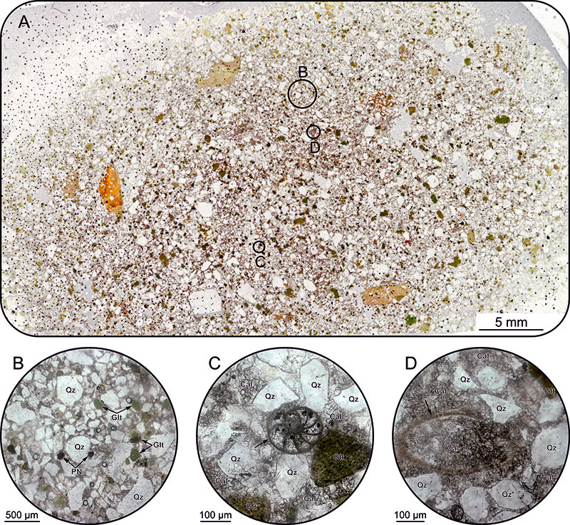
FIGURE 5. Selected calcareous nannofossils from the samples GG 501-N1 and G 501-N2 (cross-polarized light) in alphabetical order. A Biscutum constans; B Broinsonia matalosa; C Calciosolenia fossilis; D Eiffellithus equibiramus; E Eiffellithus monechiae; F Eiffellithus paragogus; G Eiffellithus turriseiffelii; H Gartnerago theta; I Gorkaea operio; J Manivitella pemmatoidea; K Prediscosphaera columnata; L Radiolithus planus; M Radiolithus undosus; N Repagulum parvidentatum; O Rhagodiscus hamptonii; P Rotelapillus crenulatus; Q Staurolithites gausorhethium; R Staurolithites laffittei; S Tegumentum stradneri; T Tranolithus orionatus; U Tubirhabdus burnettiae; V Watznaueria barnesiae; W Zeugrhabdotus erectus; X Zeugrhabdotus howei; Y Zeugrhabdotus xenotus. Scale bar valid for all specimens.
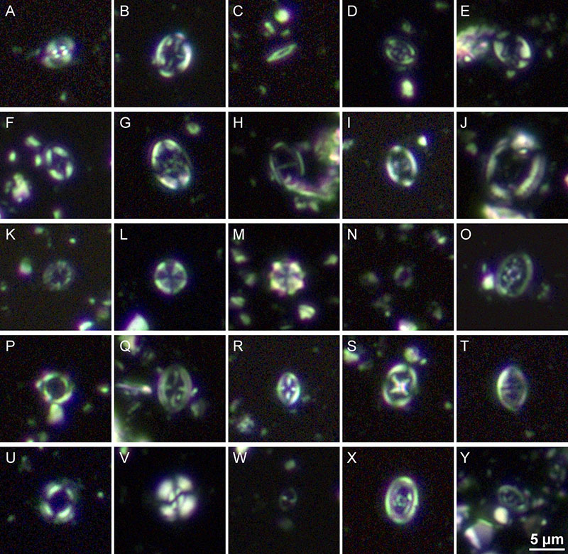
FIGURE 6. Selected calcareous nannofossils from the sample MV40005-N2 (cross-polarized light) in alphabetical order. A Axopodorhabdus albianus; B Broinsonia signata; C Chiastozygus litterarius; D, E Eiffellithus turriseiffelii; F Eprolithus apertior; G Gartnerago theta; H Helicolithus compactus; I Lithraphidites cf. acutus; J Staurolithites laffittei; K Tranolithus orionatus; L Watznaueria barnesiae; M Zeugrhabdotus acanthus; N Zeugrhabdotus bicrescenticus; O Zeugrhabdotus embergeri. Scale bar valid for all specimens.
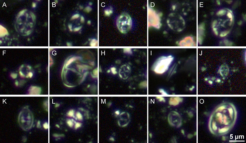
FIGURE 7. Stratigraphy of the outcropping strata at the Arnager and Madsegrav cliff section on the island of Bornholm (Denmark; see Figure 1). Based on the comparative provenance analysis of thin sections GG 501-TS and MV400005-TS, the sediment in which GG 501 was embedded, could be either part of the base of the Arnager Limestone or the base of the Arnager Greensand (black arrows). With the additional results of biostratigraphic interpretations of the outcrop sample MV400005 (nannofossils of MV400005-N1 and MV400005-N2) and the sample of adherent sediment of specimen GG 501 (nannofossils of GG 501-N1 and GG 501-N2), indicated by the red bars, an unambiguous stratigraphic classification of GG 501 to the Lower Cenomanian is established (shaded in red to the right; compiled and adapted from Hart, 1979; Hart et al., 2012; Cohen et al., 2013; Hajny, 2016; Svennevig and Surlyk, 2019).
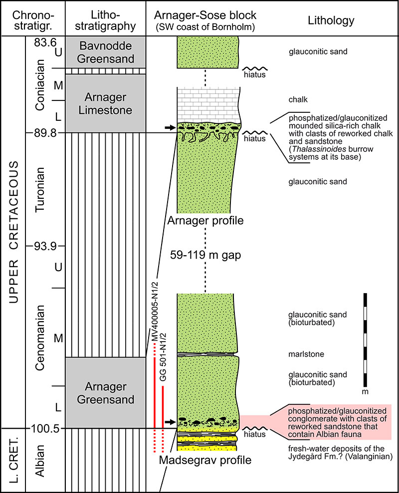
FIGURE 8. GG 502, photographs A, B of (?posterior pectoral) vertebral centrum in A antero/posterodorsal and B ventral view. CT renderings C, D of GG 502 in C anterior/posterior view and D lateral view. Abbreviations: af, articular facet; f, foramen; in, internal network; ncaf, neurocentral articular facet; ncf, floor of neural canal; p, process. Arrows point where the largely intact heart-shaped articular facet faces.
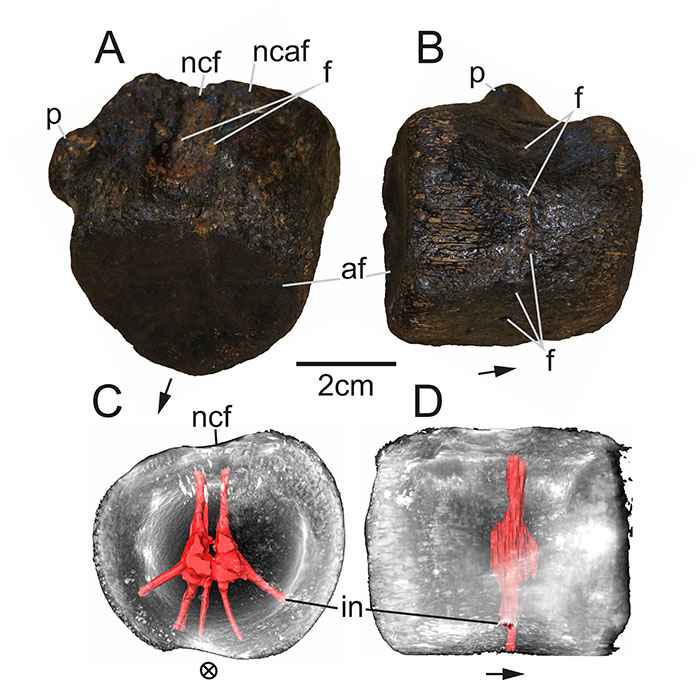
FIGURE 9. GG 422/2, a posterior cervical vertebra from the lower Toarcian of Grimmen (A-D) in A anterior, B posterior, C left lateral and D ventral view. GG 422/3, a caudal vertebral centrum from the lower Toarcian of Grimmen (E-J) in E anterior, F posterior, G left lateral, H right lateral, I ventral and J dorsal view. Abbreviations: f, foramen; haf, haemal arch facet; naf, neural arch facet; nc, neural canal; ncf, neural canal floor; rf, rib facet.
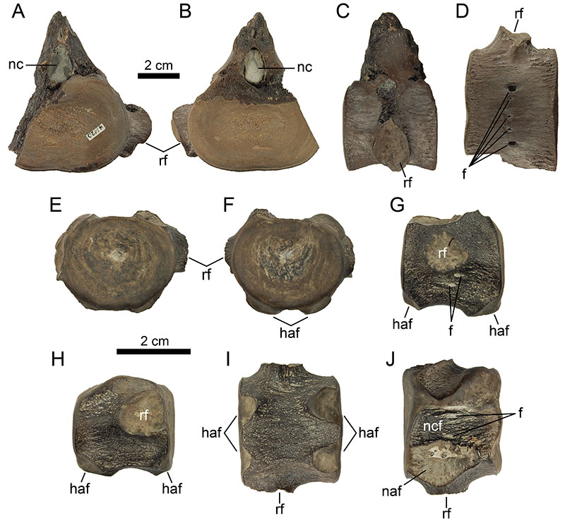
FIGURE 10. GG 422/2, CT renderings A-D of (posterior cervical) vertebra in A,B anterior and C, D anteroventrolateral view. Abbreviations: aaf, anterior articular facet; f, foramen; in, internal network; nc, neural canal.
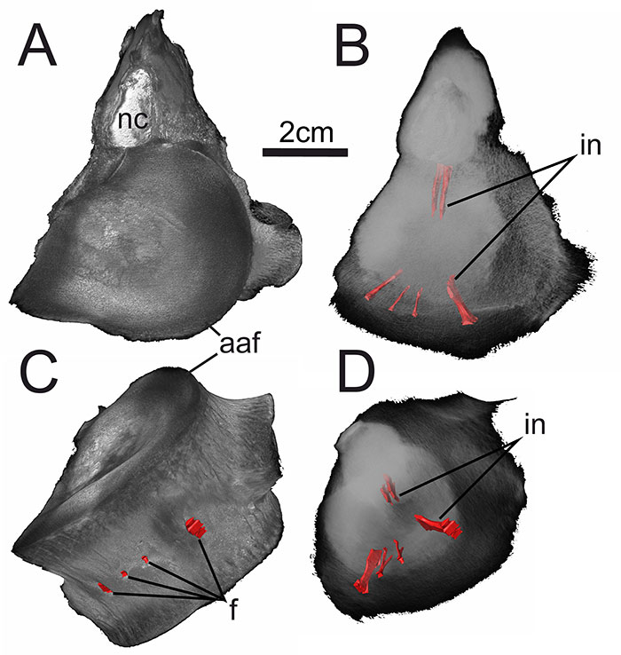
FIGURE 11. GG 422/3, CT renderings A-D of (caudal) vertebral centrum in A, B anterodorsolateral and C, D posteroventrolateral view. Abbreviations: aaf, anterior articular facet; f, foramen; in, internal network.
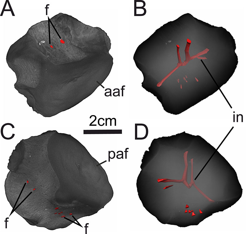
FIGURE 12. UHKD500005, photographs A, B, C of (?dorsal) vertebral centrum in A antero/posterodorsolateral, B antero/posteroventrolateral and C ventrolateral view. CT renderings D-G of UHKD500005 in D, F anterior/posterior view and E, G ventrolateral view. Abbreviations: af, articular facet; f, foramen; in, internal network; ncaf, neurocentral articular facet; tp, transverse process. Arrows point where the articular facet with the sediment cover faces.
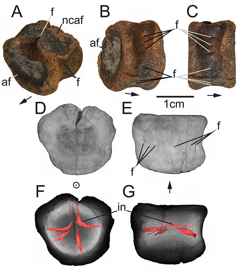
FIGURE 13. UHKD500005, CT image of mid-section. Abbreviations: ddc, distally diffuse cavity; f, foramen; in, internal network.
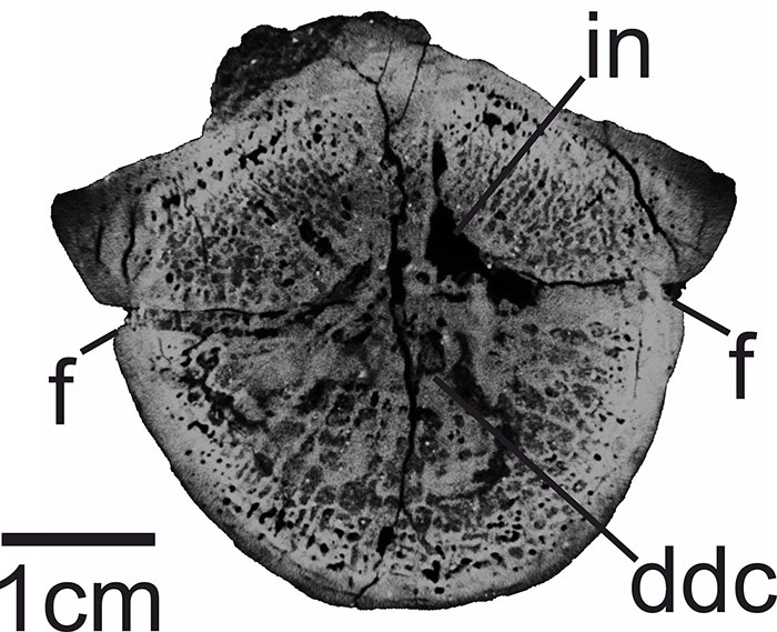
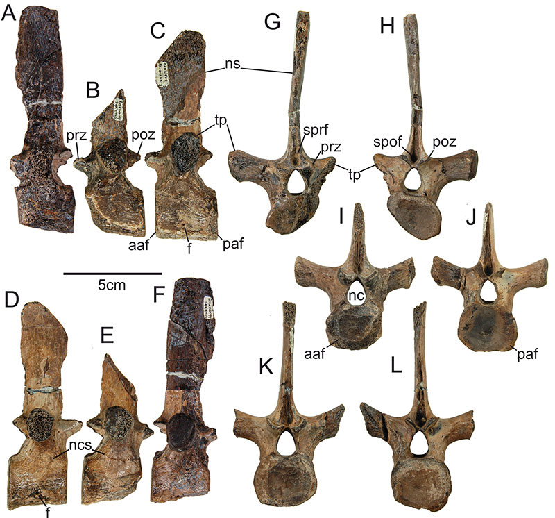
FIGURE 14. GPIH, unregistered, photographs of three dorsal vertebrae in A-C left lateral, D-F right lateral, G, I, K anterior and H, J, L posterior view. Abbreviations: aaf, anterior articular facet; f, foramen; nc, neural canal; ncs, neurocentral suture; ns, neural spine; paf, posterior articular facet; poz, postzygapophysis; prz, prezygapophysis; spof, spinopostzygaophyseal fossa; sprf, spinoprezygapophyseal fossa; tp, transverse process. Sachs et al. (2016a) show another order the individual vertebrae.