FIGURE 1. Location of the Deutsch Altenburg site complex in the territory of Austria.
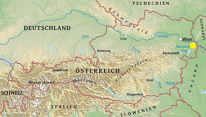
FIGURE 2. Skull of Martes vetus from Deutsch Altenburg 2AC. A, left view. B, ventral view. C, right view. D, dorsal view. Scale bar is 10 mm.
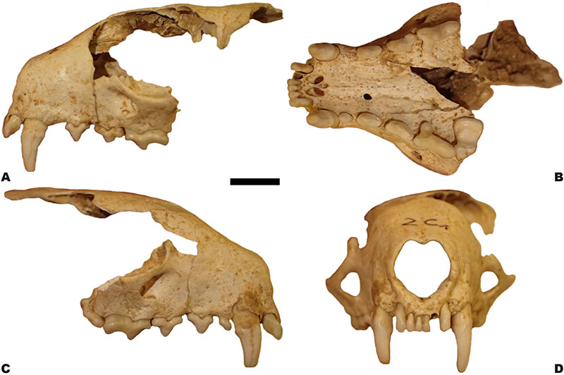
FIGURE 3. Mandibles of Martes vetus from Deutsch Altenburg 2AC. A, right mandible. B, left mandible. C, right mandible. 1, buccal view; 2, lingual view; 3, occlusal view. Scale bar is 10 mm.
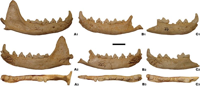
FIGURE 4. Comparison of the morphology of the skull (1) and upper dentition (2) of European martens: A, Martes vetus (Deutsch Altenburg 2AC). B, Martes vetus (Sackdilling Cave). C, extant Martes foina. D, extant Martes zibellina. E, extant Martes martes. All individuals are drawn on the same scale; skulls are shown in lateral view; dentition is shown in occlusal view. Scale bar is 10 mm for skulls and 4 mm for dentition.
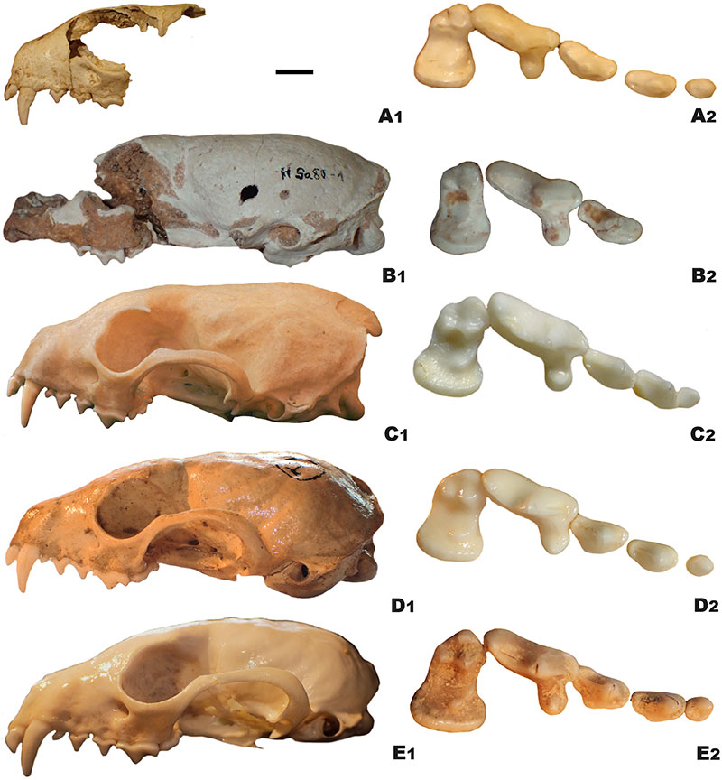
FIGURE 5. Graph showing the relationship between the height of the mandibular body measured behind m1 (H/m1, no. 14) and the spacing of mental foramens (FOR, no. 8) in European martens. For references see the Materials and Methods section.
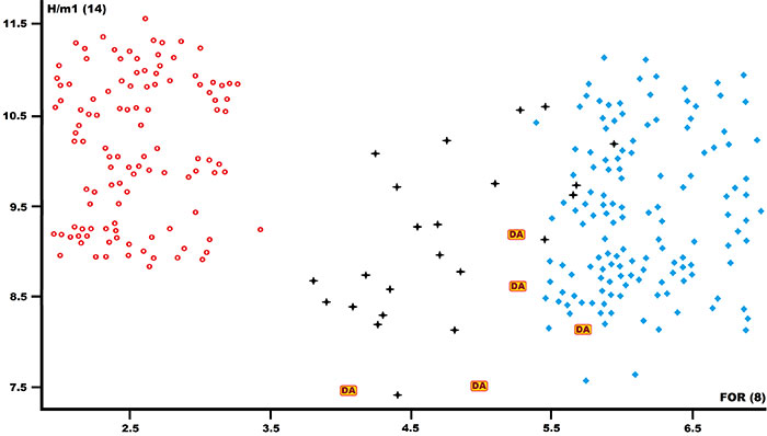
FIGURE 6. Comparison of the morphology of the mandible (1), lower dentition (2), and m1 (3) of European martens. A, Martes vetus from Deutsch Altenburg 2AC. B, extant Martes foina. C, extant Martes martes. D, extant Martes zibellina. All individuals are drawn on the same scale; mandibles are shown in the lateral view; dentition is shown in occlusal view. Scale bar is 10 mm for skulls and 4 mm for dentition.
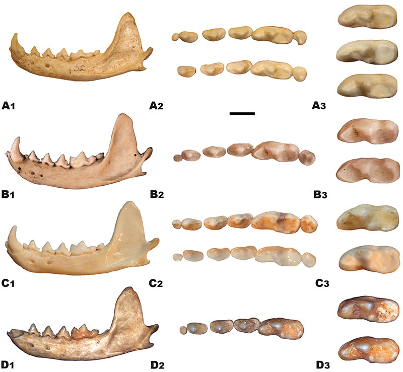
FIGURE 7. A list of selected features of the morphology of European martens, shown as a percentage of the base population of Mates vetus (purple line), for which the values of all features are presented as 100%. Black line -- Martes vetus from Deutsch Altenburg; red line -- extant Martes martes; blue line -- extant Martes foina. For references see the Materials and Methods section.
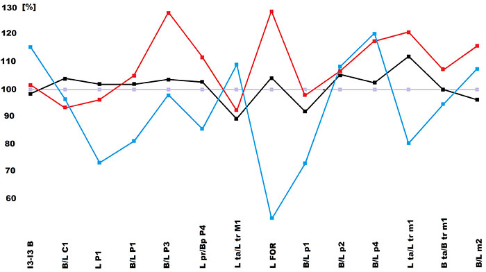
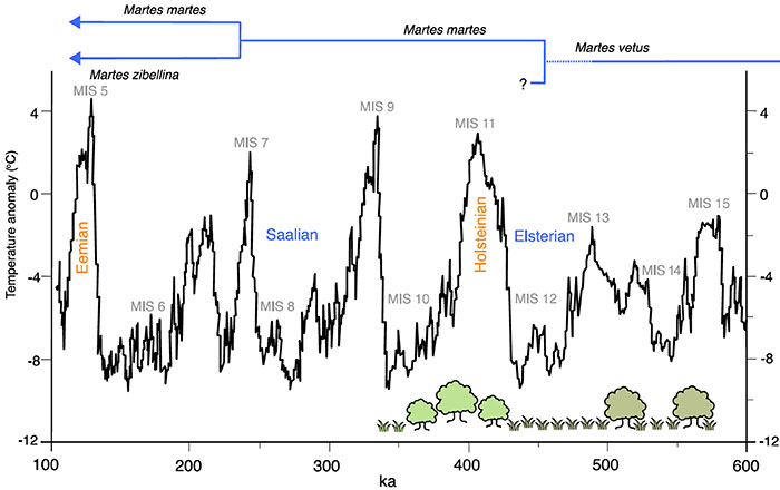
FIGURE 8. Schematic representation of the time of separation of martens from the Martes genus and the environmental conditions accompanying them.