FIGURE 1. Skeletal reconstruction of Dunkleosteus terrelli, showing the distribution of dermal bone (blue) and perichondrally ossified cartilage (yellow). Light blue = cranial elements; dark blue = postcranial trunk armor. Skull is partially transparent to show locations of neurocranium and gill arches, highlighting how the head anatomy of Dunkleosteus compares to other fishes. The scapulocoracoid and cleithrum/anterior lateral plate of the trunk armor are highlighted to show location of head-trunk boundary. Neurocranium and branchial skeleton reconstructed after osteological correlates and other arthrodires in Heintz (1932), Stensiö (1963), Miles and Westoll (1968), Johanson (2003), and Carr et al. (2009). Other cranial cartilages (e.g., palatoquadrate, Meckel's cartilage) omitted for clarity. For homologies between placoderm plates and the bones of other gnathostomes see Johanson (2003), Zhu et al. (2013), and Zhu et al. (2016a).
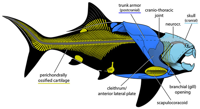
FIGURE 2. Common errors in reconstructions of Dunkleosteus. None of the depicted features are currently supported by Dunkleosteus fossils or comparative patterns in arthrodires. Errors relating to oral tissues or armor integument not depicted for clarity.
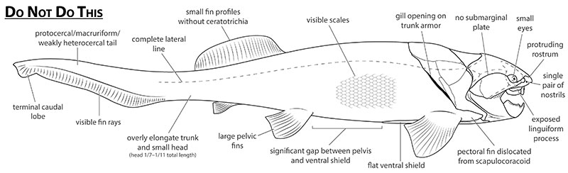
FIGURE 3. Reconstructions of the head and thoracic armor in Dunkleosteus terrelli. A, reconstruction in Heintz (1931b, 1932). B, CMNH 5768, with some plates (i.e., anterior lateral) restored after other specimens. The ventral shield is slightly retrodeformed to account for crushing. Head shield sutures not depicted as they are mostly obliterated by retrodeformation. Dashed lines represent lateral line canals. C, closeup of B denoting major plates and anatomical structures discussed in this study. Scale bar applies only to C. Abbreviations: ADL, anterior dorsolateral; AL, anterior lateral; ASG, anterior supragnathal; AVL, anterior ventrolateral; branch., branchial opening; IG, infragnathal; ling. proc., linguiform process of suborbital MD, median dorsal; obst. proc., obstantic process of anterior lateral; PMV, posterior median ventral; PVL, posterior ventrolateral; SM, submarginal plate; SO, suborbital.
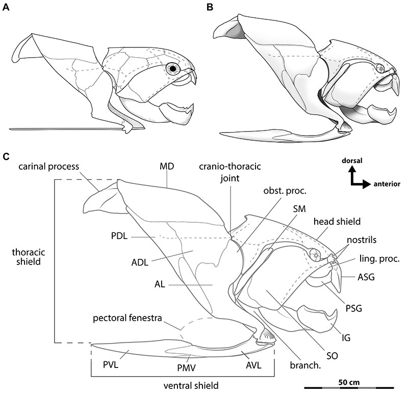
FIGURE 4. Life reconstruction of an adult Dunkleosteus terrelli, based primarily on CMNH 5768 with missing elements restored after CMNH 6090 and CMNH 7054.
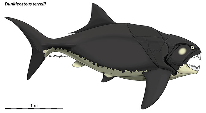
FIGURE 5. Ontogenetic series of near-complete mounted specimens of Dunkleosteus terrelli (progressively older/larger individuals from left to right) showing increasing trunk height, mouth size, and pectoral fenestra size with age. Sutures of head shield elements and between spinal/anterior lateral plates not shown because they have generally been obliterated through historical retrodeformation or otherwise obscured. Specimens depicted as close to the original mounts as possible, except for retrodeformation of the unnaturally flattened ventral shield, fixing the anterior lateral of D, and occasionally restoring damage on the plates from their bilateral counterpart. Hatched areas represent plates absent bilaterally and reconstructed after their sutures/interactions with other elements and other individuals.
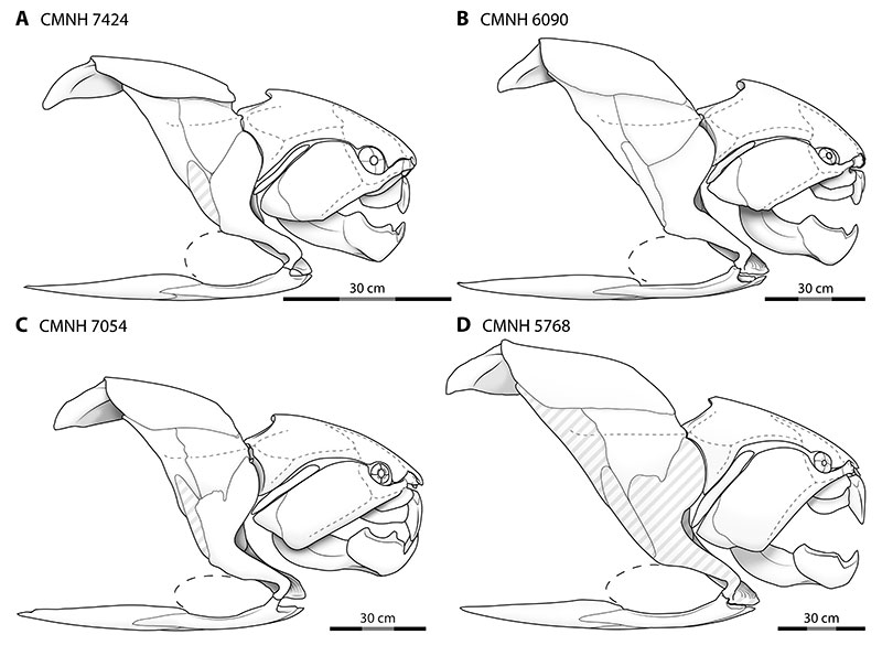
FIGURE 6. Phylogenetic position of Dunkleosteus within Arthrodira. Underlined taxa are represented by complete body outlines or extensive post-thoracic remains. Eubrachythoraci, the main clade of interest, is highlighted in gray. Phylogeny based on Dupret et al. (2007), Zhu and Zhu (2013), Zhu et al. (2016b), Qiao et al. (2016), Boyle and Ryan (2017), Jobbins et al. (2022), and Zhu et al. (2022).
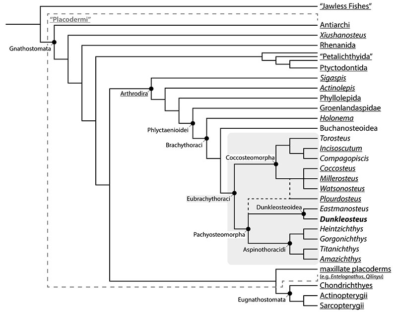
FIGURE 7. A, reconstruction of Coccosteus cuspidatus. B, photograph of ROM VP 52664, the specimen used to create A, Soft tissue outline of the caudal fin in B partially modeled after Squalus and Cephaloscyllium, two extant sharks with analogous life habits. B provided courtesy of the Royal Ontario Museum, used with permission.
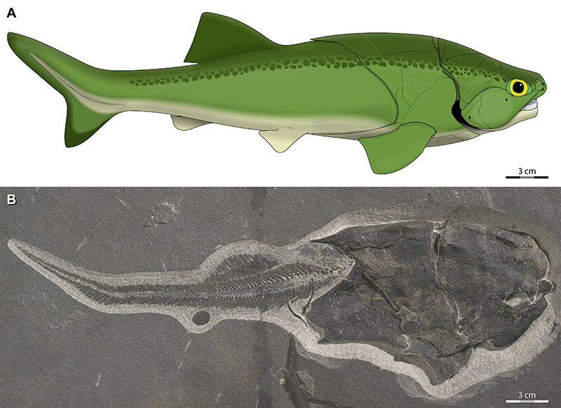
FIGURE 8. Endoskeletons of a eubrachythoracid arthrodire (A) and a eugnathostome (B), showing how anteroposterior locations of major features are conserved despite the presence of dermal armor. Dotted lines represent orbit-opercular length. A based on Miles and Westoll (1968) and Young (2010: fig. 1a), as well as specimens of Coccosteus cuspidatus examined directly; B after Compagno (1999) with caudal skeleton modified from Little and Bemis (2004) and Moreira et al. (2019). Abbreviations: branch. arches, branchial arches; hy. arch, hyoid arch; man. arch, mandibular arch; pec. girdle, pectoral girdle; pelv. girdle, pelvic girdle; post. exp., posterior expansion of the neurocranium over the branchial chamber.
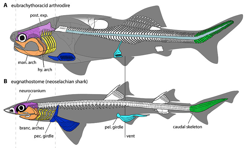
FIGURE 9. Uncertainties in reconstructions of Dunkleosteus terrelli. A, range of possible sizes in the dorsal lobe of the caudal fin, based on extant fishes. B, possible range of body lengths based on ± percent error in OOL, modified from Engelman (2023b: supplementary file S5).
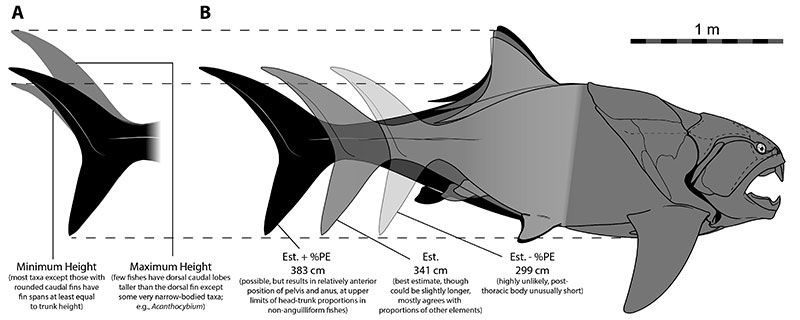
FIGURE 10. Trunk armors of eubrachythoracid arthrodires scaled to the same ventral shield length, showing the proportionally deeper trunk armor of Dunkleosteus. A, Coccosteus cuspidatus (drawn from Miles and Westoll, 1968 and ROM VP 52664); B, Compagopiscis croucheri (modified from Gardiner and Miles, 1994); C, Plourdosteus canadensis (modified from Vézina, 1988); D, Eastmanosteus calliapsis (modified from Dennis-Bryan, 1987); E, Dunkleosteus terrelli (CMNH 7054, a small adult); F, Heintzichthys gouldii (modified from Carr, 1991); G, Bungartius perissus (modified from Dunkle, 1947); H, Amazichthys trinajsticae (modified from Jobbins et al., 2022). Dotted line in D–H, represents posterior margin of pectoral fenestra. Lines at far right represent trunk height of D. terrelli for easy comparison. The posterior dorsolateral is slightly restored for crushing in D to better represent the typical morphology of D. terrelli. Trunk canal in Bungartius omitted due to uncertain morphology (Dunkle, 1947: p. 110). Scale equals 10 cm.
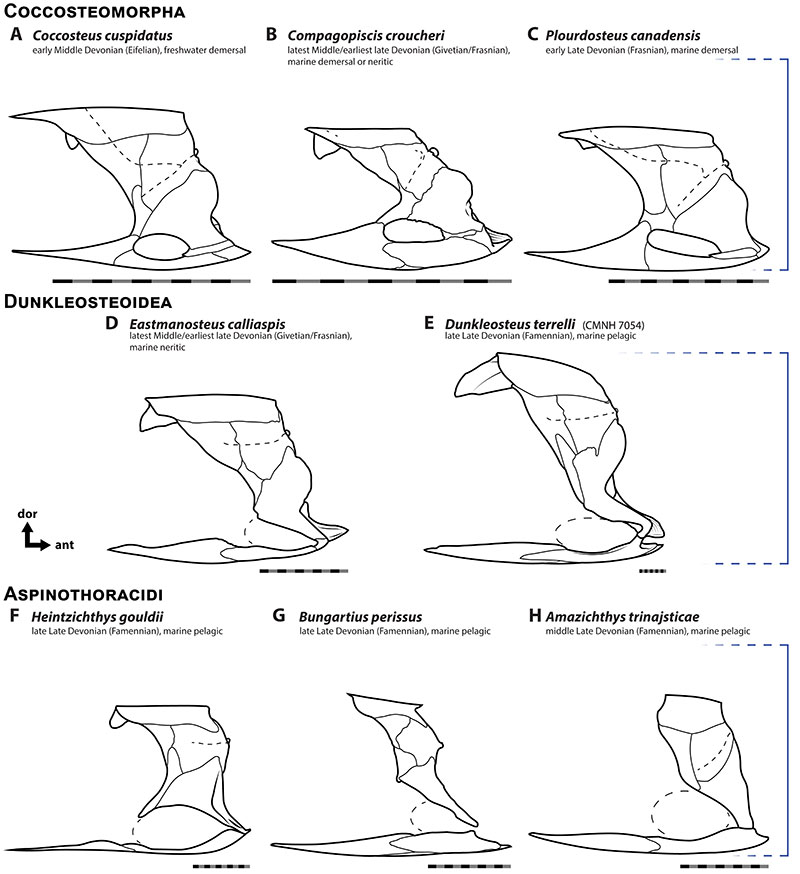
FIGURE 11. Relative pre-pectoral (A) and pre-pelvic (B) length as a proportion of total length in Dunkleosteus, other arthrodires, extant fishes, and the extinct, hyper-elongate Ichthyodectiformes. Estimated arthrodire lengths in A based on OOL, only complete arthrodires considered in B. For pre-pelvic length, values in Dunkleosteus are approximated based on the length to the end of the ventral shield given the condition in CMC VP 8294. * - data graphed using individual specimens rather than species averages, to better show the consistency in this feature across specimens. Black dashed line in A is average of all taxa, whereas in B represents average value for non-acanthopterygian, gadiform, aulopiform fishes. Modified from graphs and analyses in Appendix 3, raw data in Appendix 4. Silhouette of Rhizoprionodon terranovae provided under CC0 by Nathan Hermann on Phylopic.
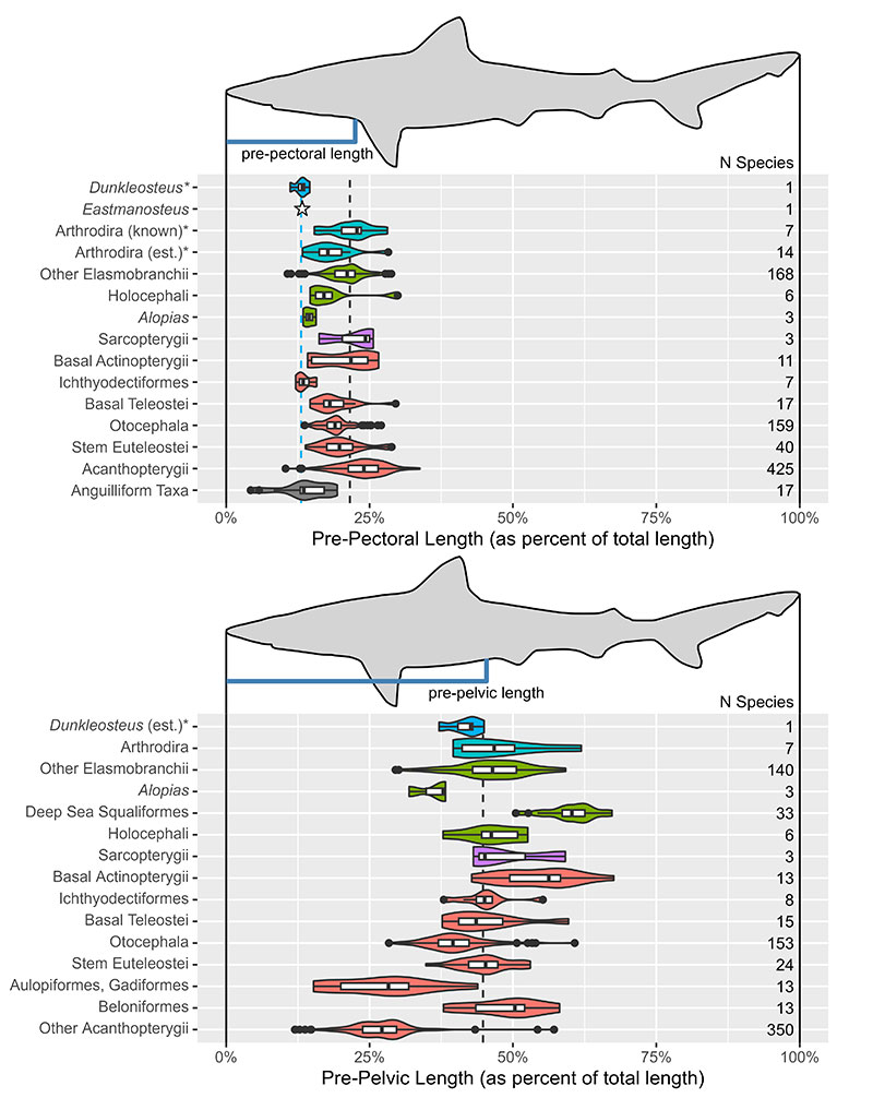
FIGURE 12. Life reconstructions of the ontogenetic series of Dunkleosteus terrelli in Figure 5, showing how fineness ratio increases with body size and results in adults having a more thunniform body plan.
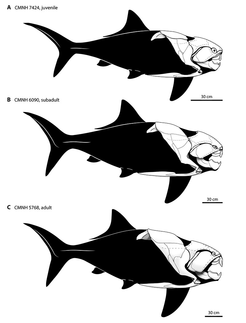
FIGURE 13. Cross-sectional reconstruction of the trunk of Dunkleosteus in posterior view, showing how the position of the spine and horizontal septum (based on the location of the cranio-thoracic joint and internal ridge on the posterior dorsolateral) results in extremely large lateral trunk musculature. Neural and haemal arch morphology modeled after Johanson et al. (2019) and van Mesdag et al. (2020). Presence and extent of transverse abdominal musculature follows Trinajstic et al. (2013).
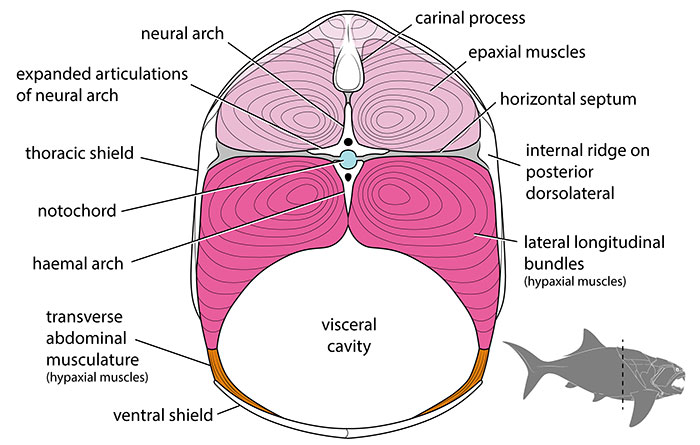
FIGURE 14. Skeletal reconstruction of Dunkleosteus, showing how the morphology of the anterior trunk constrains the path of the spinal cord to the level of the cranio-thoracic joint (red line). Other post-thoracic structures of interest are also denoted. Elements known in D. terrelli are in white whereas elements reconstructed after more complete arthrodires, particularly Coccosteus cuspidatus (see Miles and Westoll, 1968), Heintzichthys gouldii (see Dean, 1896) and Eastmanosteus calliaspis (see van Mesdag et al., 2020), in gray. Size of the neural and haemal arches based on their relative size in CMNH 50322 (see Johanson et al., 2019), which results in the reconstructed spinal column having a similar post-thoracic vertebral count to Coccosteus (see Appendix 3: section 5.1).
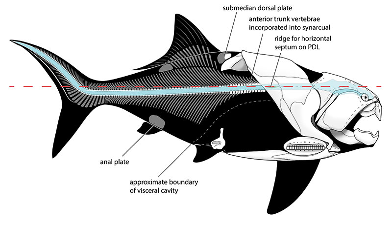
FIGURE 15. Dunkleosteus terrelli (CMNH 7054, with posterior lateral plate and ventral shield partially restored after CMNH 6090) in internal view, showing the internal ridge on the posterior dorsolateral for the horizontal septum and rough boundaries of trunk musculature. Boundary between lateral longitudinal bundles and ventral longitudinal bundles/abdominal musculature set at the dorsal border of the pectoral fin base/pectoral fenestra, based on the condition in chondrichthyans and coccosteomorphs (De Iuliis and Pulerà, 2011; Trinajstic et al., 2013).
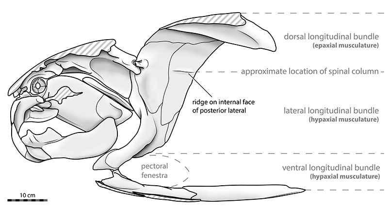
FIGURE 16. Thoracic armors of non-eubrachythoracid arthrodires scaled to the same armor length, showing the lateral line extending beyond the posterior margin of the thoracic armor (contrast with Figure 10). A, Groenlandaspis riniensis (Groenlandaspidae), redrawn from Long et al. (1997); B, Parabuchanosteus murrumbidgeensis (“buchanosteoid”), redrawn from Long et al. (2014); C, Holonema westolli (Holonematidae), redrawn from Miles and White (1971). Anterior is to the right in all images. Scale equals 10 cm.
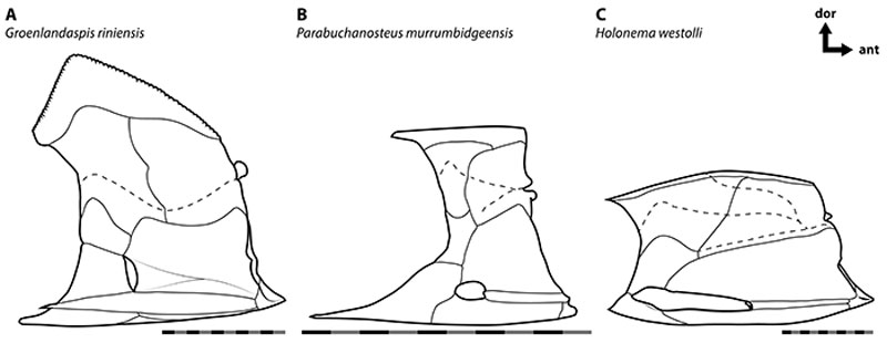
FIGURE 17. A–B, Sclerotic rings of aspinothoracidan arthrodires in external view, showing dermal ornamentation. A, Selenosteus angustipectus (CMNH 8055); B, Gymnotrachelus hydei (CMNH 8042). C, uncrushed sclerotic ring of Dunkleosteus (CMNH 6194) in side view, showing the natural curvature of this element. Scale equals 1 cm.
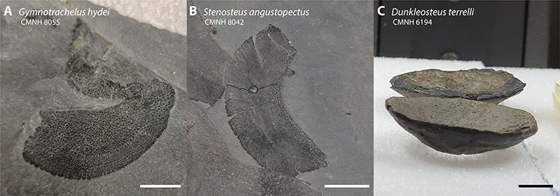
FIGURE 18. Photo (A) and line drawing (B) of dorsal fin outline in CMC VP8545, an unidentified aspinothoracidan from the Late Devonian (Famennian) Chagrin Shale of Ohio. Abbreviations as in Figure 3.
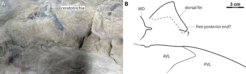
FIGURE 19. CMC VP8294, nearly complete juvenile specimen of Dunkleosteus terrelli preserving the pelvic basals and a partial submedian dorsal plate in situ. A, complete specimen; B close-up of the pelvic basals; C, exposed cross-section of the pelvic basals.
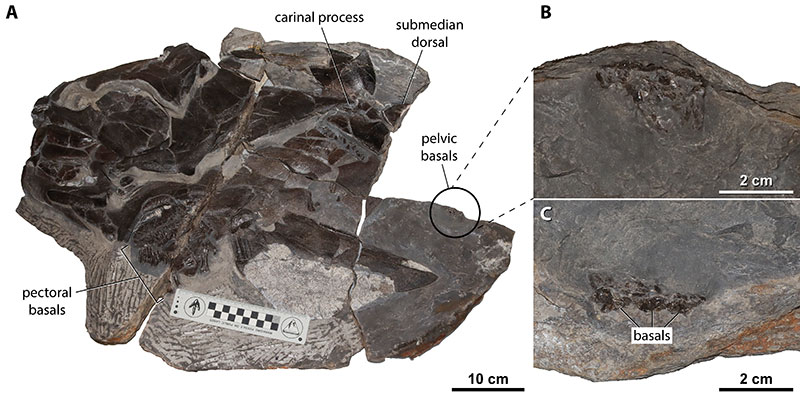
FIGURE 20. Pectoral fin base length scaled against total length (A) and body mass (B) in Dunkleosteus and extant nektonic sharks. Pectoral fin base length in Dunkleosteus measured as the length of the pectoral fenestra. B is on a log10 scale because mass increases cubically relative to pectoral fin base length; similar results are obtained if graphing against the cubic root of body mass (see Appendix 3: fig. 4.14). Graph is zoomed in omitting the outlier Rhincodon for clarity. For color legend for elasmobranchs, see Appendix 3: fig. 4.12. For additional analyses including other arthrodires, see Appendix 3: section 4.6. Sources of lengths and weights for Dunkleosteus in Appendix 3 and raw data in Appendix 4.
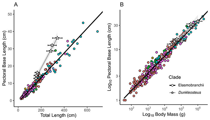
FIGURE 21. A, pelvic girdle of Dunkleosteus terrelli (CMNH 7054, reversed) in lateral(?) view. B, pelvic girdle of CMNH 7054 in approximate life position scaled to the rest of the specimen.
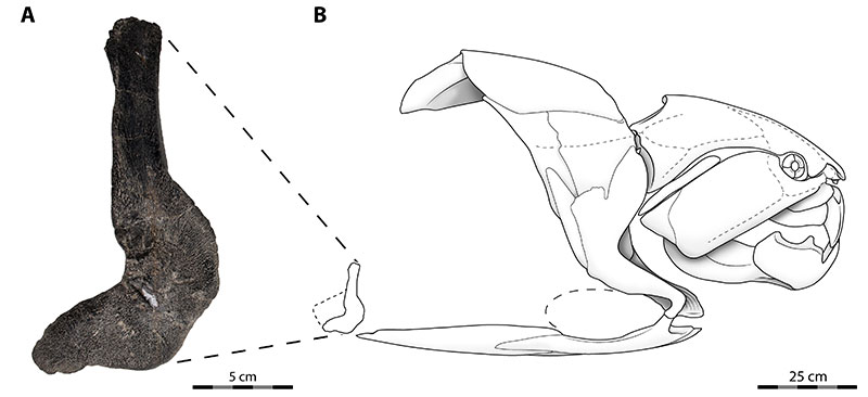
FIGURE 22. Parallel trends of body fineness evolution in pelagic Arthrodira, Ichthyopterygia, Scombridae, and Chondrichthyes (Lamniformes). A–B, Arthrodira; C–D, Ichthyopterygia; E–F, Scombridae; G–H, Lamniformes. In the case of E, all scombrids are open water fishes to some degree but there is a distinct difference in body shape between coastal (neritic) non-thunnins like Scomber and oceanic (pelagic) thunnins. Note that while some features in Dunkleosteus are well-supported (fineness ratio, pelvic fin size) and others have some anatomical constraints (dorsal fin position, caudal fin span), others are inferred approximations requiring further study (caudal peduncle height); see Table 1. A–B, present study; C–D modified from McGowan and Motani (2003); E, drawn from USNM 25256 in Goode (1884), published in the public domain by NOAA; F drawn from measurements in Rivas (1955) and Russell (1934); G redrawn from 153 TL specimen collected by F.H. Mollen (ERB 1183; Mollen, 2019); H drawn from measurements of MZL 23981 in De Maddalena et al. (2003).
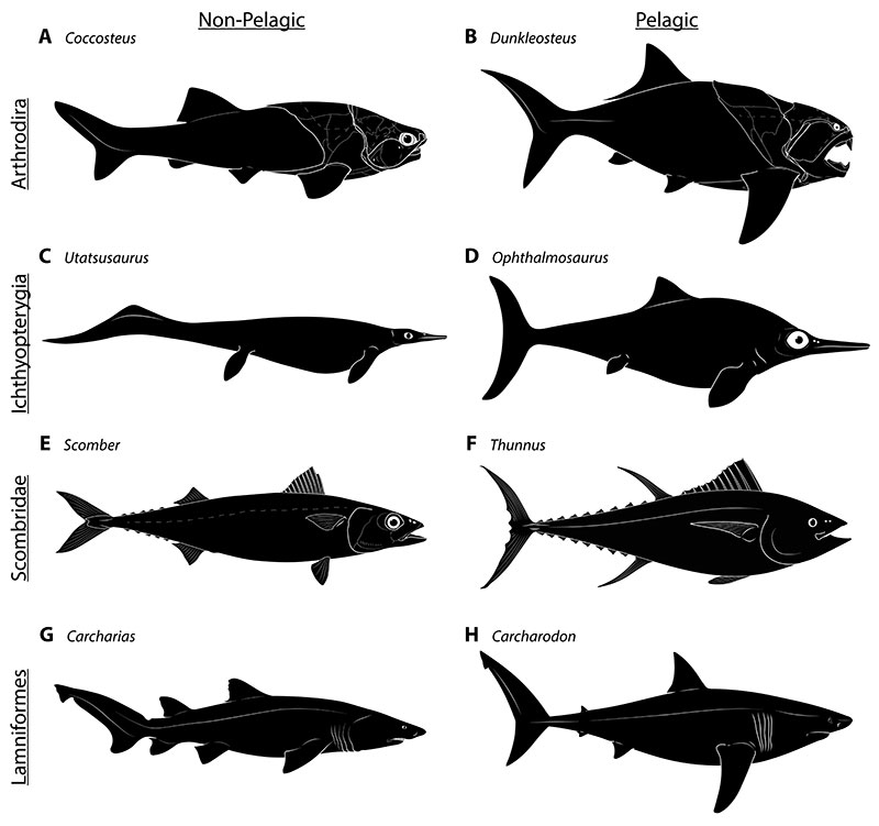
FIGURE 23. Two morphological optima of pelagic fishes compared to a generalized neritic relative, scaled to the same total length. A, Scomber scombrus, a generalized coastal species; B, Acanthocybium solandrei, an ectothermic pelagic taxon; C, Thunnus thynnus, an endothermic pelagic taxon. A and C as in Figure 22, B drawn from FSBC 6267.
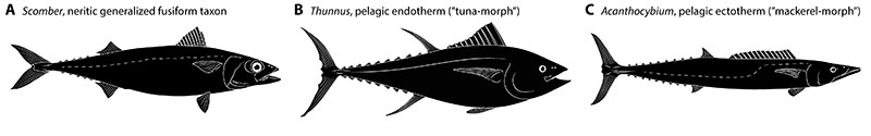
FIGURE 24. Ventral shields of arthrodires in external view. A–C, ventral shields of non-eubrachythoracid (A–B) and coccosteid (C) arthrodires, showing the absence of a posteriorly-projecting triangular process of the anterior ventrolateral plate. D–F, ventral shields of incisoscutoid (D) and aspinothoracidan (E–F), showing the prominent, posteriorly-projecting process of the anterior ventrolateral plate. G–I, ventral shield of Dunkleosteus terrelli, showing the very large process. A, Phlyctaenius acadicus (modified from Heintz, 1933); B, Bryantolepis brachycephala (modified from Denison, 1962); C, Coccosteus cuspidatus (redrawn from Miles and Westoll, 1968); D, Compagopiscis croucheri (redrawn from Gardiner and Miles, 1994); E, Heintzichthys gouldii (following Carr, 1991); F, Gymnotrachelus hydei (redrawn from Carr, 1994); G, composite juvenile, after Hussakof and Kepler (1905) and CMNH 7424 and CMNH 8982; H, CMNH 6090; I, CMNH 5768. Dashed lines represent overlap areas between plates, with grey shaded area representing the overlap between anterior and posterior ventrolateral plates. In D, G, and I, precise internal shapes were not available, so overlap is approximated.