FIGURE 1. Overview of the geology and stratigraphy of fossiliferous localities (modified from Garrido, 2010). A-B, map of South America (A) and Argentina (B) highlighting the Neuquén Group in violet. C, stratigraphic column showing the subgroups and formations comprising the Neuquén Group, with the Portezuelo Formation and Cerro Lisandro Formation indicated by red arrows. D, geological map illustrating the main outcrops and localities in the area.
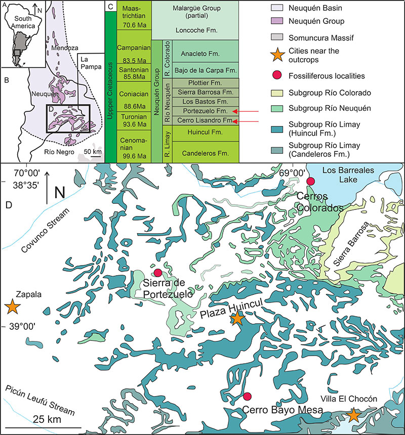
FIGURE 2. Terminology of tooth plates and histological section planes. A, occlusal view of the pterygopalatine tooth plate (adapted from Panzeri et al., 2022b). The labial edge shows denticulations and clefts; on the occlusal surface, are the ridges, the furrows (dotted lines), and the gray area indicates the plateau zone. B, lateral view of a denticulation, showing cusps along the crest. C, tooth plate in a medial view, vertical plane (a) and horizontal plane (b). Abbreviations: cl, cleft; cu, cusps; dn, denticulation; fu, furrow; ri, ridge.
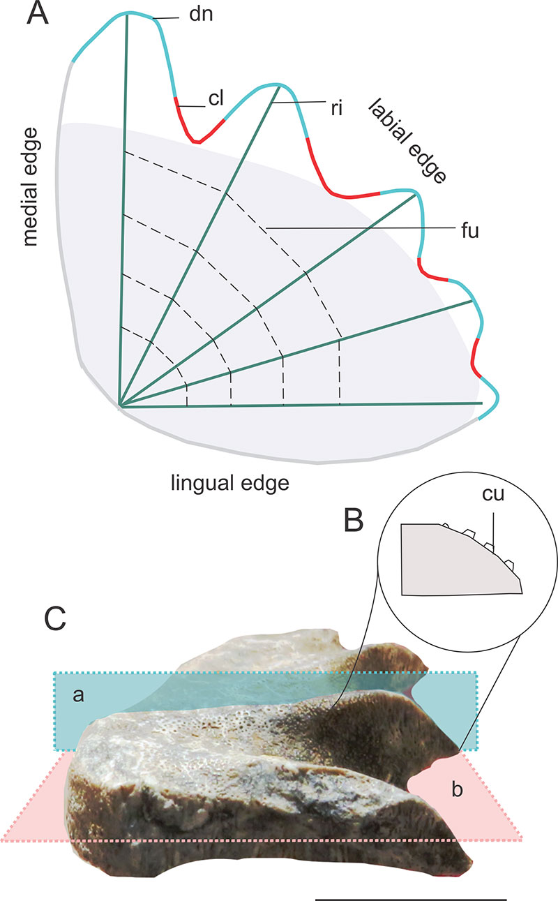
FIGURE 3. Pterygopalatine tooth plates and upper jaw bones of Chaoceratodus portezuelensis. A-B, MCF-PVPH-374, pterygopalatine bone illustrating the ascending process in dorsal (A) and labial (B) views. C, MCF-PVPH-427, position of the ascending process. D, MCF-PVPH-425, tooth plate showing the prominence in the base of the first denticulation (arrow), the position of the inner angle (dashed line), and the life wear pattern. E-F, MCF-PVPH-427, pterygopalatine tooth plate showing the position of the inner angle (E, dashed line) and the posterior direction of the ridges (F, arrows). G, MCF-PVPH-425, tooth plate illustrating life wear pattern and step. H, MCF-PVPH-437, tooth plate illustrating the wear pattern and the last small-sized denticulation (arrow). I, MCF-PVPH-s/n, tooth plate with cusps and a prominence in the base of the first denticulation (arrow). J, MCF-PVPH-s/n, small-sized tooth plate with wear pattern. K, MCF-PVPH-438, tooth plate with inclination of the denticulations and flat plateau area. L-M, MCF-PVPH-437, tooth plate with posterior wear facets, with the flat plateau area tending to be concave. Abbreviations: ap, ascending process of the pterygopalatine bone; cu, cusps; en, enamel; st, step; tx, bone texture; wp, wear pattern. Scale bar: A-B, 0.5 cm; C-N, 1 cm.
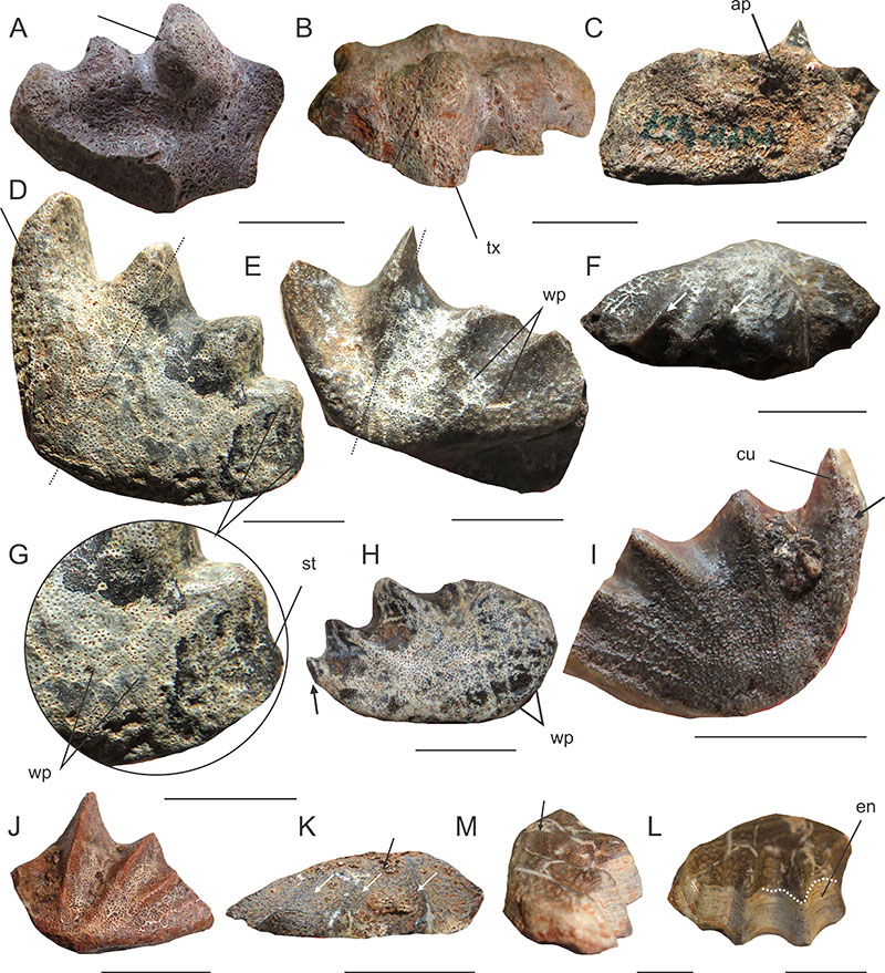
FIGURE 4. Prearticular tooth plates and lower jaw bones of Chaoceratodus portezuelensis. A-B, MCF PVPH 373, holotype of Chaoceratodus portezuelensis in occlusal (A) and medial view (B), the dashed line marks the position of the inner angle. C, MCF-PVPH-572, tooth plate fused to the prearticular bone in lingual view, note the texture. D, MCF-PVPH-542, prearticular tooth plate fused to bone illustrating prearticular bone in labial and ventral view with Ruge’s canal divided by a ridge. E, MCF-PVPH-542, tooth plate showing the punctuations. F, MCF-PVPH-572, prearticular tooth plate with bilobated spur. G, MCF-PVPH-542, tooth plate showing the position of the inner angle and anterior facet of the first denticulation. H, MCF-PVPH-436, prearticular tooth plate, the arrow marks the proximity to the inner angle. I, MCF-PVPH-542, prearticular tooth plate in labial view showing the elevation of the plateau (arrow, convex plateau). J, MCF-PVP-424, prearticular tooth plate illustrating bilobated spur. K-L, MCF-PVPH-436, prearticular tooth plate with marked anterior wear facet, the arrow marks the proximity to the inner angle (J), and cusps on the labial edge (K, L). M, MCF-PVPH-572, tooth plate with posterior wear facet. Abbreviations: spu, spur; af, anterior facet; pf, posterior facet; pun, punctuations; ru, Ruge’s canal; tx, bone texture. Scale bar: 1 cm.
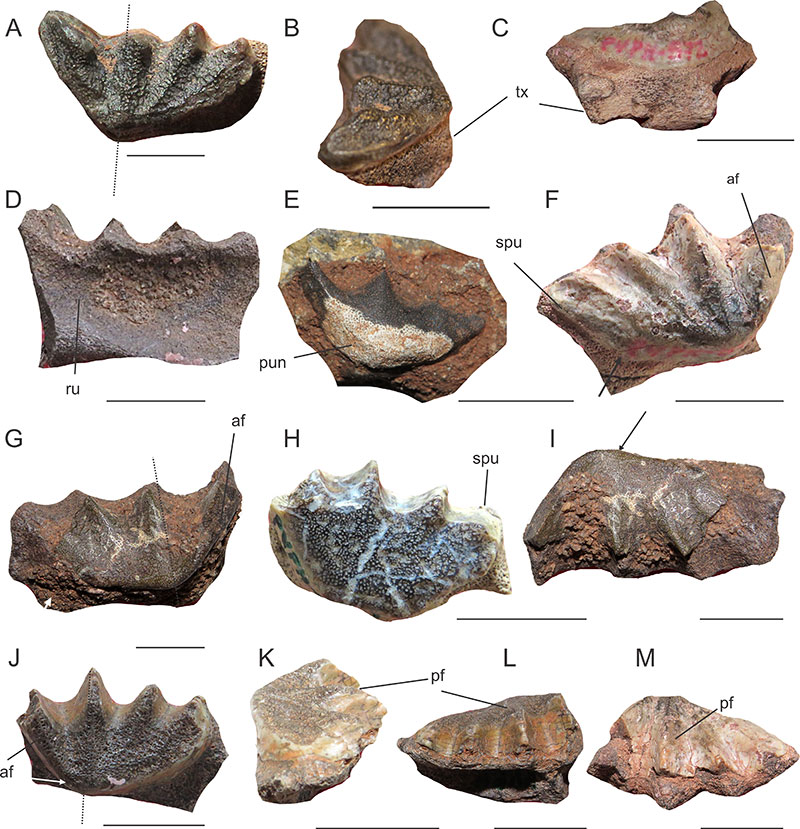
FIGURE 5. Dental histology of the species Chaoceratodus portezuelensis, MCF-PVPH-915A (Cerro Lisandro Formation), horizontal cross-section. A, section under stereoscope. B, denteons immersed in disorganized birefringent interdenteonal dentine and arranged in a row with a monorefringent center. C, area of the plateau where interdenteonal dentine arrangements are more evident. D, detail of the arrangement. E, denticulation with a central row of monorefringent interdenteonal dentine. F, detail of E. G, birefringent circumdenteonal dentine and dentinal tubules. H, circumdenteonal dentine under lambda filter. I, circumdenteonal dentine under polarized light. J, transitional denteons toward the mediolingual edge, mantle dentine is not evident. K, furrow area with clusters of denteons. L, band of birefringent enamel. Sections under a petrographic microscope (B-L), polarized light (I), and polarized light with a lambda filter (B-F, H-L). Abbreviations: bid, birefringent interdenteonal dentine; cd, circumdenteonal dentine; dt, dentinal tubules; en, enamel; gr, granules; mid, monorefringent interdenteonal dentine; trd, transitional denteons. Scale bar: A, 1 cm; B-C, E, J-K, 1 mm; D-F, 500 μm; G-I, L, 250 μm.
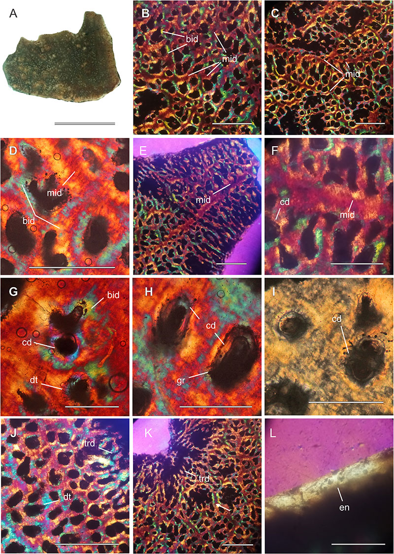
FIGURE 6. Dental histology of the species Chaoceratodus portezuelensis, MCF-PVPH-915B (Cerro Lisandro Formation). A, continuous denteons: linear and branched in an occluso-pulpal direction. B, linear pulp canals, incremental lines are evident. C, discontinuous pulp canals, disorganized birefringent interdenteonal dentine is observed, with monorefringent interdenteonal dentine in between. D, arrangement of interdenteonal dentine, and surrounding the pulp canal is the circumdenteonal dentine. E-F, same details as D but under polarized light (E) and normal light (F). Sections under petrographic microscope (A-F), with polarized light (E), polarized light with lambda filter (A-D), and normal light (F). Abbreviations: bid, birefringent interdenteonal dentine; cd, circumdenteonal dentine; il, incremental line; ld, linear denteon; mid, monorefringent interdenteonal dentine; pc, pulp canal. Scale bar: A-C, 1mm; D-F, 500 μm.
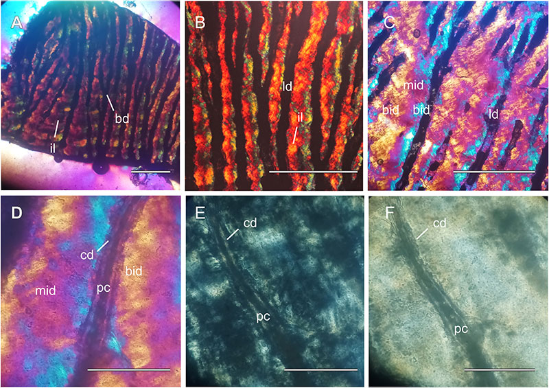
FIGURE 7. Cretaceous Dipnoans from Patagonia. A-B, tooth plate (MPCA-AT 11) of cf. Chaoceratodus portezuelensis from El Anfiteatro area in oclussal (A) and pulpar (B) views, the arrow indicates the first incomplete denticulation. C-D, same tooth plate in labial view, the arrow indicates the concave plateau area. E-G, pterygopalatine tooth plates of Metaceratodus kaopen, showing varying degrees (MPCN-PV-SN 7, E and MPCN-PV-1-3, F) of wear and a prearticular tooth plate (MPCN-PV-SN 4, G). H-J, tooth plates of Metaceratodus wichmanni, displaying different wear levels (MACN-Pv RN 350/2H and MACN-Pv RN 157A I) and a prearticular tooth plate (MML 202, J). K-M, tooth plates of Metaceratodus baibianorum, with varying wear (MPEF-PV 11425 and MPEF-PV 11422, L) and a prearticular tooth plate (MPEF-PV 11422 M). N-P, pterygopalatine tooth plates assigned to Atlantoceratodus iheringi (N-O), showing different wear levels (MPM-PV-1872.11.1, N and MPMPV-1872.11.3, O) and a prearticular tooth plate (MPM-PV-1874.22.1, P). Q-S, pterygopalatine tooth plates of Rinconodus salvadori (MAU-Pv-N-478/3, Q and MAU-PV-LI-613, R) and prearticular tooth plate (MAU-PV-LI-613, S). Scale bar: 1 cm.
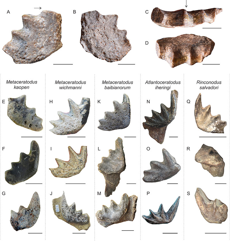
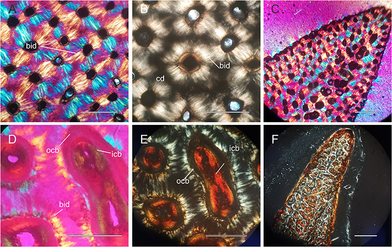
FIGURE 8. Histological cross-sections of tooth plates of Metaceratodus baibianorum and Atlantoceratodus iheringi. A-C, sections of M. baibianorum tooth plates illustrating dentine arrangement in detail and at the first denticulation level (MPEF-PV 11419). D-F, histological cross-sections of tooth plates of Atlantoceratodus iheringi (MPM-PV-1882), illustrating dentine arrangement (D, E) and the first denticulation under normal light (F). Abbreviations: bid, birefringent interdenteonal dentine; cd, circumdenteonal dentine; icb, inner circumdenteonal dentine; ocb, outer circumdenteonal dentine. Polarized light: B, E; lambda filter: A, C-D. Scale bar: B, D-E, 250 µm; A, C, F, 500 µm.