FIGURE 1. Locality of the Howe-Stephens Quarry. A) Map of the United States. Grey box outlines Wyoming, in which the Howe Ranch is located. B) Position of the Howe Ranch in Wyoming, just north of Shell, is indicated by the red star. C) The Howe Ranch, and the positions of the quarries on the site exposing the Morrison Formation. Figure 1B is based on Tschopp et al. (2015b) and information from the SMA, and 1C is based on Siber and Möckli (2009).
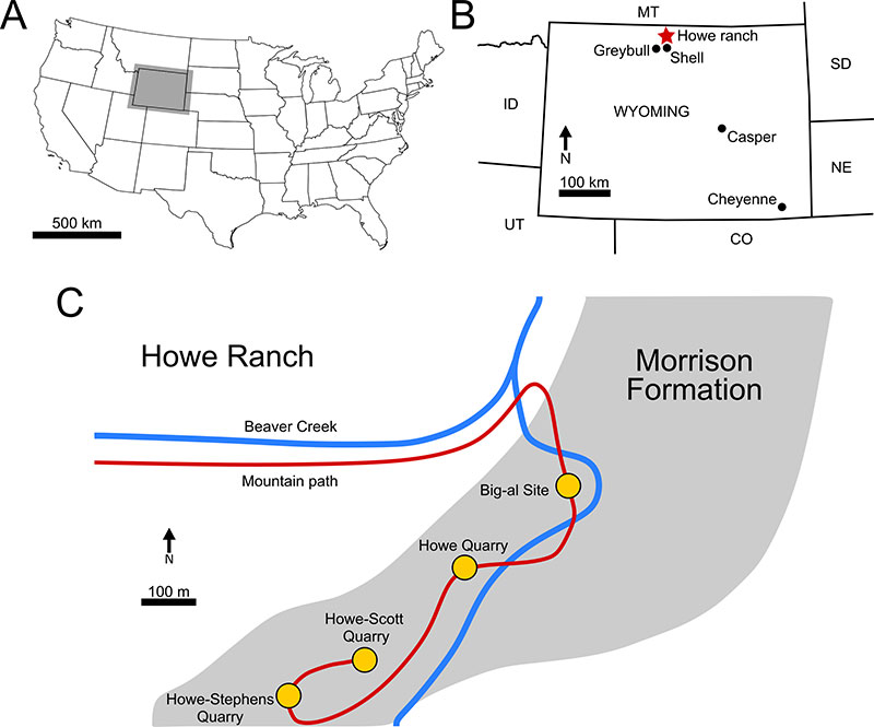
FIGURE 2. Quarry map of Ardetosaurus viator MAB011899. Excavation map of the Howe-Stephens Quarry, indicating the major finds from 1992-2000. Individual dinosaurs are color coded, and MAB011899 is coded with dark blue, and named ‘Diplodocus Brösmeli’ herein. The red crosses indicate the missing/lost cervical vertebrae. Note the relatively similar color for ‘Brösmeli’ and ‘David’ (SMA 0086), but their significant separation in the quarry. Figure is courtesy of the SMA. Quarry sections equal 1 by 0.5 m.
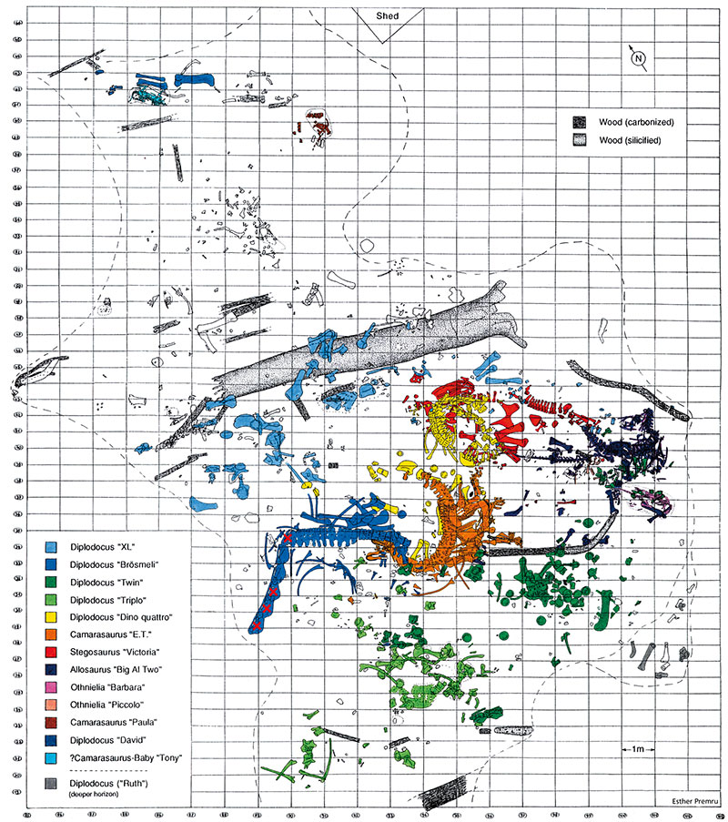
FIGURE 3. Stratigraphic positions of the quarries located on the Howe Ranch. Figure is based on the stratigraphy log made by the SMA in 2003.
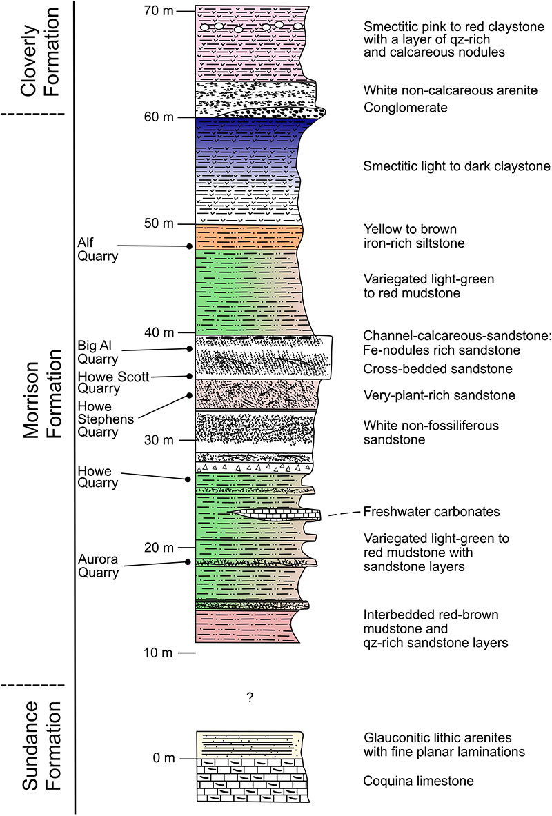
FIGURE 4. Skeletal reconstruction of Ardetosaurus viator MAB011899. Skeletal reconstruction indicating preserved bones (white), excavated bones but subsequently lost (light gray) and not preserved (dark gray). Unknown elements are based on other diplodocines. Scale bar equals 1 m. Reconstruction by Ole Zant.
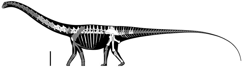
FIGURE 5. Cervical vertebrae 13, 14 and 15 of Ardetosaurus viator MAB011899, as photographed by the SMA. Cervical vertebrae 13 and 14 in A) left lateral and B) anterolateral view, with the preserved cervical ribs still present. Cervical vertebra 15 is shown in C) posterolateral and D) anterolateral view. Photographs are courtesy of the SMA. Scale bar is only applicable for figures A and B, as there is no measurable reference for CV15 due to the oblique views. Abbreviations: ap, anterior process; CPRL, centroprezygapophyseal lamina; pl, pleurocoel; PRDL, prezygodiapophyseal lamina; prz, prezygapophysis; SPRL, spinoprezygapophyseal lamina.
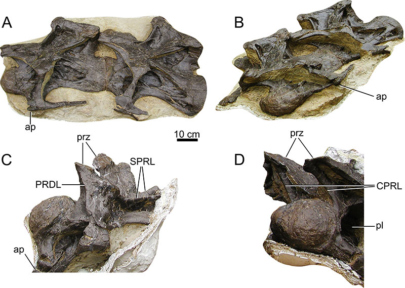
FIGURE 6. Cervical vertebra 13 of Ardetosaurus viator MAB011899. CV13 is shown in A) ventral, B) dorsal, C) left lateral, D) right lateral, E) posterior, and F) anterior view. A close up of the white box in F is provided of the accessory laminae in the SPRF, shown in anterodorsal view. White shaded areas indicate reconstructed parts. The left cervical rib loop was obscured in ventral view for support and therefore roughly outlined here. White dotted lines in A indicate the remnants of the ventral keel. 1 indicates the triangular projections on the diapophysis. Abbreviations: al, accessory lamina; CPRL, centroprezygapophyseal lamina; epi, epipophysis; pap, parapophysis; PCDL, posterior centrodiapophyseal lamina; pre, pre-epipophysis; PRSL, prespinal lamina; pvf, posteroventral flange; SPOL, spinopostzygapophyseal lamina; SPRL, spinoprezygapophyseal lamina; TPOL, interpostzygapophyseal lamina.
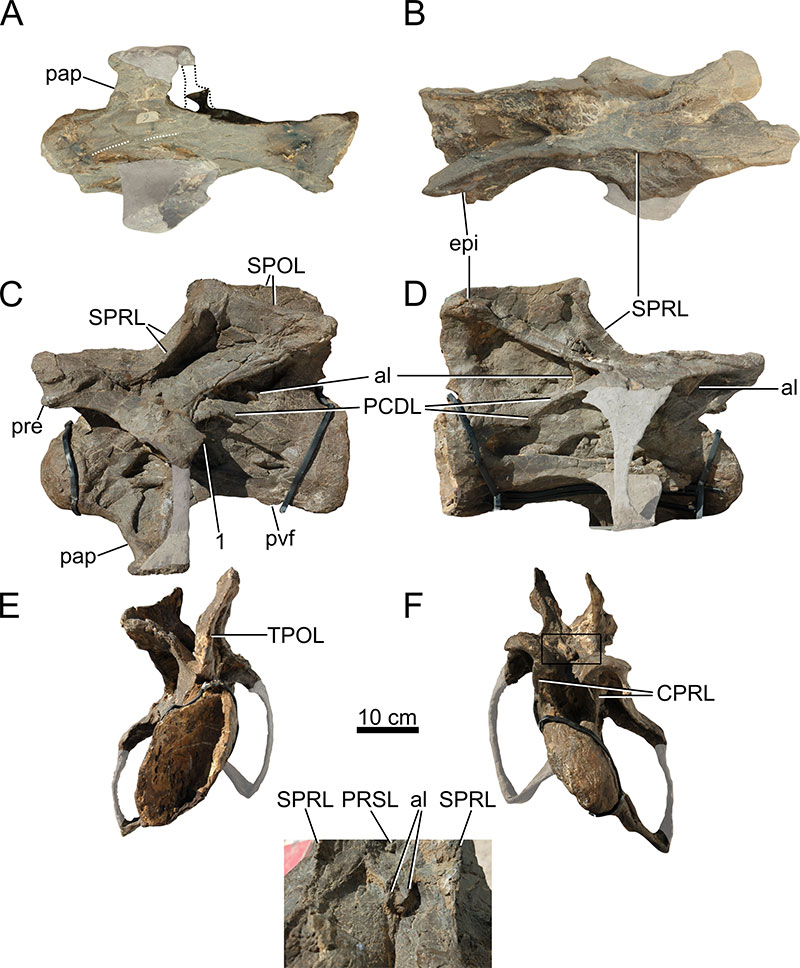
FIGURE 7. Cervical vertebra 14 of Ardetosaurus viator MAB011899. CV14 is shown in A) ventral, B) dorsal, C) left lateral, D) right lateral, E) posterior, and F) anterior view. A close up of the white box in F is provided of the accessory laminae in the SPRF, shown in anterodorsal view. Note the transition of these laminae to a more posterior position in the SPRF. White shaded areas indicate reconstructed parts. White dotted line indicates the remnant of the ventral keel. 1 indicates the triangular projections on the diapophysis. Abbreviations: al, accessory lamina; CPOL, centropostzygapophyseal lamina; CPRL, centroprezygapophyseal lamina; epi, epipophysis; pap, parapophysis; PCDL, posterior centrodiapophyseal lamina; pre, pre-epipophysis; PRSL, prespinal lamina; pvf, posteroventral flange; SPOL, spinopostzygapophyseal lamina; SPRL, spinoprezygapophyseal lamina; TPOL, interpostzygapophyseal lamina.
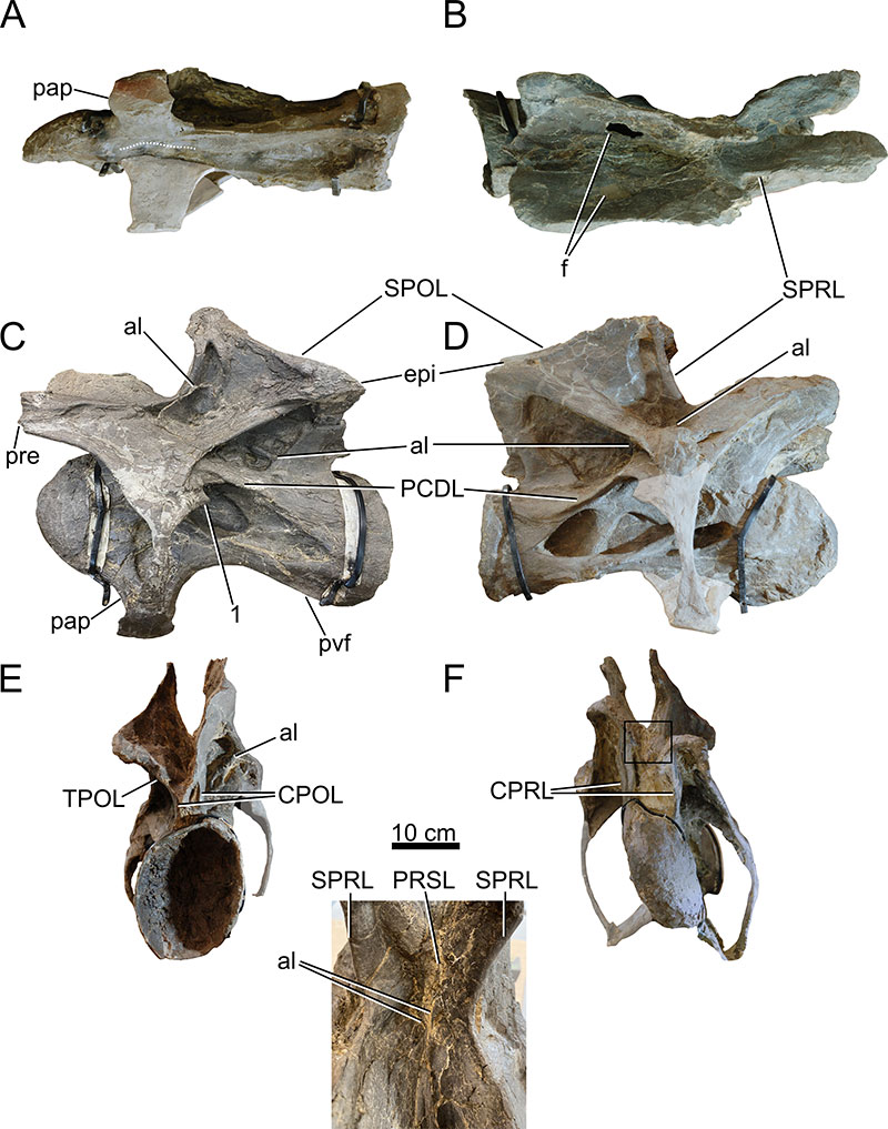
FIGURE 8. Dorsal vertebra 1 of Ardetosaurus viator MAB011899. DV1 is shown in A) ventral, B) dorsal, C) left lateral, D) right lateral, E) posterior, and F) anterior view. White shaded areas indicate reconstructed parts. Note the bifurcated ACDLs. Abbreviations: ACDL, anterior centrodiapophyseal lamina; al, accessory lamina; CPOL, centropostzygapophyseal lamina; CPRL, centroprezygapophyseal lamina; pap, parapophysis; PCDL, posterior centrodiapophyseal lamina; PODL, postzygodiapophyseal lamina; poz, postzygapophysis; prz, prezygapophysis; SPRL, spinoprezygapophyseal lamina; TPOL, interpostzygapophyseal lamina.
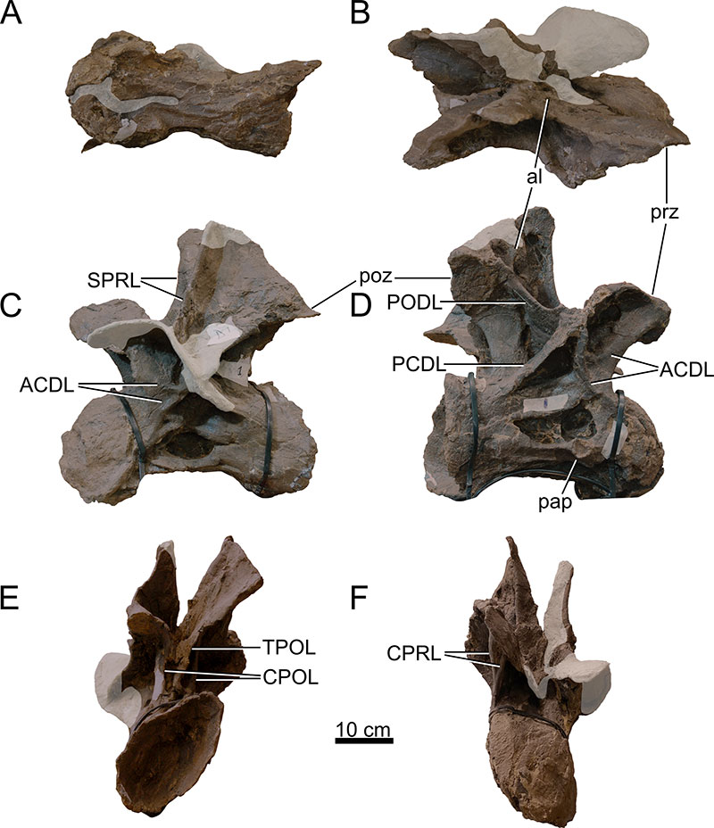
FIGURE 9. Dorsal vertebra 2 of Ardetosaurus viator MAB011899. DV2 is shown in A) ventral, B) dorsal, C) left lateral, D) right lateral, E) posterior, and F) anterior view. Note the shallow fossae medial to the ventrally bifurcating CPOLs. White shaded areas indicate reconstructed parts. Note the bifurcated ACDLs. Abbreviations: ACDL, anterior centrodiapophyseal lamina; al, accessory lamina; CPOL, centropostzygapophyseal lamina; CPOL-f, centropostzygapophyseal lamina fossa; pap, parapophysis; PCDL, posterior centrodiapophyseal lamina; SPRL, spinoprezygapophyseal lamina; TPOL, interpostzygapophyseal lamina; TPRL, interprezygapophyseal lamina.
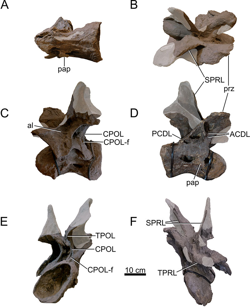
FIGURE 10. Dorsal vertebra 3 of Ardetosaurus viator MAB011899. DV3 is shown in A) ventral, B) dorsal, C) left lateral, D) right lateral, E) posterior, and F) anterior view. Note the displacement of the SPRL/SPDL, as well as the first appearance of the hyposphene, albeit crushed. White shaded areas indicate reconstructed parts. Abbreviations: ACDL, anterior centrodiapophyseal lamina; al, accessory lamina; CPRL, centroprezygapophyseal lamina; hyp, hyposphene; pap, parapophysis; PCDL, posterior centrodiapophyseal lamina; prz, prezygapophysis; SPDL, spinodiapophyseal lamina; SPRL, spinoprezygapophyseal lamina.
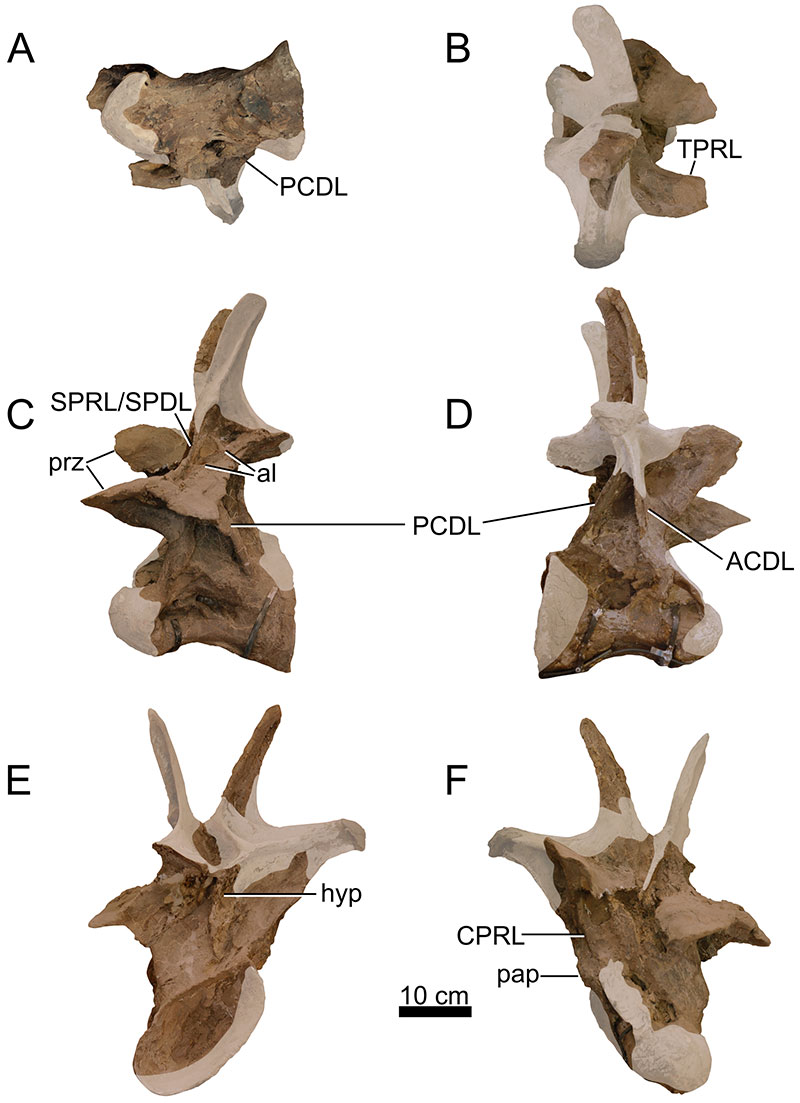
FIGURE 11. Dorsal vertebra 4 of Ardetosaurus viator MAB011899. DV4 is shown in A) ventral, B) dorsal, C) left lateral, D) right lateral, E) posterior, and F) anterior view. White shaded areas indicate reconstructed parts. Abbreviations: hyp, hyposphene; pap, parapophysis; PCDL, posterior centrodiapophyseal lamina; PODL, postzygodiapophyseal lamina; SPDL, spinodiapophyseal lamina; SPOL, spinopostzygapophyseal lamina; SPRL, spinoprezygapophyseal lamina; tap, triangular aliform process.
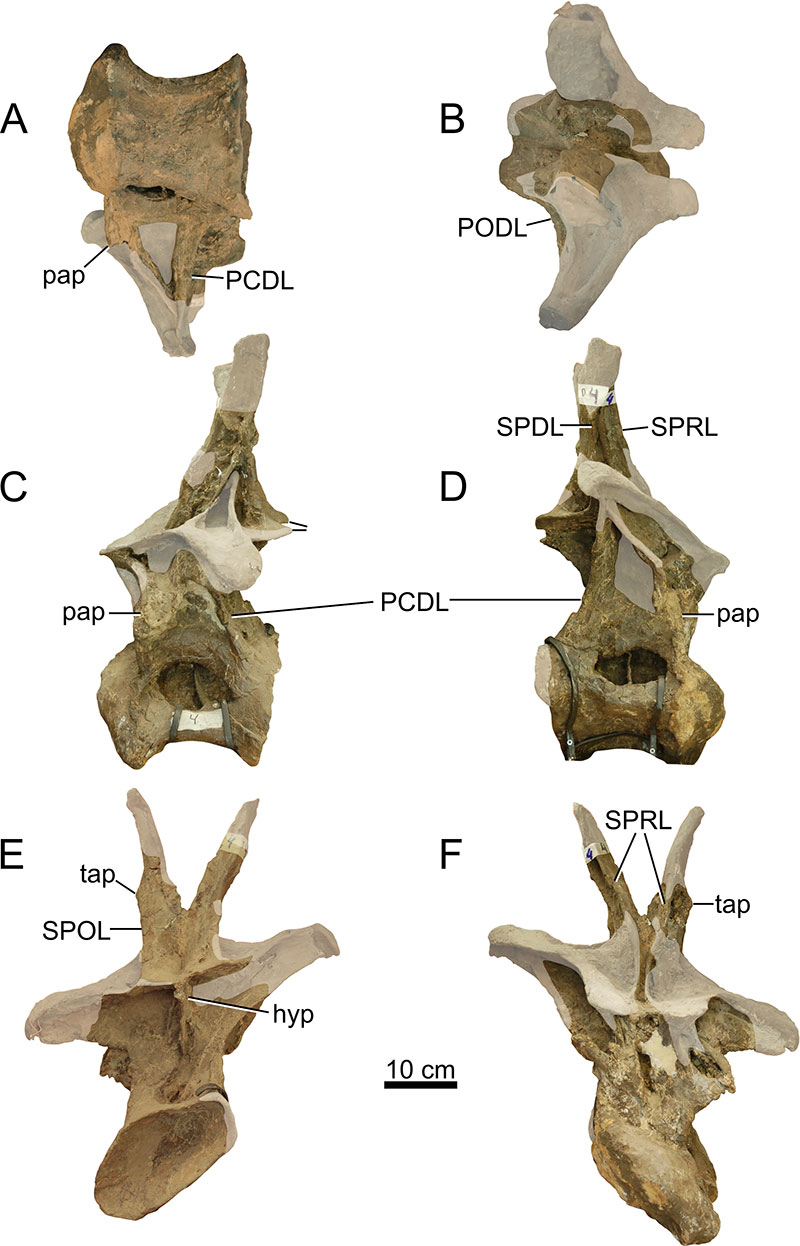
FIGURE 12. Dorsal vertebra 5 of Ardetosaurus viator MAB011899. DV5 is shown in A) ventral, B) dorsal, C) left lateral, D) right lateral, E) posterior, and F) anterior view. Note the complex morphology of the left parapophysis and the surrounding laminae. White shaded areas indicate reconstructed parts. Abbreviations: ACPL, anterior centroparapophyseal lamina; al, accessory lamina; mSPOL, medial spinopostzygapophyseal lamina; lSPOL, lateral spinopostzygapophyseal lamina; pap, parapophysis; PCDL, posterior centrodiapophyseal lamina; PCPL. posterior centroparapophyseal lamina; PODL, postzygodiapophyseal lamina; PPDL, paradiapophyseal lamina; PRPL, prezgyoparapophyseal lamina; SPDL, spinodiapophyseal lamina; SPRL, spinoprezygapophyseal lamina.
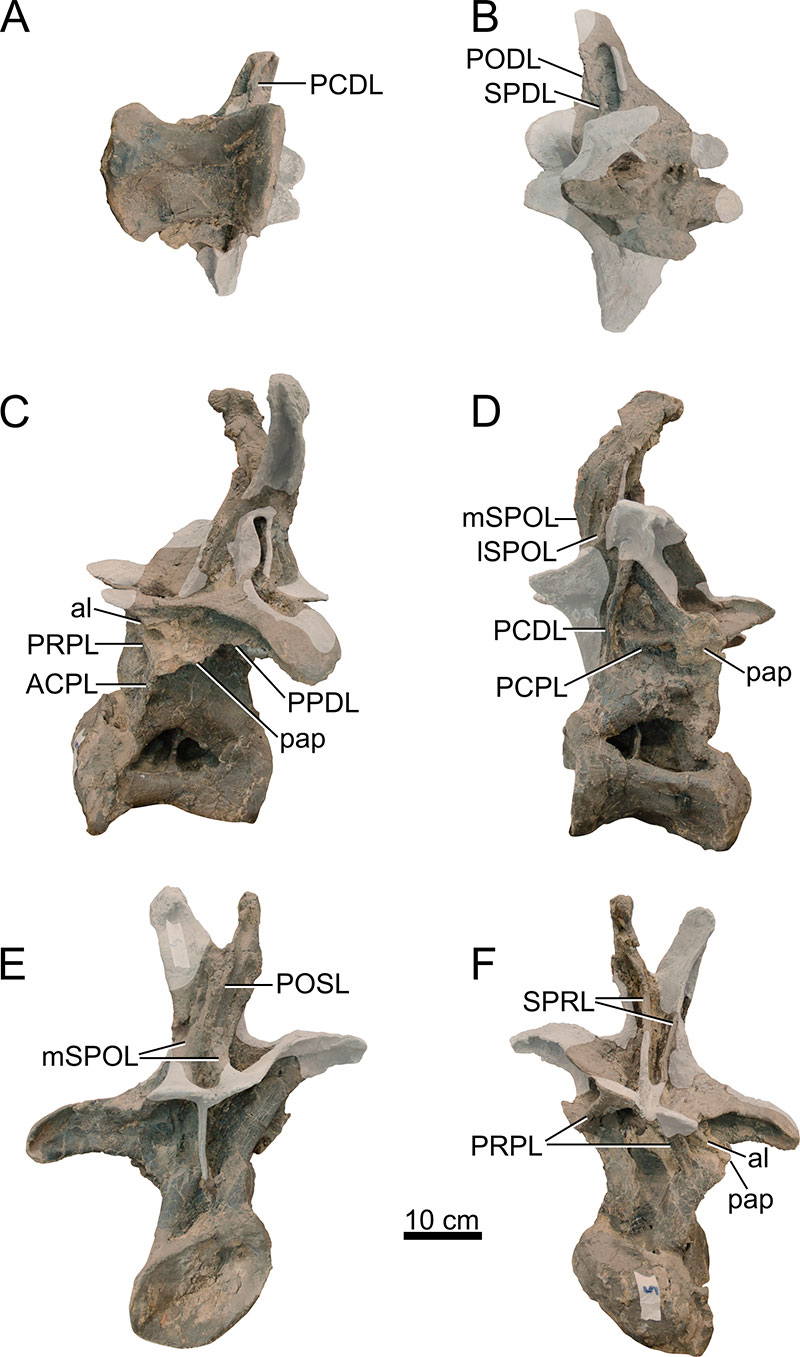
FIGURE 13. Dorsal vertebra 6 of Ardetosaurus viator MAB011899. DV6 is shown in A) ventral, B) dorsal, C) left lateral, D) right lateral, E) posterior, and F) anterior view. White shaded areas indicate reconstructed parts. Abbreviations: hyp, hyposphene; mCPOL, medial centropostzygapophyseal lamina; mSPOL, medial spinopostzygapophyseal lamina; lCPOL, lateral centropostzygapophyseal lamina; lSPOL, lateral spinopostzygapophyseal lamina; PCDL, posterior centrodiapophyseal lamina; PODL, postzygodiapophyseal lamina; poz, postzygapophysis; PRDL, prezygodiapophyseal lamina; PRSL, prespinal lamina; SPDL, spinodiapophyseal lamina; SPRL, spinoprezygapophyseal lamina; tap, triangular aliform process.
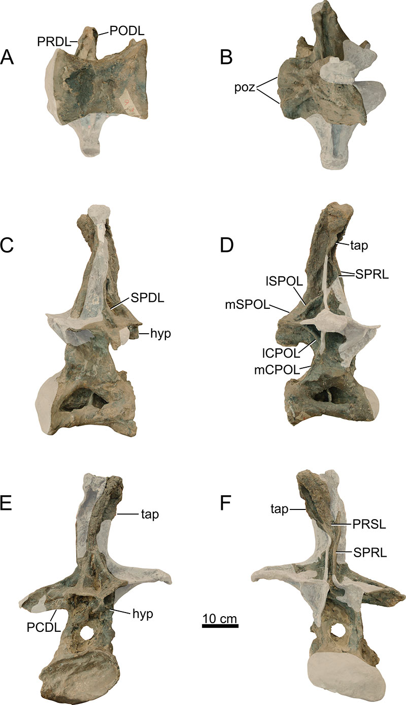
FIGURE 14. Dorsal vertebra 7 of Ardetosaurus viator MAB011899. DV7 is shown in A) ventral, B) dorsal, C) left lateral, D) right lateral, E) posterior, and F) anterior view. White shaded areas indicate reconstructed parts. Abbreviations: hyp, hyposphene; mCPOL, medial centropostzygapophyseal lamina; mSPOL, medial spinopostzygapophyseal lamina; lCPOL, lateral centropostzygapophyseal lamina; lSPOL, lateral spinopostzygapophyseal lamina; PCDL, posterior centrodiapophyseal lamina; PCPL, posterior centroparapophyseal lamina; PODL, postzygodiapophyseal lamina; poz, postzygapophysis; PRSL, prespinal lamina; SPRL, spinoprezygapophyseal lamina; tap, triangular aliform process.
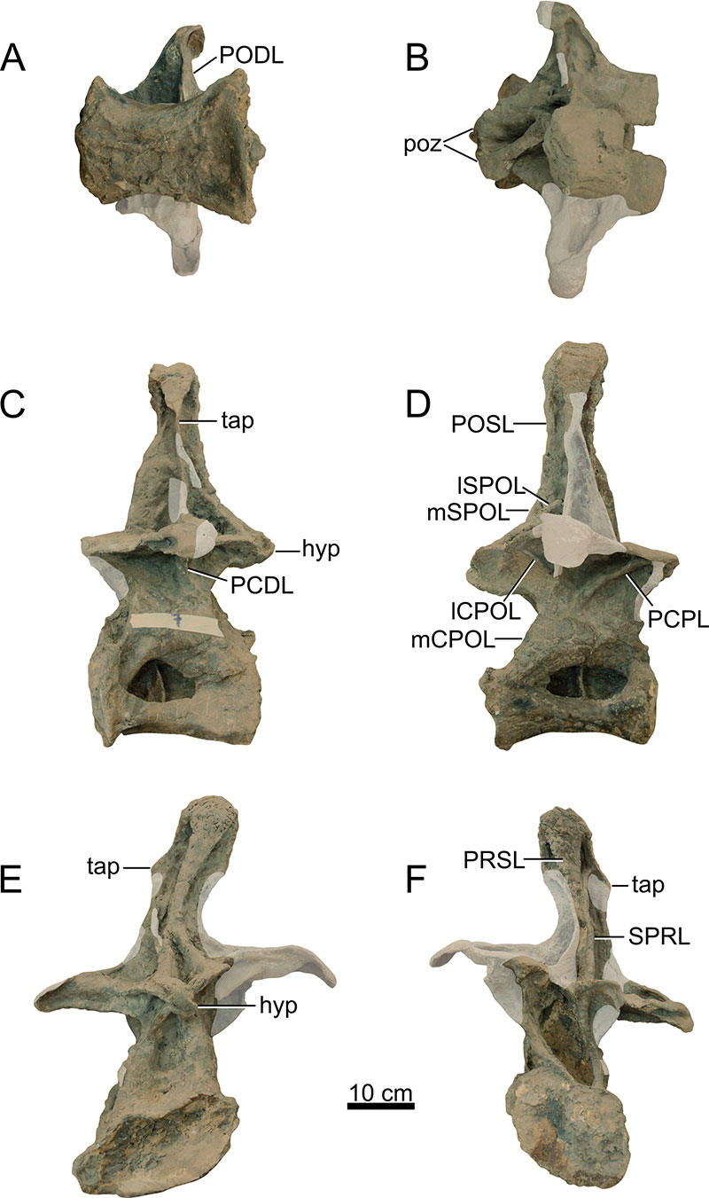
FIGURE 15. Dorsal vertebra 8 of Ardetosaurus viator MAB011899. DV8 is shown in A) ventral, B) dorsal, C) left lateral, D) right lateral, E) posterior, and F) anterior view. Note the attached piece of bone (1) to the lateral surface of the neural arch. White shaded areas indicate reconstructed parts. Abbreviations: hyp, hyposphene; mCPOL, medial centropostzygapophyseal lamina; PCDL, posterior centrodiapophyseal lamina; PCPL, posterior centroparapophyseal lamina; PODL, postzygodiapophyseal lamina; POSL, postspinal lamina; poz, postzygapophysis; prz, prezygapophysis; SPDL, spinodiapophyseal lamina; SPOL, spinopostzygapophyseal lamina; SPRL, spinoprezygapophyseal lamina.
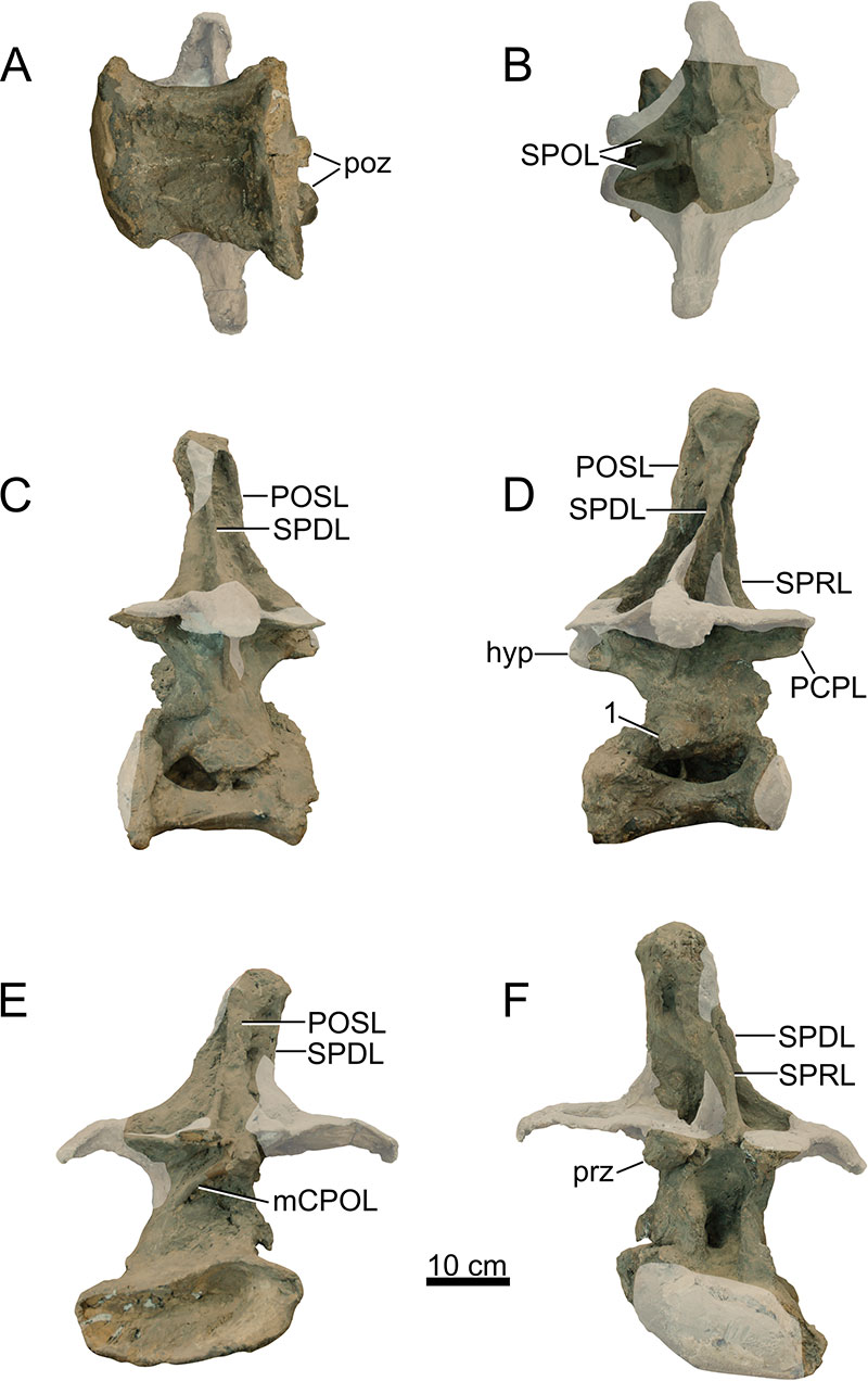
FIGURE 16. Dorsal vertebra 9 of Ardetosaurus viator MAB011899. DV9 is shown in A) ventral, B) dorsal, C) left lateral, D) right lateral, E) posterior, and F) anterior view. White shaded areas indicate reconstructed parts. Abbreviations: CPRL, centroprezygapophyseal lamina; mCPOL, medial centropostzygapophyseal lamina; lCPOL, lateral centropostzygapophyseal lamina; PCDL, posterior centrodiapophyseal lamina; PCPL, posterior centroparapophyseal lamina; POSL, postspinal lamina; poz, postzygapophysis.
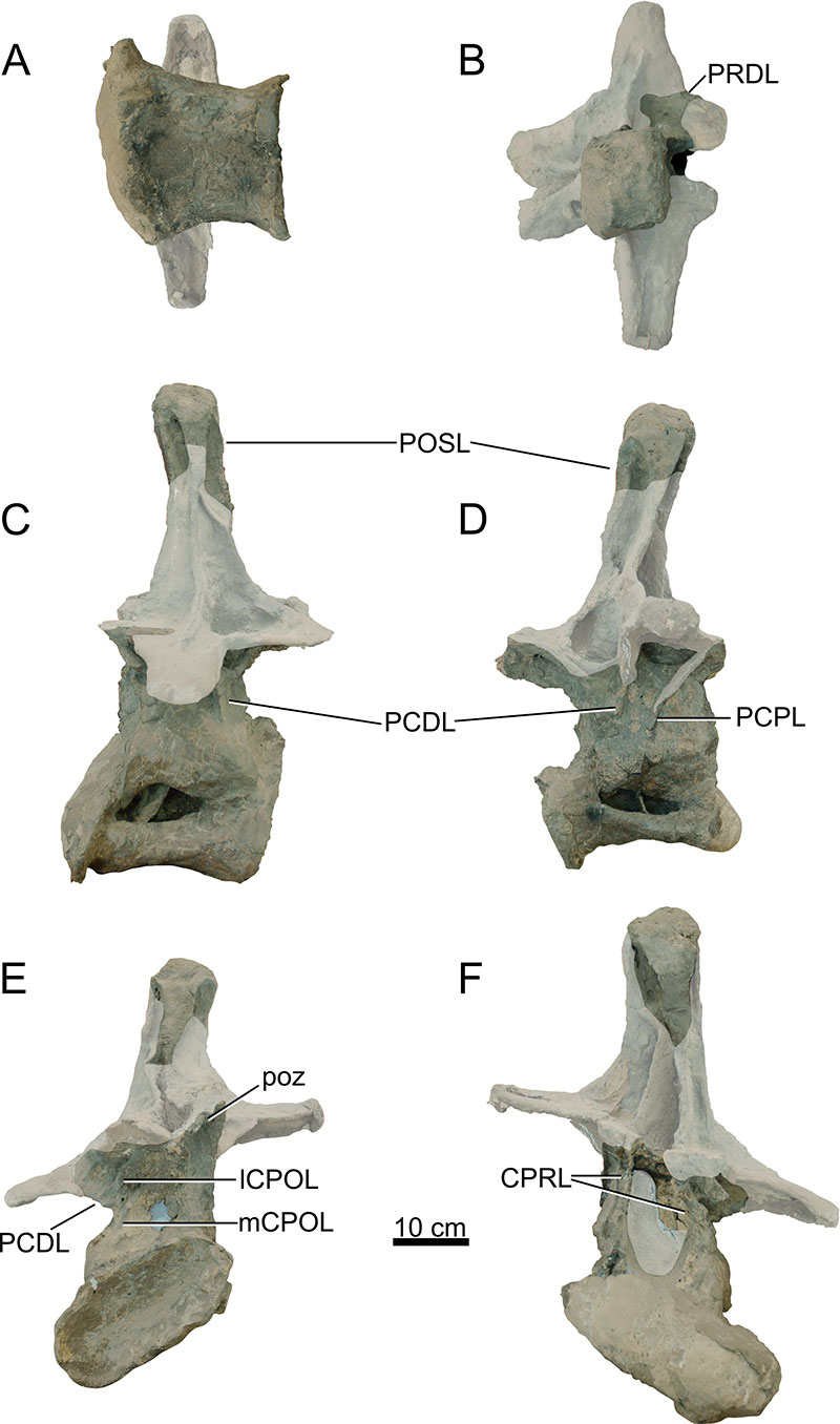
FIGURE 17. Dorsal vertebra 10 of Ardetosaurus viator MAB011899. Outline of DV10 is drawn on a left dorsolateral photograph of the sacrum. Photograph is courtesy of René Fraaije. Abbreviations: CPRL, centroprezygapophyseal lamina; lCPOL, lateral centropostzygapophyseal lamina; mCPOL, medial centropostzygapophyseal lamina; PCDL, posterior centrodiapophyseal lamina; PCPL, posterior centroparapophyseal lamina; prz, prezygapophysis; SPDL, spinodiapophyseal lamina; SPOL, spinopostzygapophyseal lamina; SPRL, spinoprezygapophyseal lamina.
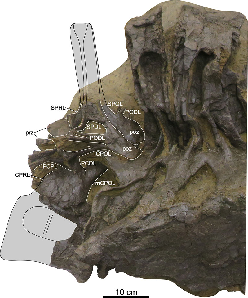
FIGURE 18. Dorsal ribs of Ardetosaurus viator MAB011899. Left ribs: A) RL1 in anterolateral view, B) RL?4 in lateral view, and C) RL?8 in anterolateral view. Right ribs: D) RR?3 in lateral view, E) RR?5 in posterior view, and F) RR?6 in lateral view. Rib heads: H) rib head of RR?3 in anterior view, I) rib head of RR?4 in anterior view, and J) rib head of RR?6 in anterior view. Note the pronounced ridges (1) on the rib heads of RL1, RR?3 and RR?6. White shaded areas indicate reconstructed parts. Abbreviations: cap, capitulum; tub, tuberculum.
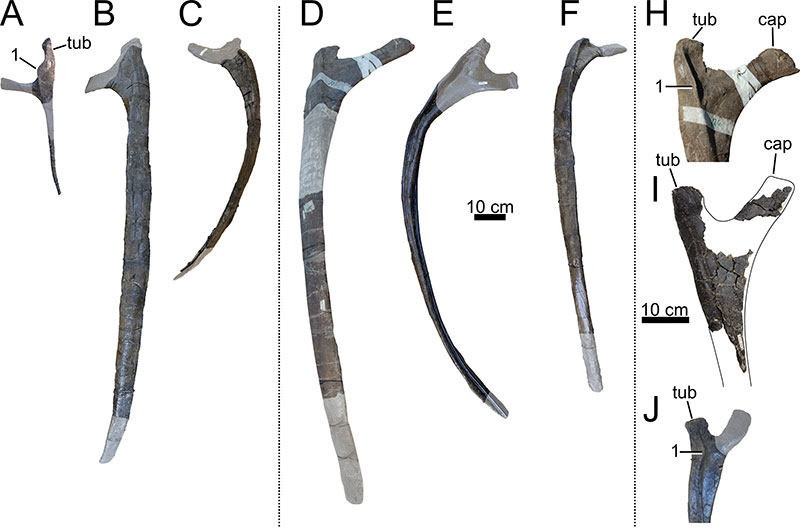
FIGURE 19. Sacrum, dorsal vertebra 10 and ilium of Ardetosaurus viator MAB011899. Composite figure of the sacral elements. A) Fifth sacral vertebrae in left anterolateral view. B) Left ilium in lateral view. C) Sacrum in posterodorsal view. D) Sacrum and outline of DV10 in left dorsolateral view. Markings 1, 2, 3, 4, and 5 indicate the sacral ribs in anteroposterior order. Note that in Figure D, the anterior part of the preacetabular process is missing, which had broken off during transport. Figures A, C, and D are courtesy of René Fraaije, made during the preparation and mounting process. Abbreviations: CPRL, centroprezygapophyseal lamina; CPOL, centropostzygapophyseal lamina; PODL, postzygodiapophyseal lamina; POSL, postspinal lamina; poz, postzygapophysis; prap, preacetabular process; PRDL, prezygodiapophyseal lamina; prz, prezygapophysis; pup, pubic peduncle; SPDL, spinodiapophyseal lamina; SPOL, spinopostzygapophyseal lamina.
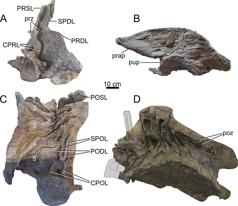
FIGURE 20. Caudal vertebra Cd1 of Ardetosaurus viator MAB011899. Cd1 is shown in A) left lateral, B) right lateral, C) posterior, and D) anterior view. White shaded areas indicate reconstructed parts.
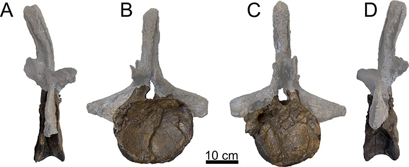
FIGURE 21. Caudal vertebra Cd2 of Ardetosaurus viator MAB011899. Cd2 is shown in A) left lateral, B) right lateral, C) posterior, and D) anterior view. White shaded areas indicate reconstructed parts.
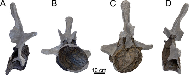
FIGURE 22. Caudal vertebra Cd3 of Ardetosaurus viator MAB011899. Cd3 is shown in A) left lateral, B) right lateral, C) posterior, and D) anterior view. White shaded areas indicate reconstructed parts. Note the presence of distinct CPRLs ventral to the prezygapophyseal rami. Abbreviations: CPRL, centroprezygapophyseal lamina; POSL, postspinal lamina.
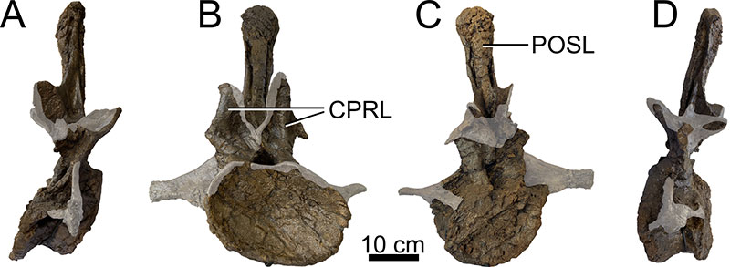
FIGURE 23. Caudal vertebra Cd?4-6 of Ardetosaurus viator MAB011899. Cd?4-6 is shown in A) left lateral, B) right lateral, C) posterior, and D) anterior view. Note the absence of distinct CPRLs ventral to the prezygapophyseal rami. Abbreviations: ACDL, anterior centrodiapophyseal lamina; PCDL, posterior centrodiapophyseal lamina; PODL, postzygodiapophyseal lamina; POSL, postspinal lamina; poz, postzygapophysis; prz, prezygapophysis; SPOL, spinopostzygapophyseal lamina; SPRL, spinoprezygapophyseal lamina.
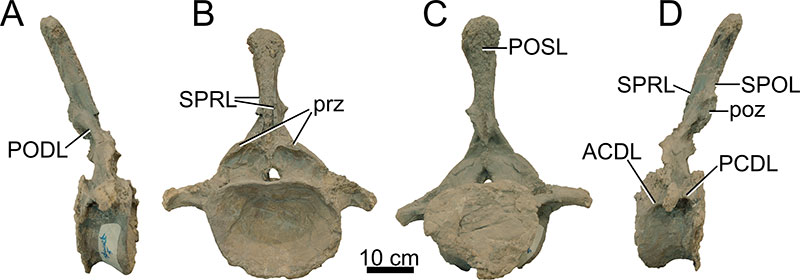
FIGURE 24. Caudal vertebra Cd?5-8 of Ardetosaurus viator MAB011899. Cd?5-8 is shown in A) left lateral, B) right lateral, C) posterior, and D) anterior view. Note the absence of distinct CPRLs ventral to the prezygapophyseal rami. Abbreviations: PODL, postzygodiapophyseal lamina; POSL, postspinal lamina; poz, postzygapophysis; prz, prezygapophysis; SPRL, spinoprezygapophyseal lamina.
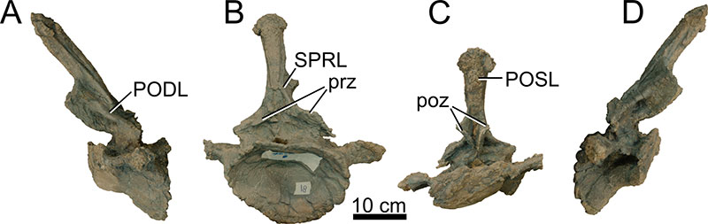
FIGURE 25. Chevrons of Ardetosaurus viator MAB011899. First chevron in A) anterior, B) right lateral and C) posterior view. Third chevron in D) anterior, E) left lateral, and F) posterior view. Note the (1) vertical striations on the anterior surface of the blade in Figure A. The rectangle in Figure A shows the chevron portion shown left of the chevron, wherein the striations are highlighted in white. Figure B includes the outline of the first chevron angled as it would be in vivo. Figure E already shows the third chevron angled as in vivo. Abbreviation: hc, haemal canal.
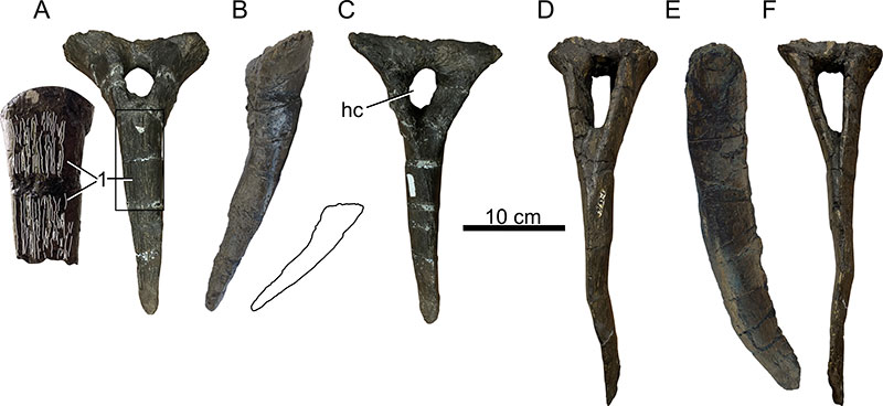
FIGURE 26. Left coracoid of Ardetosaurus viator MAB011899. Coracoid in shown in A) medial and B) lateral view. White shaded areas indicate reconstructed parts. Abbreviations: cof, coracoid foramen; gl, glenoid.
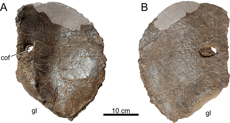
FIGURE 27. Pubes of Ardetosaurus viator MAB011899. Left pubis is shown in A) lateral and B) medial view. Right pubis is shown in C) lateral and D) medial view. Abbreviations: ac, acetabular surface; isa, ischial articular surface; of, obturator foramen.
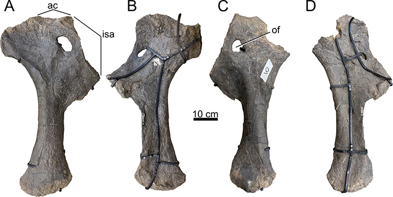
FIGURE 28. Ischia of Ardetosaurus viator MAB011899. Ischia are shown in A) right lateral (left ischium cut out), B) left lateral (right ischium cut out), C) ventral, and D) dorsal view. Abbreviations: ac, acetabular surface; pua, pubic articular surface.
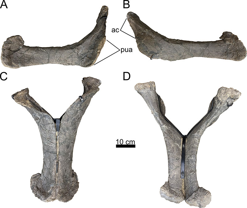
FIGURE 29. Left femur of Ardetosaurus viator MAB011899. Femur is shown in A) anterolateral, B) anteromedial, and C) distal views. Femur could be photographed only from the mount during the mounting process, hence the awkward position and angles. White shaded areas indicate reconstructed parts. Abbreviations: fic, fibular condyle; tic, tibial condyle.
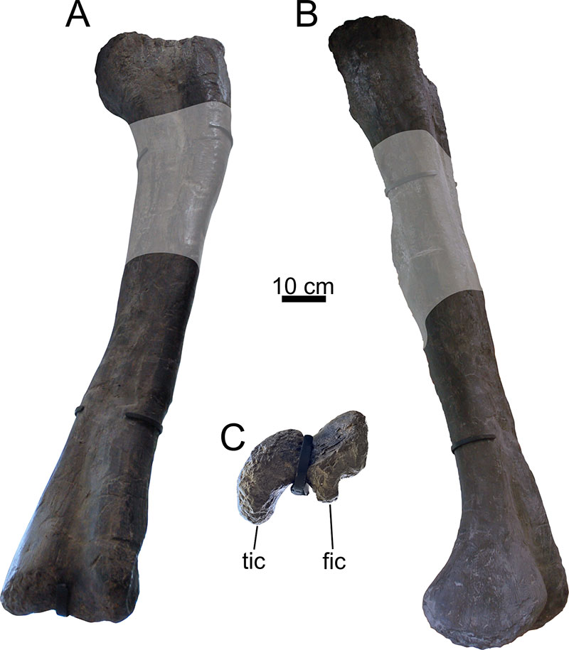
FIGURE 30. Left tibia of Ardetosaurus viator MAB011899. Tibia is shown in A) proximal, B) lateral, C) anterior, D) posterior, E) medial, and F) distal views. Abbreviations: cc, cnemial crest; fbtr, fibular trochanter.
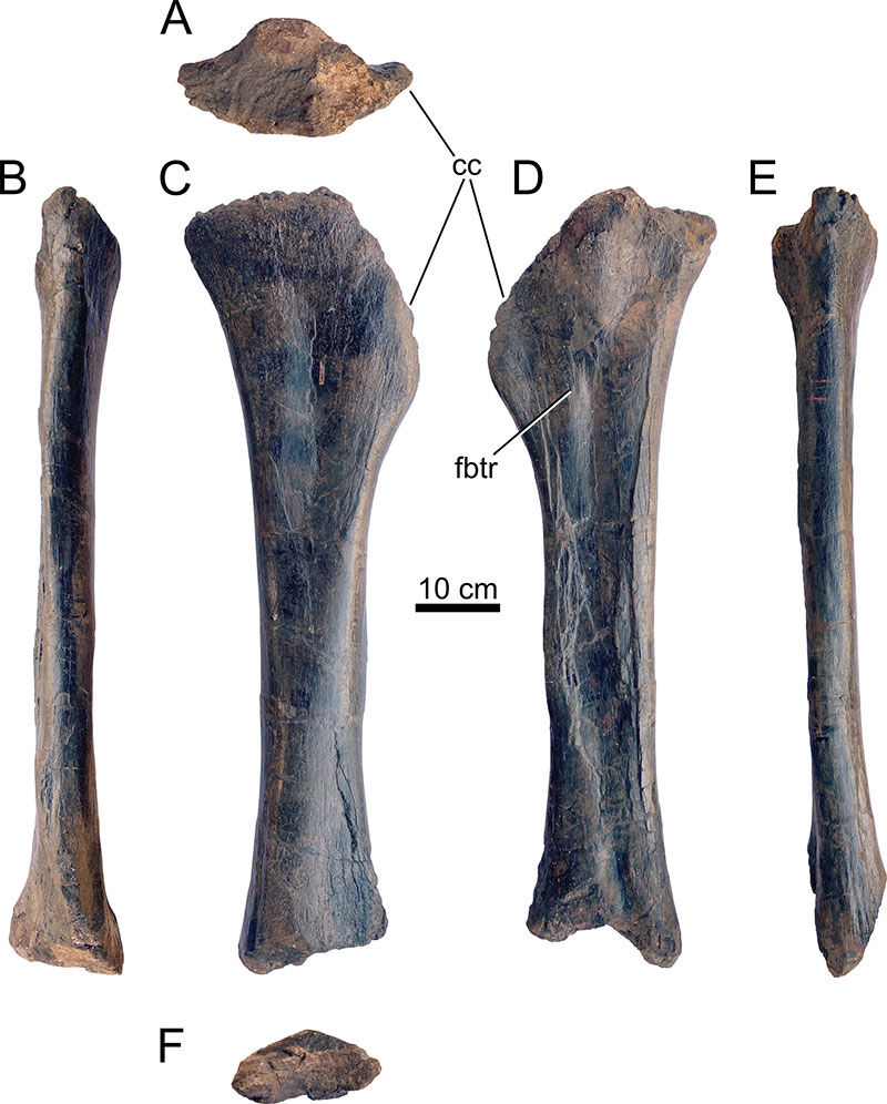
FIGURE 31. Left fibula of Ardetosaurus viator MAB011899. Fibula is shown in A) lateral, B) anterior, C) medial, D), posterior view. The proximal end is lacking. The number ‘13’ attached with tape to the lateral surface of the fibular is an old catalogue number of the Oertijdmuseum. Abbreviation: tilf, M. iliofibularis trochanter.
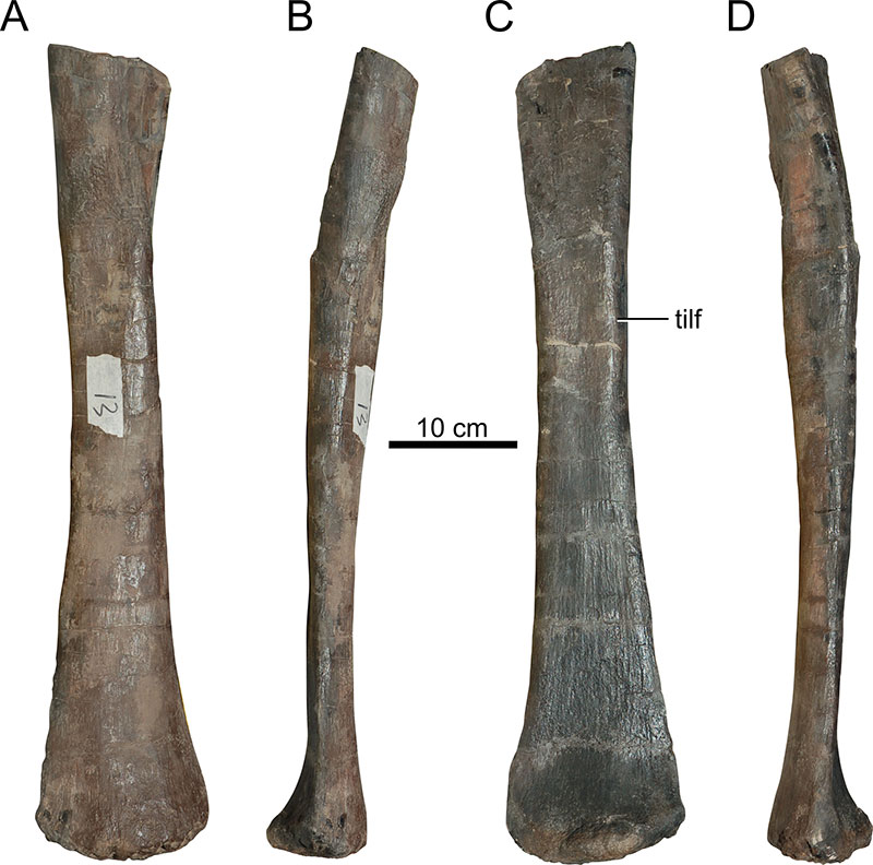
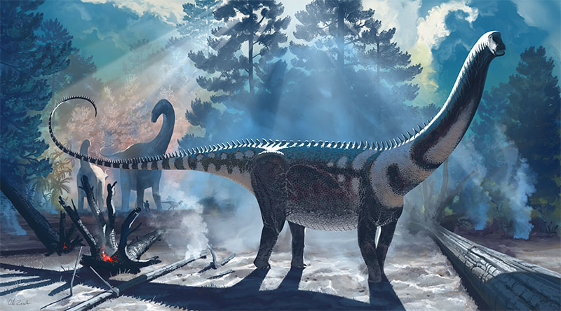
FIGURE 32. Life reconstruction of Ardetosaurus viator MAB011899. Illustration by Ole Zant.