FIGURE 1. Animation of the soaking process of a rib of Aenocyon dirus (LACMP23-46765) with 200 mL Novec 73DE.
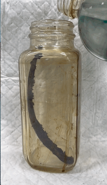
FIGURE 2. Chemical structures of solvents used in the experiment. Trade name in bold, with chemical nomenclature for each below. Undisclosed proprietary components are listed in italics.
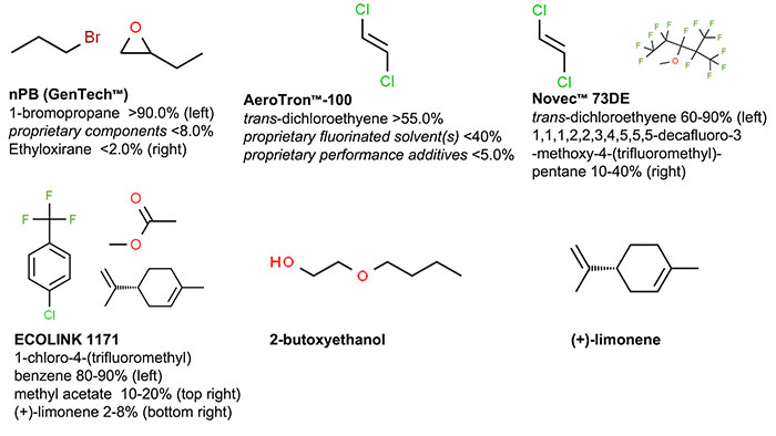
FIGURE 3. Animation of manual preparation of an Aves synsacrum (LACMP23-45449) at 8x speed.
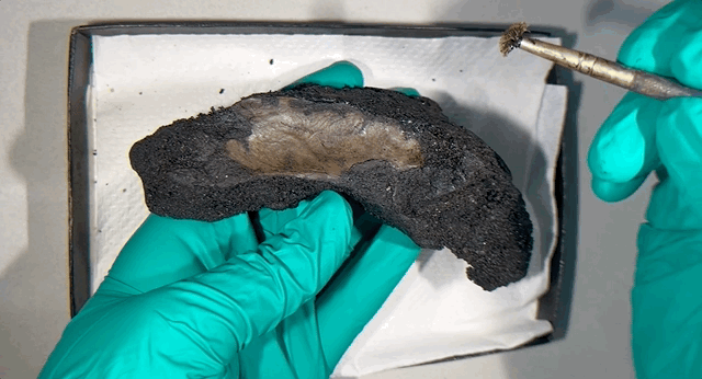
FIGURE 4. Manual preparation example with a thoracic vertebra of a juvenile Aenocyon dirus (LACMP23-38126). Scale bars are 1 cm. A) Before preparation, B) Example of manual preparation techniques on an Aves synsacrum (LACMP23-45449), using a foam swab on targeted area of matrix, C) After manual preparation.
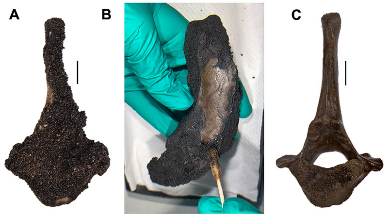
FIGURE 5. Hand tools used in preparation, from left to right: bamboo toothpicks, double tipped cotton swabs, wood manicure sticks, synthetic filament paint brushes, polyethylene bulb droppers, and polyurethane foam swabs.
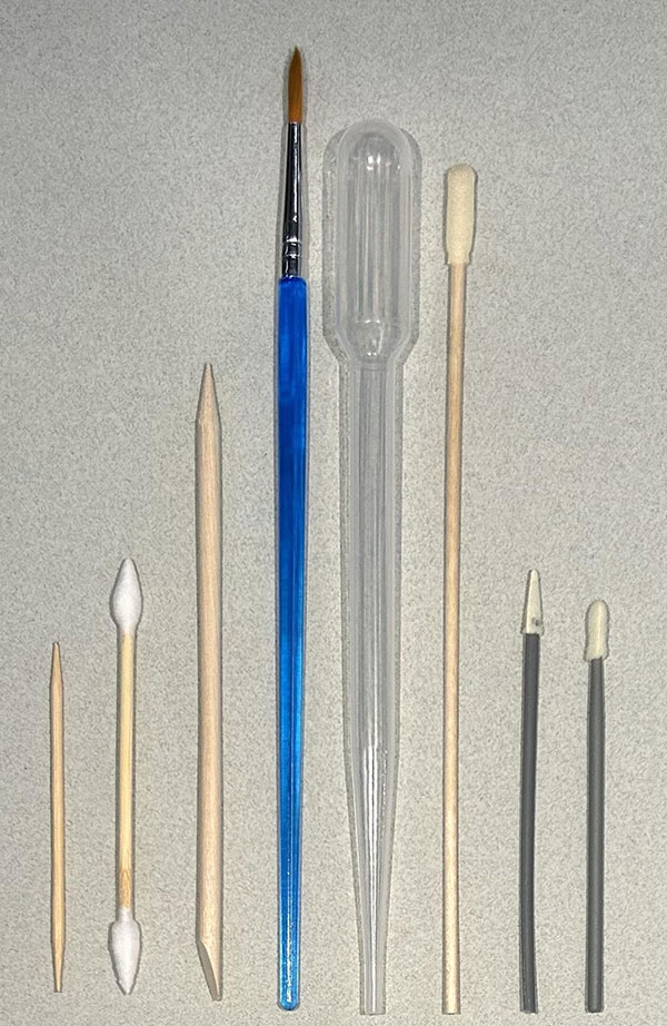
FIGURE 6. Soaked preparation example with a thoracic vertebra of Canis latrans (LACMP23-33690). Scale bars are 1 cm. A) Before preparation, B) Submerged in solvent, C) After soaking and off-gassing, D) After removing residual matrix with hand tools. The red arrow points to a spot of residual matrix present after soaking, and the red ovals highlight an exposed cavity that lost supportive interior matrix after soaking.
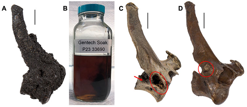
FIGURE 7. Crania manual preparation. Scale bars are 2 cm. A) Before (top) and after (bottom) preparation of a crania of Canis latrans LACMP23-41926. B) Before (top) and after (bottom) preparation of a crania of Aenocyon dirus LACMP23-41940.
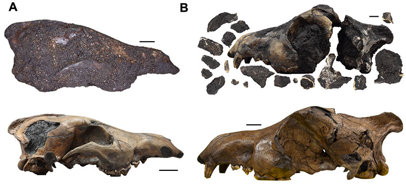
FIGURE 8. In-progress specimen soaking examples, approximately five minutes after submersion and backlit with lamps to highlight color and translucency, demonstrating the relative efficacy of each solvent to dissolve asphalt. From left to right, nPB (Gentech), AeroTron™-100, Novec™ 73DE, Ecolink™ 1171, 2-butoxyethanol (Butyl Cellosolve), (+)-limonene.
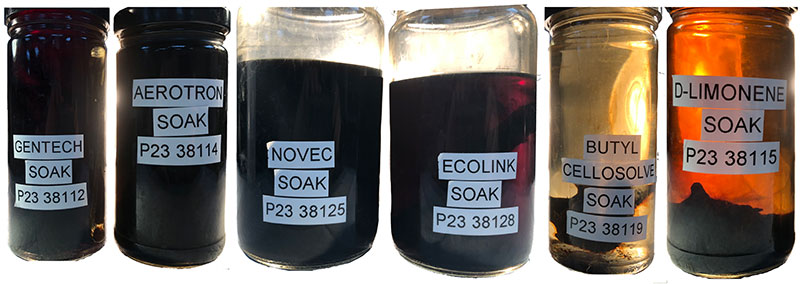
FIGURE 9. Surface damage examples. Scale bars are 1 cm. A) Thoracic vertebra of Aenocyon dirus LACMP23-38125 and B) Metapodial of A. dirus LACMP23-38116 with no damage. C) Aves femur LACMP23-34952 and D) Thoracic vertebra of A. dirus LACMP23-38128 with scratches (red arrows). E) Metapodial of A. dirus LACMP23-33686 with a chip (red arrow). F) Aves femur LACMP23-34955 with exposed and degraded cancellous bone.
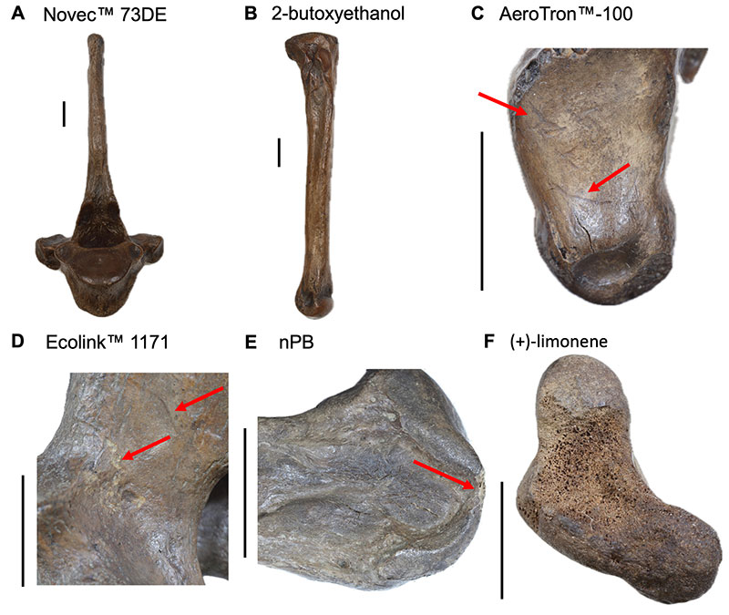
FIGURE 10. Surface appearance examples. Scale bars are 1 cm. A) Stable cancellous bone and natural shine in Aves femur LACMP23-34948. B) Soaked thoracic vertebra of Aenocyon dirus LACMP23-38120 and C) Cervical vertebra of Canis latrans LACMP23-34944 with dehydrated cancellous bone. D) Metapodial of A. dirus LACMP23-34932 with dehydrated end. E) Thoracic vertebra of A. dirus LACMP23-38123 with dark glossy exterior. F) Thoracic vertebra of A. dirus LACMP23-38129 with wet-seeming asphalt in cancellous bone.
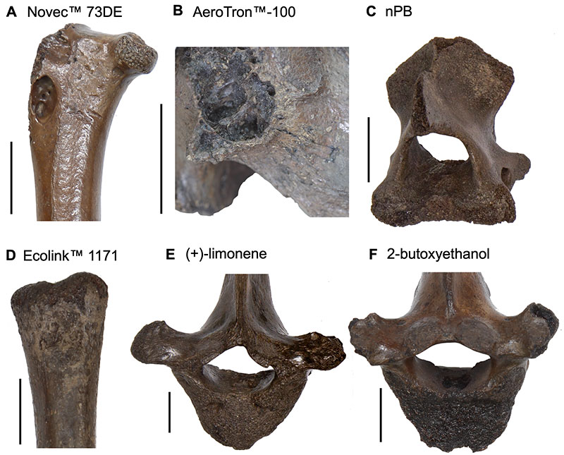
FIGURE 11. Exudation examples, with red arrows indicating exposed asphalt. Scale bars are 1 cm. A) Aves femur LACMP23-34952, B) Thoracic vertebra of Aenocyon dirus LACMP23-38129 and C) Metapodial of A. dirus LACMP23-34936 showed minor signs of asphalt exudation that stabilized within the first month after treatment. D) Thoracic vertebra of A. dirus LACMP23-38124 and E) Aves femur LACMP23-34956 had moderate instances of asphalt exudation which stabilized by the end of the assessment period. F) Metapodial of A. dirus LACMP23-34942 revealed possible continued asphalt instability throughout the study.
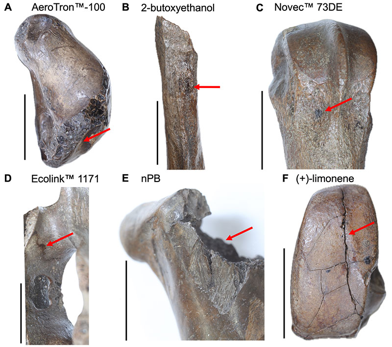
FIGURE 12. Visible degradation examples. Top is the overall specimen with green box highlighting inset areas, middle is inset after preparation, and bottom is inset after nine months. Red arrows and circles highlight changes. Scale bars are 1 cm. A) Cervical vertebra of Canis latrans LACMP23-38114, B) Aves femur LACMP23-38330 and C) Aves femur LACMP23-34951 with no change in original cracks. D) Metapodial of Aenocyon dirus LACMP23-38118 with crack growth and new cracks. E) Metapodial of A. dirus LACMP23-34934 with crack growth and continued asphalt exudation. F) Metapodial of A. dirus LACMP23-33686 with new cracks.
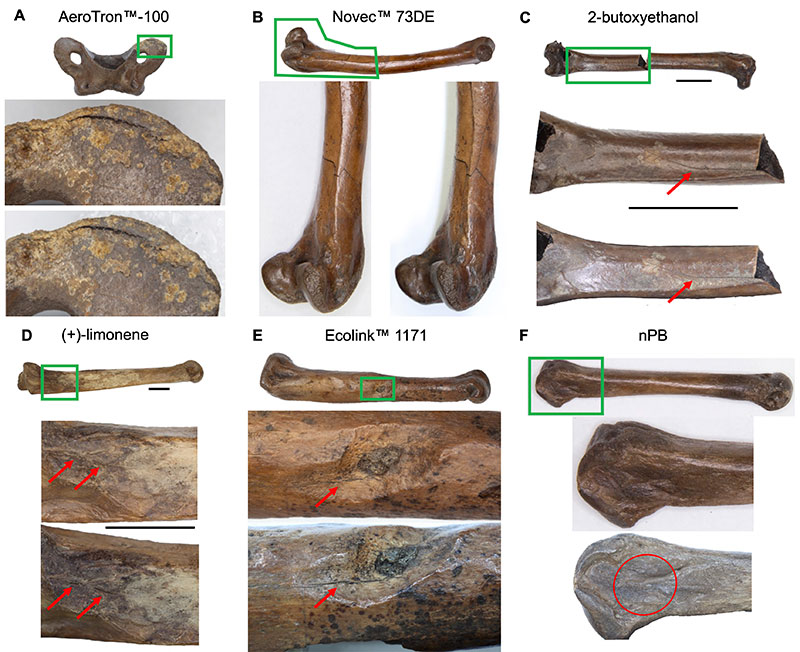
FIGURE 13. Cranial fragmentation examples. White asterisks highlight repaired fragments and blue circles highlight heavily cracked areas that did not disassociate during preparation. Red arrows highlight warping. Scale bars are 2 cm. A) Soaked specimens, top Aenocyon dirus LACMP23-18912 and bottom, Canis latrans LACMP23-42850. B) Manually prepared specimen A. dirus LACMP23-41940. Scale bars are 2 cm.
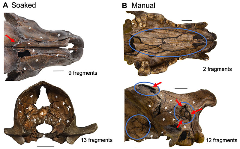
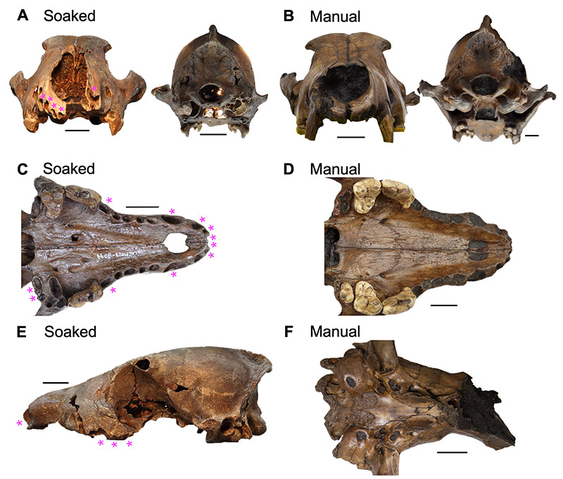
FIGURE 14. Cranial matrix retention examples. Scale bars are 2 cm. Pink asterisks highlight dehydrated dental alveoli. A) Loss of internal matrix in major cavities for left, Aenocyon dirus LACMP23-8580 and right, Canis latrans LACMP23-5351. B) Retention of internal matrix in A. dirus LACMP23-41940 (left) and C. latrans LACMP23-41926 (right). C) Loss of matrix in dental alveoli in C. latrans LACMP23-8099. D) A. dirus LACMP23-41930 with matrix retained in dental alveoli. E) Matrix lost in foramina and non-morphological cavities in C. latrans LACMP23-17329. F) Retained matrix in foramina and non-morphological cavities in manual A. dirus LACMP23-41929.