FIGURE 1. Maps showing the fossil locality (black square) of SMAC 2731, Megabalaena sapporoensis. The base maps (left and right) are modified from Tanaka and Kohno (2015) and the topographic map published by the Geospatial Information Authority of Japan respectively.
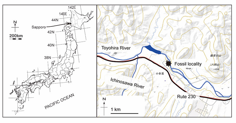
FIGURE 2. Photos taken during digging up SMAC 2731, Megabalaena sapporoensis. A photo taken at a very beginning of excavation in 2008 showing several associated vertebrae (A). A green field note is 16. 5 cm long and 9. 5 cm wide. A photo taken in 2009 showing the locality of SMAC 2731, Megabalaena sapporoensis , just beside Toyohira River (B).
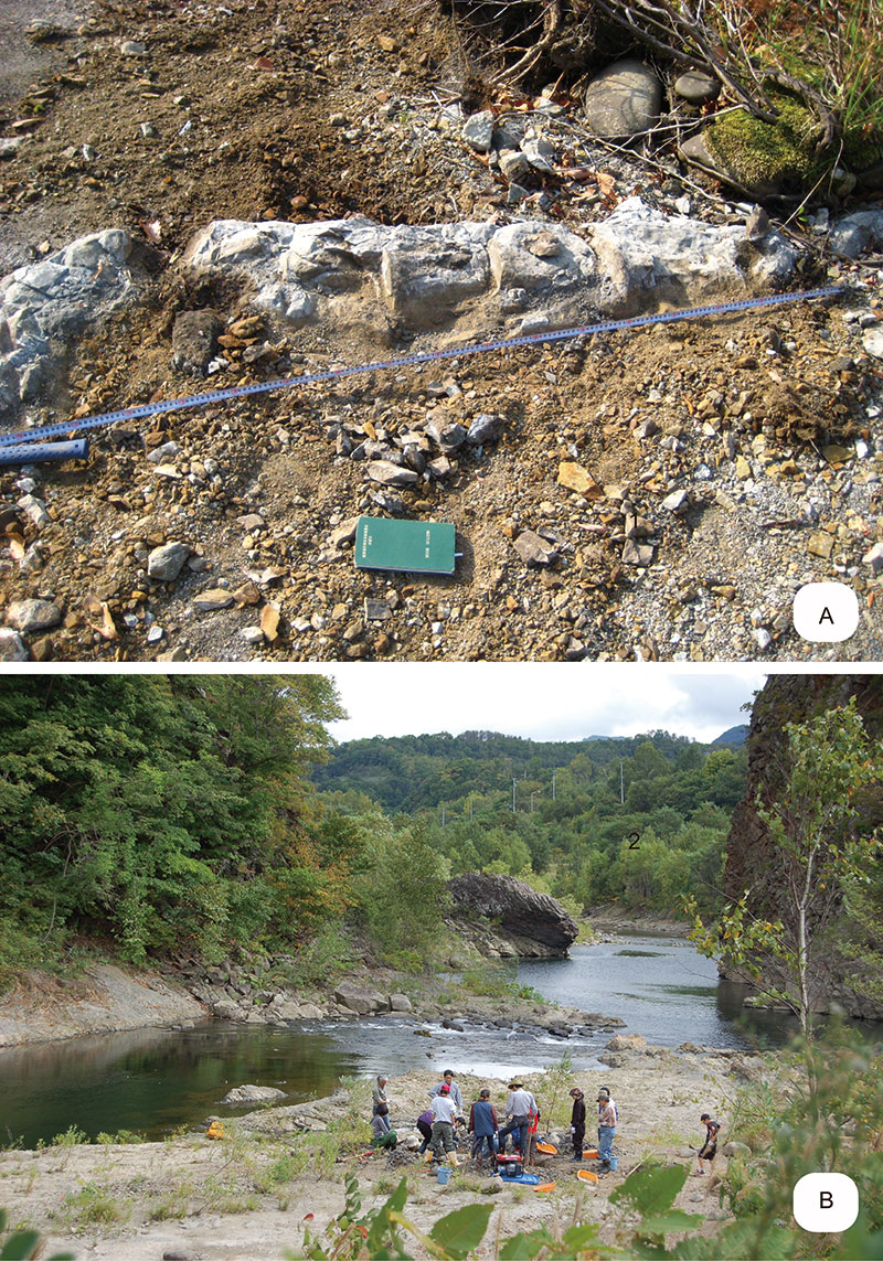
FIGURE 3. Images based on a 3D model showing preserved elements in brown of SMAC 2731, Megabalaena sapporoensis in dorsal view (A) and left lateral view (B). Missing parts were created by T. Shinmura refer to extant balaenids NMNS-M55028 and MTUF-M061A both Eubalaena japonica . The vertebrae posterior to the ninth thoracic vertebra are difficult to identify because of their limited preservation. Ribs separated into over 100 fragments. These elements' original positions are uncertain and roughly estimated based on their morphologies and the extant species Eubalaena glacialis .
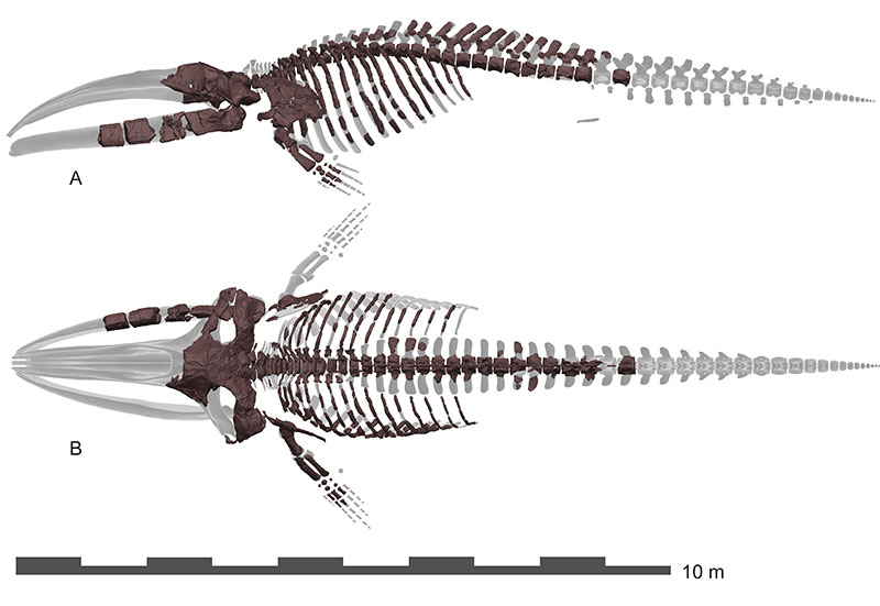
FIGURE 4. Images based on a 3D model showing skull elements of SMAC 2731, Megabalaena sapporoensis in left lateral view (A) and dorsal view (B). Deformations are restored using 3D model editor by T. Shinmura. Settings are the same to Figure 3.
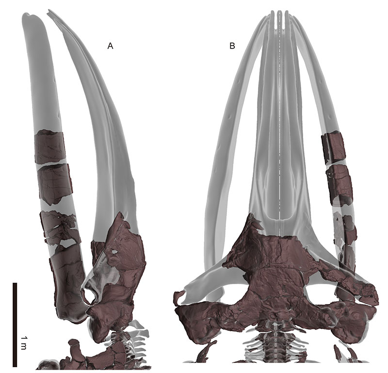
FIGURE 5. Images based on a 3D model showing preserved skull elements of SMAC 2731, Megabalaena sapporoensis in dorsal view (A), ventral view (B), and posterior view (C). Deformations are not restored.
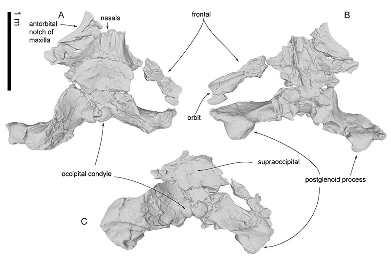
FIGURE 6. Frontal and jugals of SMAC 2731, Megabalaena sapporoensis. Right frontal in dorsal view (A, ventral view (B), and lateral view (C). Left jugal in lateral view (D). Right (E) and left jugals (F) in ventral view. Right (G) and left jugals (H) in dorsal view.
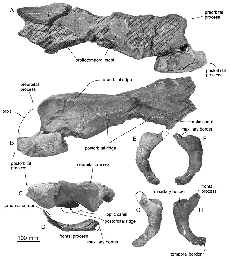
FIGURE 7. Vertex area of skull, SMAC 2731, Megabalaena sapporoensis in dorsal view (A), anterior view (B), and ventral view (C). These images are based on a 3D model.
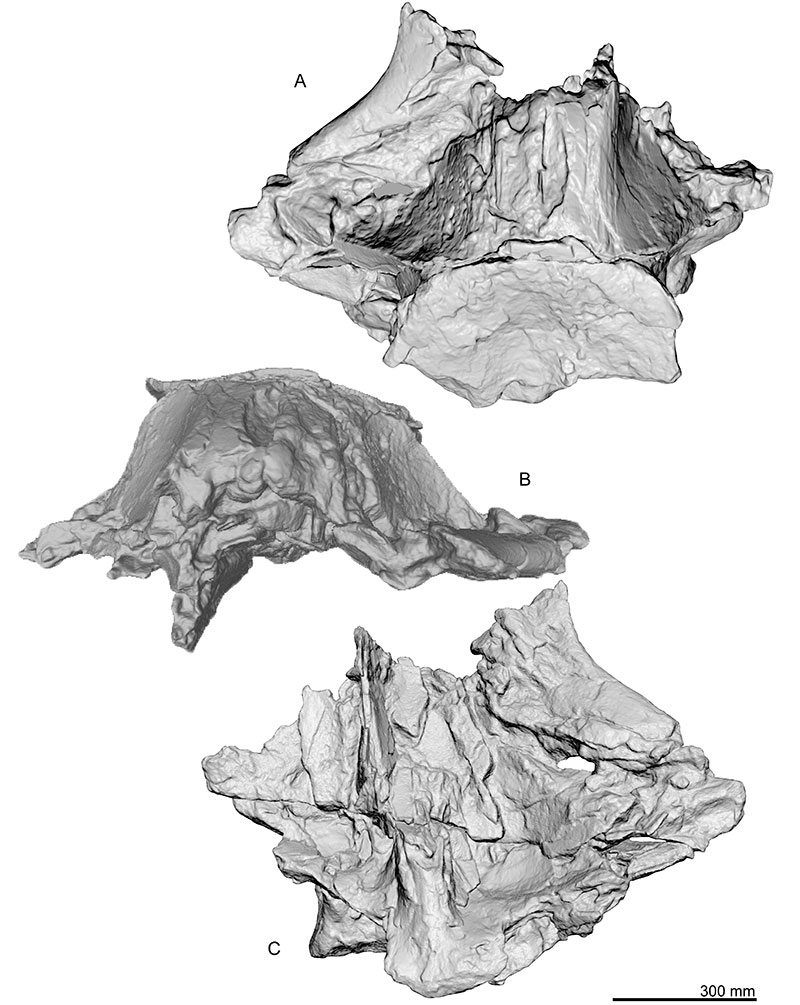
FIGURE 8. Key features on vertex area of skull, SMAC 2731, Megabalaena sapporoensis in dorsal view (A), anterior view (B), and ventral view (C). These images are based on a 3D model.
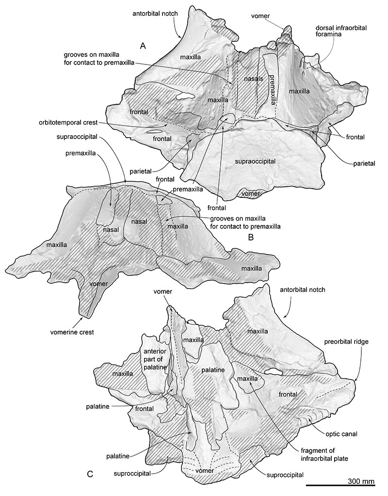
FIGURE 9. Basicranium, SMAC 2731, Megabalaena sapporoensis in ventral view (A), posterior view (B), and dorsal view (C). These images are based on a 3D model.
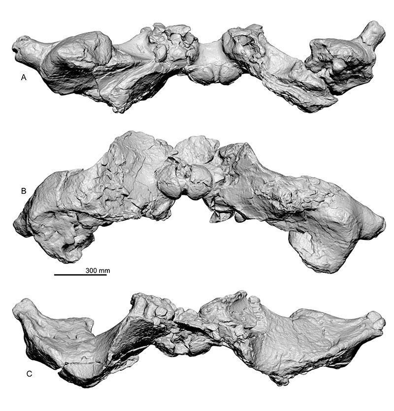
FIGURE 10. Key features on basicrania, SMAC 2731, Megabalaena sapporoensis in ventral view (A), posterior view (B), and dorsal view (C). These images are based on a 3D model.
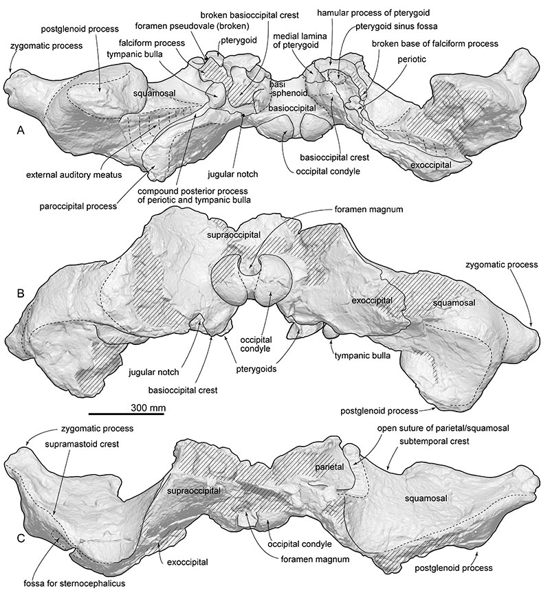
FIGURE 11. Skull, SMAC 2731, Megabalaena sapporoensis in right lateral view (A), left lateral view (B), and key features (C, D). These images are based on a 3D model.
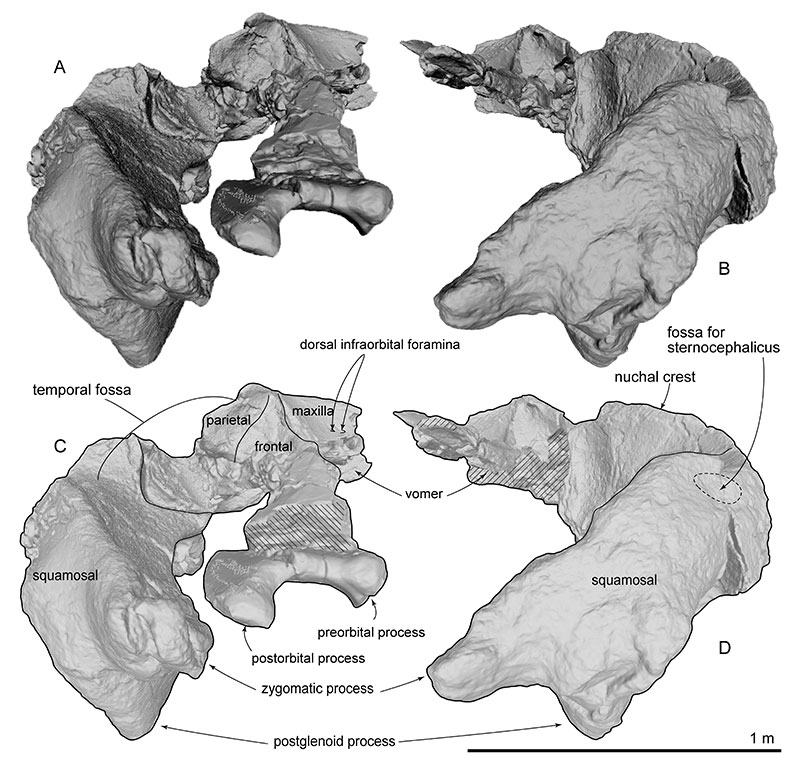
FIGURE 12. Ear bones in situ of SMAC 2731, Megabalaena sapporoensis, right periotic and tympanic bulla in ventral view (A), lateral view (B), and left periotic in ventral view (C).

FIGURE 13. Left tympanic bulla of SMAC 2731, Megabalaena sapporoensis in lateral view (A), anterior view (B), dorsal view (C), ventral view (D), posterior view (E), and medial view (F). ’
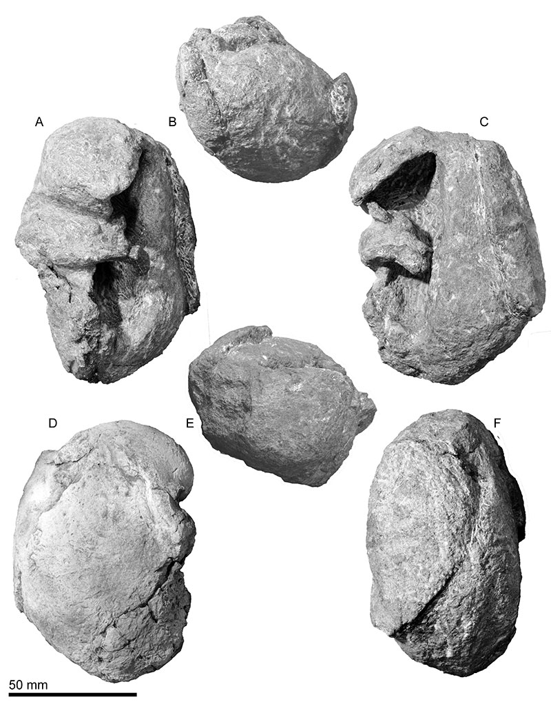
FIGURE 14. Key features of left tympanic bulla of SMAC 2731, Megabalaena sapporoensis in lateral view (A), anterior view (B), dorsal view (C), ventral view (D), posterior view (E), and medial view (F).
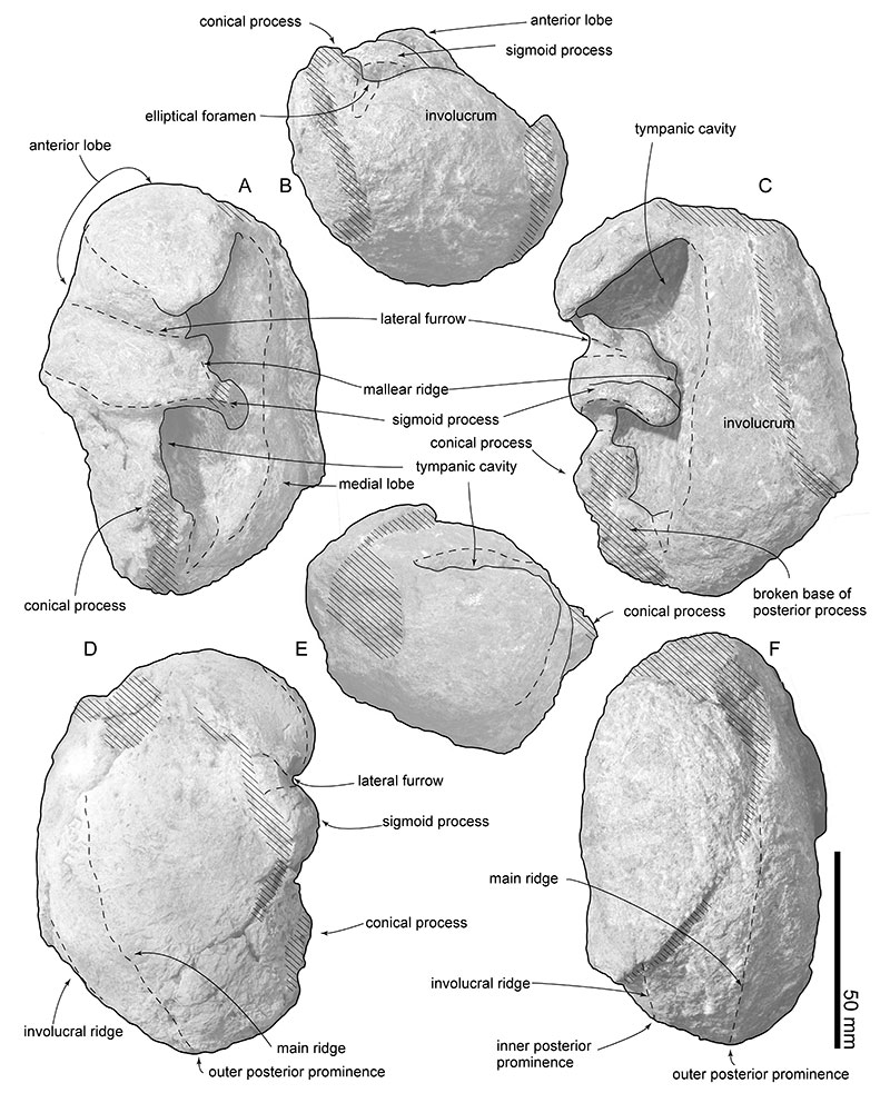
FIGURE 15. Right mandible of SMAC 2731, Megabalaena sapporoensis in posterior view (A), lateral view (B), dorsal view (C), anterior view (D), medial view (E), and ventral view (F). These images are based on a 3D model.
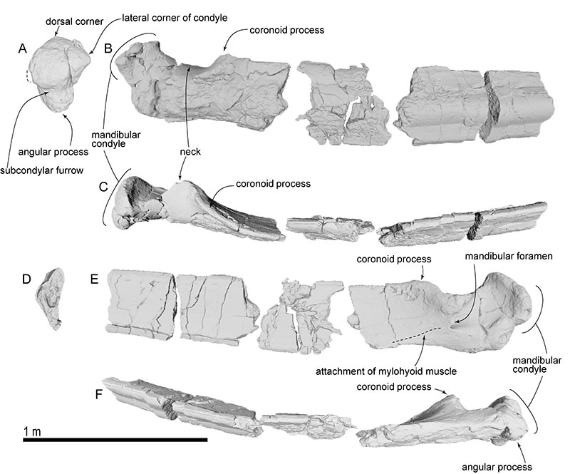
FIGURE 16. Hyoid bones of SMAC 2731, Megabalaena sapporoensis. Basihyal and thyrohyal in anterior view (A), posterior view (B), and right lateral view (D). Right stylohyal in medial view (D), posterior end (E), and lateral view (F). Hyoid bones in dorsal view (G) and ventral view (H).
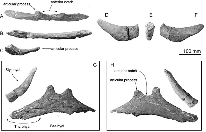
FIGURE 17. Sternum of SMAC 2731, Megabalaena sapporoensis in anterior view (A), left lateral view (B), ventral view (C), dorsal view (D), posterior view (E).
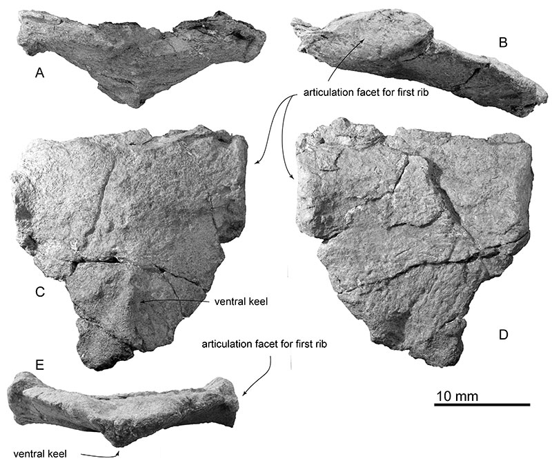
FIGURE 18. Cervical vertebrae of SMAC 2731, Megabalaena sapporoensis. In anterior view, atlas (A), axis and third cervical vertebra (B), fourth cervical vertebra (C), fifth cervical vertebra (D), sixth cervical vertebra (E), seventh cervical vertebra (F). Whole cervical vertebrae in left lateral view (G) and dorsal view (H). (G) and (H) are based on a 3D model.
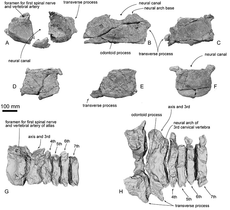
FIGURE 19. Thoracic and more posterior vertebrae of SMAC 2731, Megabalaena sapporoensis, in left lateral view based on a 3D model (A). T hird thoracic vertebra in anterior view (B), more posterior vertebrae in anterior view (C and D), third most posterior vertebra in posterior view (E).
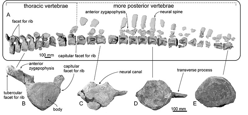
FIGURE 20. Ribs of SMAC 2731, Megabalaena sapporoensis . Ribs interpreted as from the left side (A to R), and right side (S to DD).
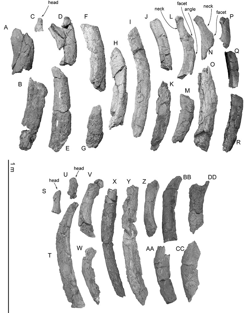
FIGURE 21. A cross-section photo of the distal end of a rib T in Figure 20.
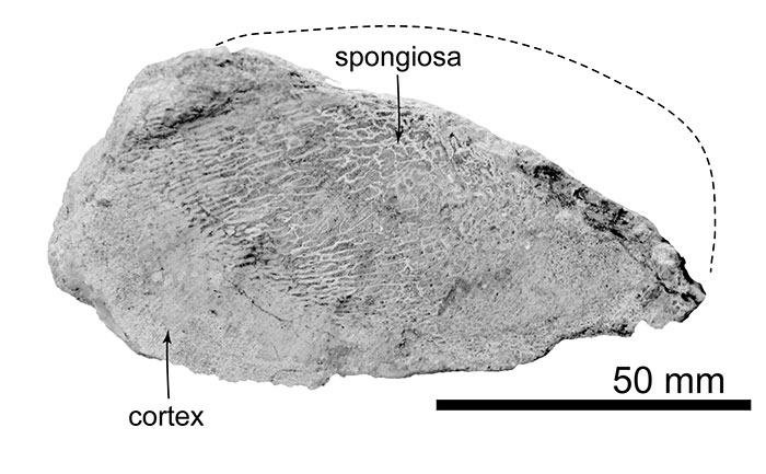
FIGURE 22. Forelimb elements of SMAC 2731, Megabalaena sapporoensis . All preserved left side elements in lateral view and a cast showing original deposited position of the ulna, radius, carpals and a metacarpal based on a 3D model (A). Left scapula in ventral view (B). Right scapula in lateral view (C). Humerus in anterior view (D), medial view (E), posterior view (F), and proximal view (G). Radius in anterior view (H), medial view (I), posterior view (J), and distal view (K). Ulna in anterior view (L), medial view (M), posterior view (N), and distal view (O).
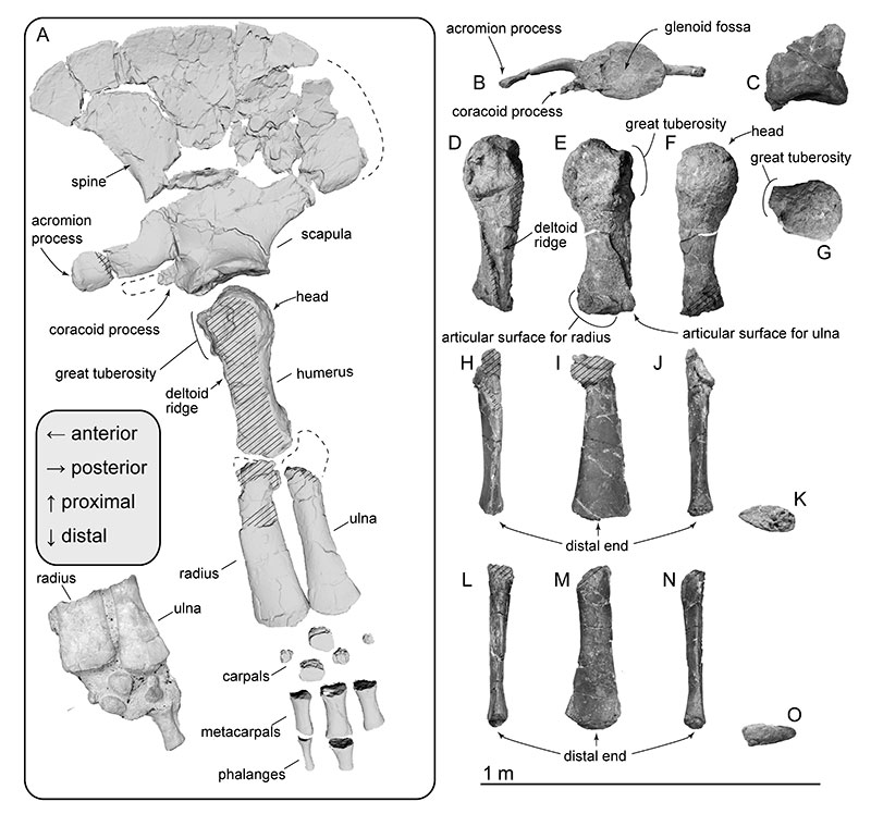
FIGURE 23. 50% majority (top) and strict (bottom) consensus trees of 53 most parsimonious trees showing the phylogenetic position of Megabalaena sapporoensis gen. et sp. nov. with branch length. The most parsimonious trees were 1003 steps. The clades Aetiocetidae, Eomysticetidae, Balaenidae, Cetotheriidae and a clade comprising Isanacetus, Parietobalaena and related taxa are collapsed. (See Appendix 5 for cladogram with all taxa shown.)
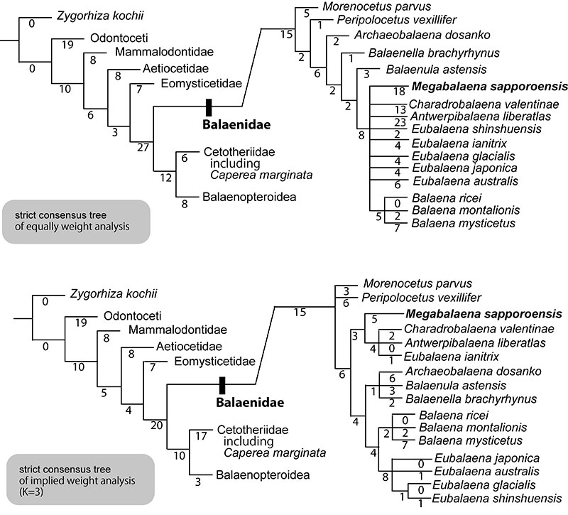
FIGURE 24. Size distribution of the Balaenidae. The topology adopted the 50% majority consensus tree in this study (top in Figure 22). Total length of balaenids were taken from Table 6.
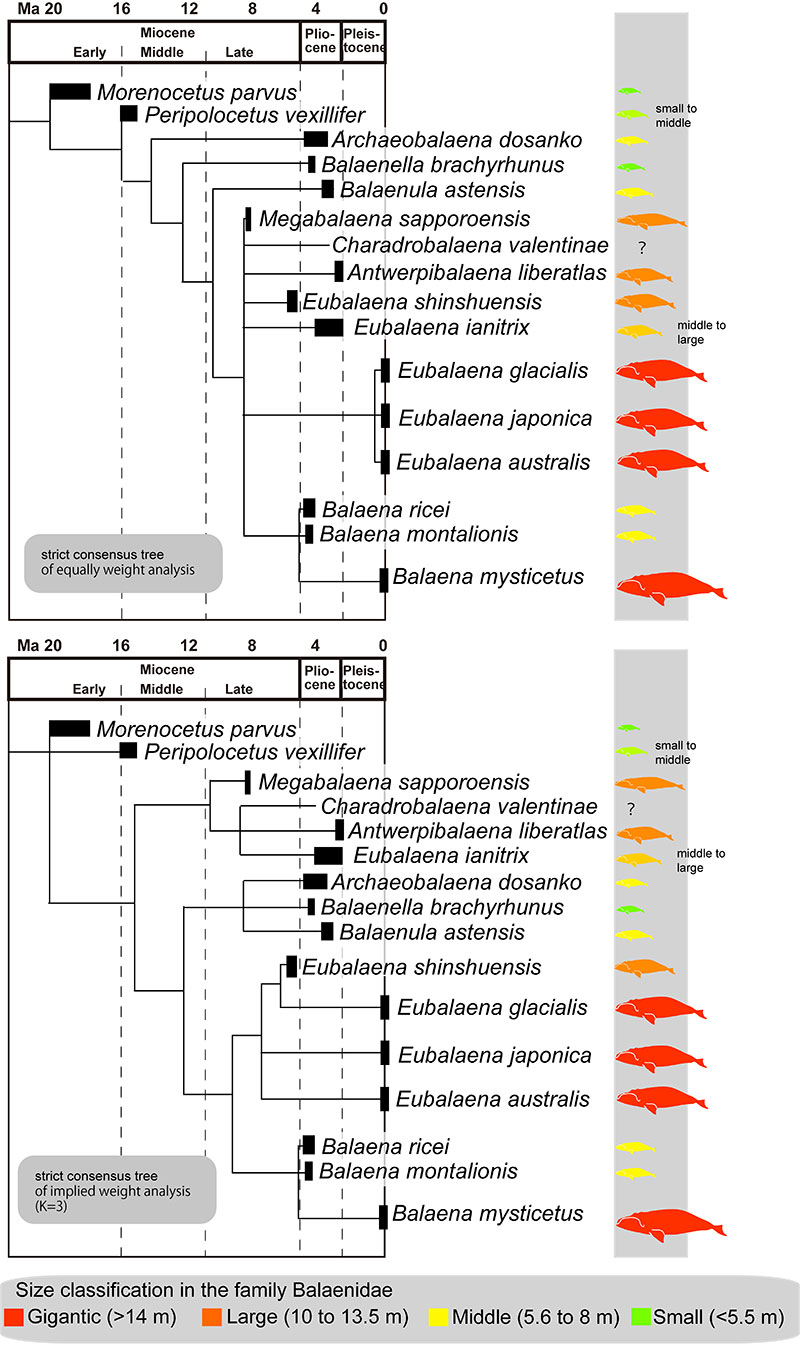
FIGURE 25. Balaenidae diversity with maximum and minimum body size of the Balaenidae over time. Total length of balaenids were taken from Table 6. Some epochs have only one record of balaenid, which can be used for body size estimation. Eubalaena ianitrix estimated as 8. 2 m of body length is omitted because it has a wide range from latest Early to Late Pliocene.
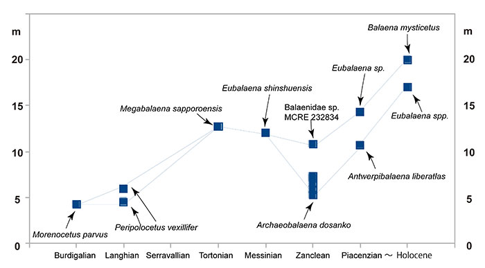
FIGURE 26. Forelimb elements of balaends. Megabalaena sapporoensis (A), Charadrobalaena valentinae, outline taken from Bisconti et al. (2023) and is a mirror image (B), Antwerpibalaena liberatlas , outline taken from Duboys de Lavigerie et al. (2020) and is a mirror image (C), Eubalaena japonica , outline taken from Omura (1958) (D), and are not to scale.
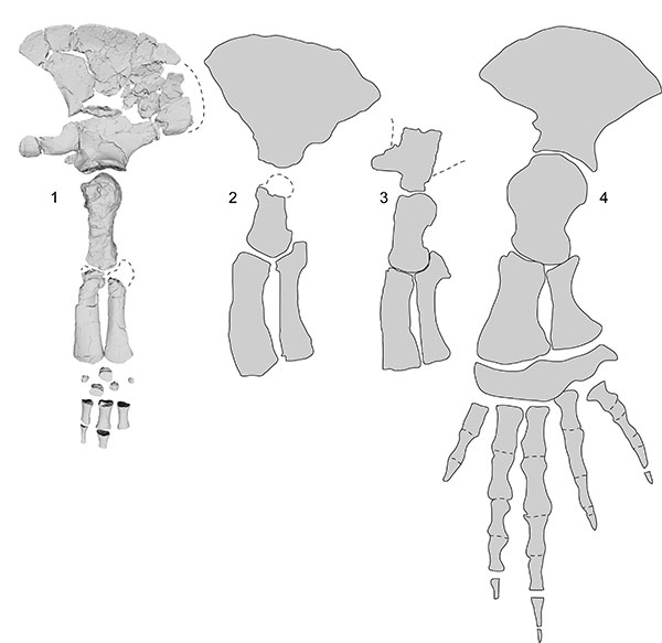
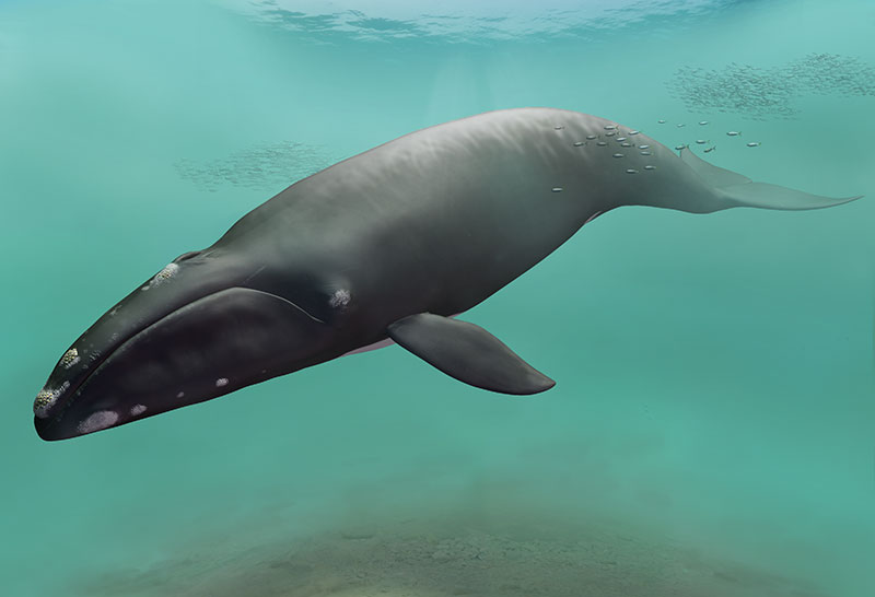
FIGURE 27. Restoration of Megabalaena sapporoensis by Tatsuya Shinmura (Ashoro Museum of Paleontology).