FIGURE 1. Location of La Rioja and the sites; A, geographical map of the Iberian Peninsula; B, geological map of the northeastern Cameros Basin and location of the El Horcajo site (modified from Suárez-Gonzalez et al., 2013 and Isasmendi et al., 2021).
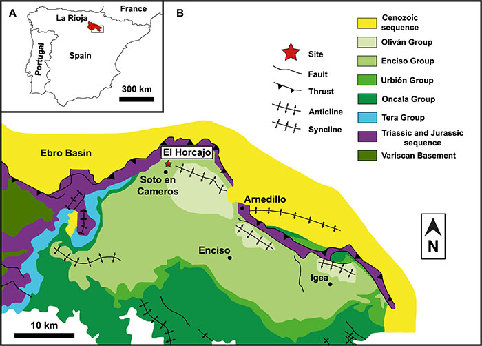
FIGURE 2. The El Horcajo site stratigraphic column (modified from Moreno-Azanza et al., 2016).
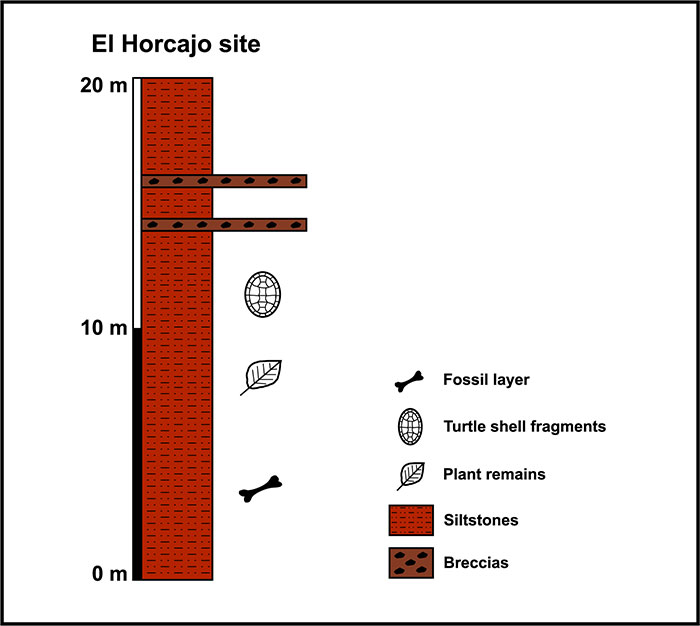
FIGURE 3. Anterior caudal vertebra CP 411: A, anterior view; B, posterior view; C, left lateral view; D, right lateral view; E, dorsal view; F, ventral view; G, position of CP 411 between the anterior caudal vertebrae showing transverse processes of the caudal series (modified from Scott Hartman, 2016). The question symbol (?) indicates the plausible precise location. Abbreviations: aaf, anterior articular facet; ahaef, anterior articular haemal facet; crib, caudal rib; fm, fusion marks; Ha, height of the anterior articular facet of the centrum; Hp, height of the posterior articular facet of the centrum; L, vertebral centrum length; narc, neural arch; nca, neural canal; nf, nutritional foramen; paf, posterior articular facet; phaef, posterior articular haemal facet; Wa, width of the anterior articular facet of the centrum; Wp, width of the posterior articular facet of the centrum. Scale bar equals 10 cm.
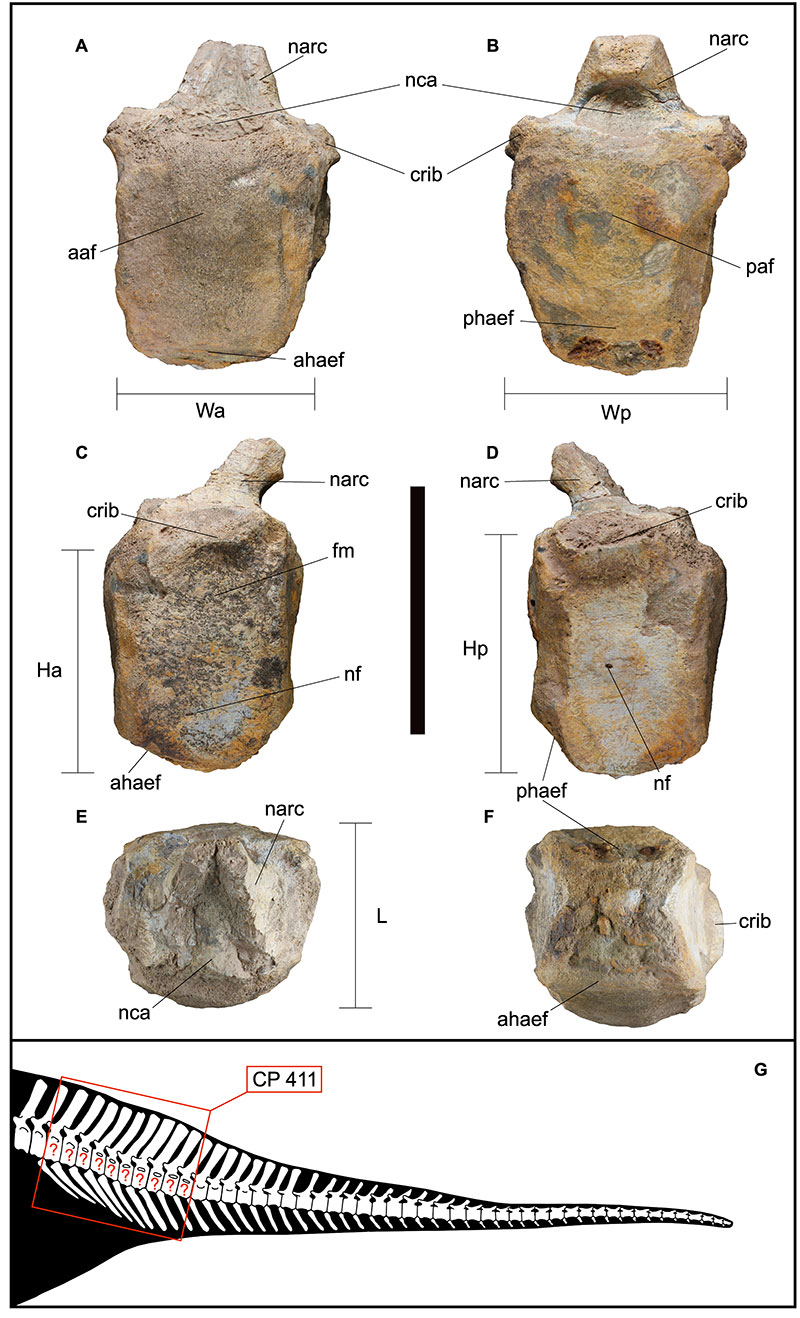
FIGURE 4. Fragment of proximal end of left humerus CP 412: A, anterior view; B, posterior view; C, proximal view; D, distal view; E, position of CP 412 on the left humerus (modified from Scott Hartman, 2016). Abbreviations: breg, bone regrowth; H, height; hh, humeral head; L, length; W, width. The morphology of the dashed lines is indicative; it does not indicate a precise morphology. Scale bar equals 10 cm.
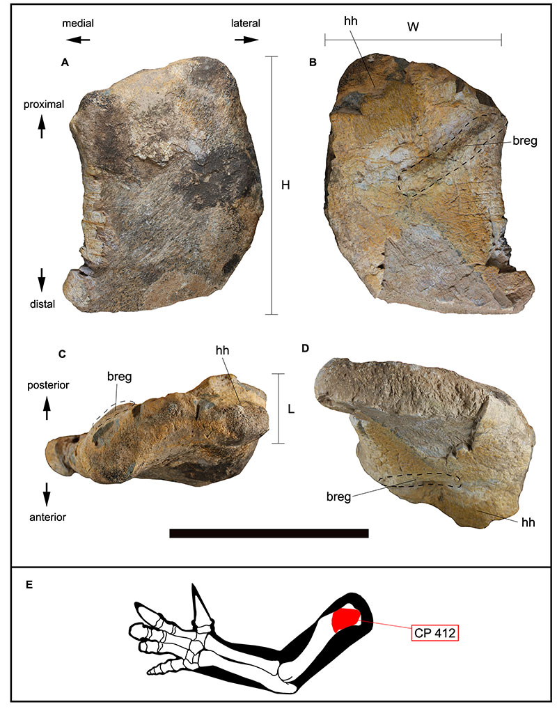
FIGURE 5. Left proximal carpal fragment (radiale) CP 414: A, anterior view; B, dorsal view; C, posterior view; D, ventral view; E, proximal view; F, distal view. Abbreviations: H, height; L, length; mcl, metacarpal l; mcllf, metacarpal ll facet; raf, radius facet; W, width. Scale bar equals 5 cm.
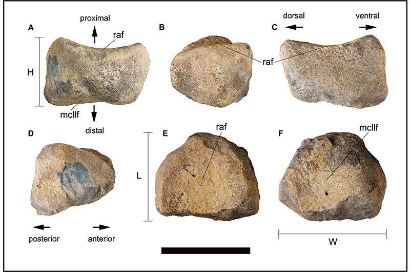
FIGURE 6. Left tibia CP 415: A, anterior view; B, posterior view; C, lateral view; D, medial view; E, proximal view; F, distal view; G, position of CP 415 on the left tibia (modified from Scott Hartman, 2016). Abbreviations: cobon, compact bone; H, height; medca, medullary cavity; L, length; W, width. Scale bar equals 5 cm.
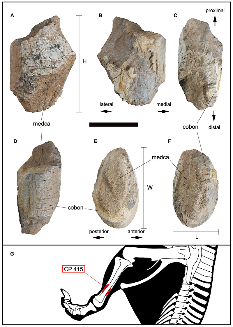
FIGURE 7. Left proximal fibula fragment CP 413: A, lateral view; B, posterior view; C, medial view; D, proximal view; E, position of CP 413 on the left fibula (modified from Scott Hartman, 2016). Abbreviations: cfi, caput fibulae; cpr, cranial process; fa.fe, facies articularis femoralis; fa.ti, facies articularis tibialis; H, height; L, length; W, width. Scale bar equals 5 cm.
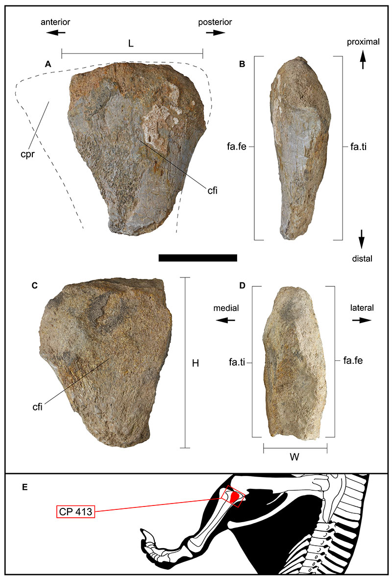
FIGURE 8. PCA diagram (PC1 Vs. PC2) of normalized linear measurements of caudal vertebral centra of CP 411, dryosauridae, hypsilophodontidae, rhabdodontomorpha, hadrosauriformes and styracosternan ornithopods.
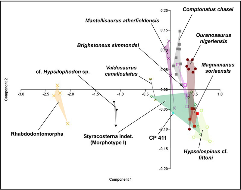
FIGURE 9. Approximate reconstruction of the indeterminate styracosternan ornithopods found in the Early Cretaceous of the eastern Cameros Basin. Illustration by Adrián Blázquez Riola.
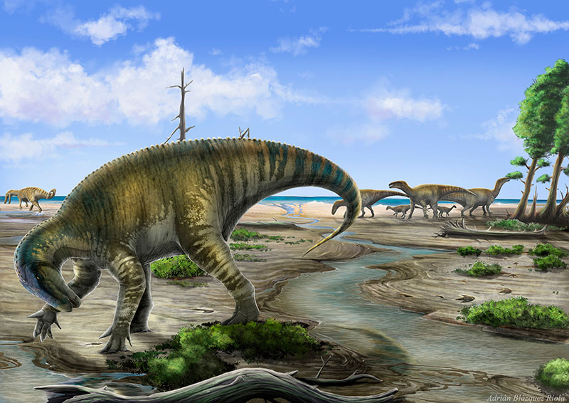
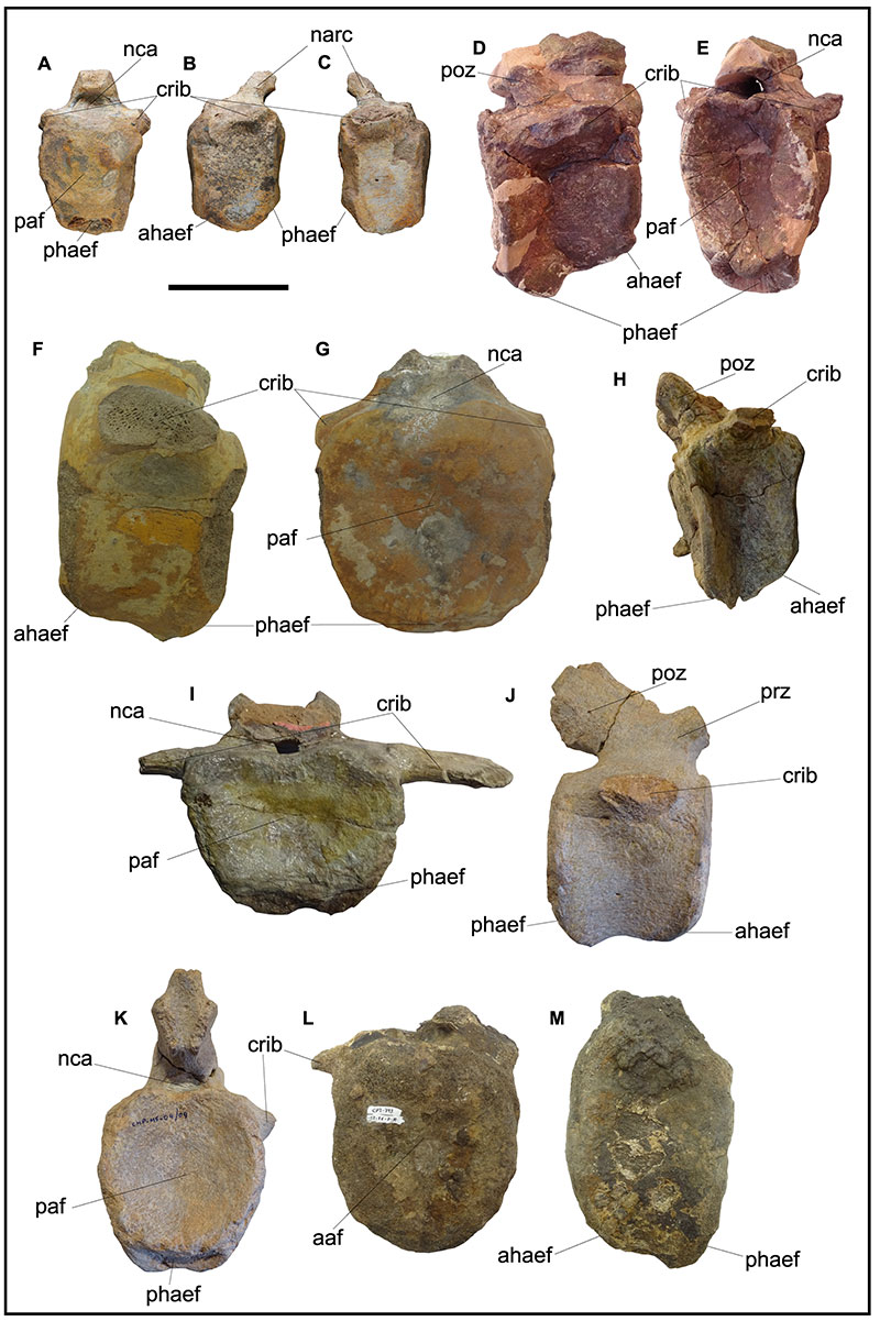
FIGURE 10. CP 411 compared to selected anterior caudal vertebrae of styracosternan ornithopods. CP 411: A, in posterior view; B, in left lateral view; C, in right lateral view. This work. Iguanodontidae indet. (MDS-TBMV, 7): D, in right lateral view; E, in posterior view. Photos taken from MDS-TBMV, 7 (Torcida Fernández-Baldor et al., 2006). Iguanodon cf. galvensis (MAP-8043): F, in left lateral view; G, in posterior view. Photos taken from MAP-8043 (García-Cobeña et al., 2022). Magnamanus soriaensis (MNS 2000/122.2): H, in right lateral view; I, in posterior view. Photos taken from MNS 2000/122.2 (Fuentes-Vidarte et al., 2016). Iguanodon bernissartensis (CMP-MS-04/04): J, in right lateral view; K, in posterior view. Photos taken from CMP-MS-04/04 (Gasulla et al., 2022). Styracosterna indet. (Morphotype I) (MAP-793): L, in anterior view; M, in left lateral view. Photos taken from MAP-793 (Verdú et al., 2019). Abbreviations: ahaef, anterior articular haemal facet; crib, caudal rib; narc, neural arch; nca, neural canal; paf, posterior articular facet; poz, postzygapophysis; phaef, posterior articular haemal facet; prz, prezygapophysis. Scale bar equals 10 cm.