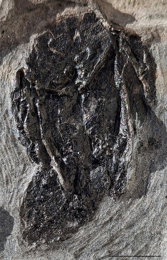
FIGURE 1. Microbrachius longi sp. nov. plates from Essi farm in South-East Estonia. Left lateral plates (L). A-B, GIT 628-9, in visceral aspect: A, Photograph of the specimen; B, Illustration of the specimen; C, GIT 628-79, in visceral aspect; D, GIT 628-81, in dorsal (external aspect). Abbreviations: cpr, preorbital angle; cpt, postorbital angle; ifc1, infraorbital sensory line; lpri, lateropremesial ridge of the headshield; pr.po, antero-lateral corner of otico-occipital depression; r, premedian ridge; rlc, rod-like crenulated anterior margin. Scale bars equal 1 mm.
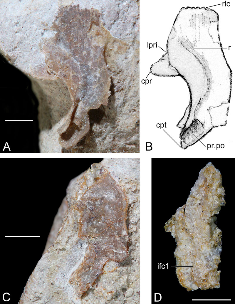
FIGURE 2. Microbrachius longi sp. nov. from Essi farm in South-East Estonia. Nuchal + postpineal plates (Nu+Pp). A-B, GIT 628-5, in dorsal (external) aspect: A, Photograph of the specimen; B, Illustration of the specimen; C-D, GIT 628-6, in dorsal (external) aspect; C, Photograph of the specimen; D, Illustration of the specimen; E-F, GIT 628-7, in visceral aspect; E, Photograph of the specimen, F, Illustration of the specimen; G-H. GIT 628-8, in visceral aspect; G, Photograph of the specimen; H, Illustration of the specimen. from Essi farm in South-East Estonia. cf.L area of overlap for the lateral plate; cf.Pn, area of overlap for the paranuchal plate; cr.pto, postorbital crista of the headshield; csl, central sensory line groove; d.end, opening of canal for endolymphatic duct; d.end1, internal opening of canal for endolymphatic duct; L, lateral plate; mc, lateral corners of the nuchal plate; mr, median ridge on the postpineal plate; nm, obtected nuchal area; Nu, nuchal plate; pc, postero-lateral corners of the nuchal plate; Pn, paranuchal plate; Pp, postpineal plate; pp.mc, lateral corners of the pineal plate; pr.nm, posterior median process of headshield; sog, supraoptic groove of headshield. Scale bars equal 1 mm.

FIGURE 3. Microbrachius longi sp. nov. from Essi farm in South-East Estonia. Submarginal plates (Sm): A-B, GIT 628-32, in dorsal (external) aspect; A, Photograph of the specimen, B, Illustration of the specimen; C-D, GIT 628-33, in visceral aspect; C, Photograph of the specimen; D, Illustration of the specimen. Abbreviation: smr, submarginal ridge. Scale bars equal 1 mm.
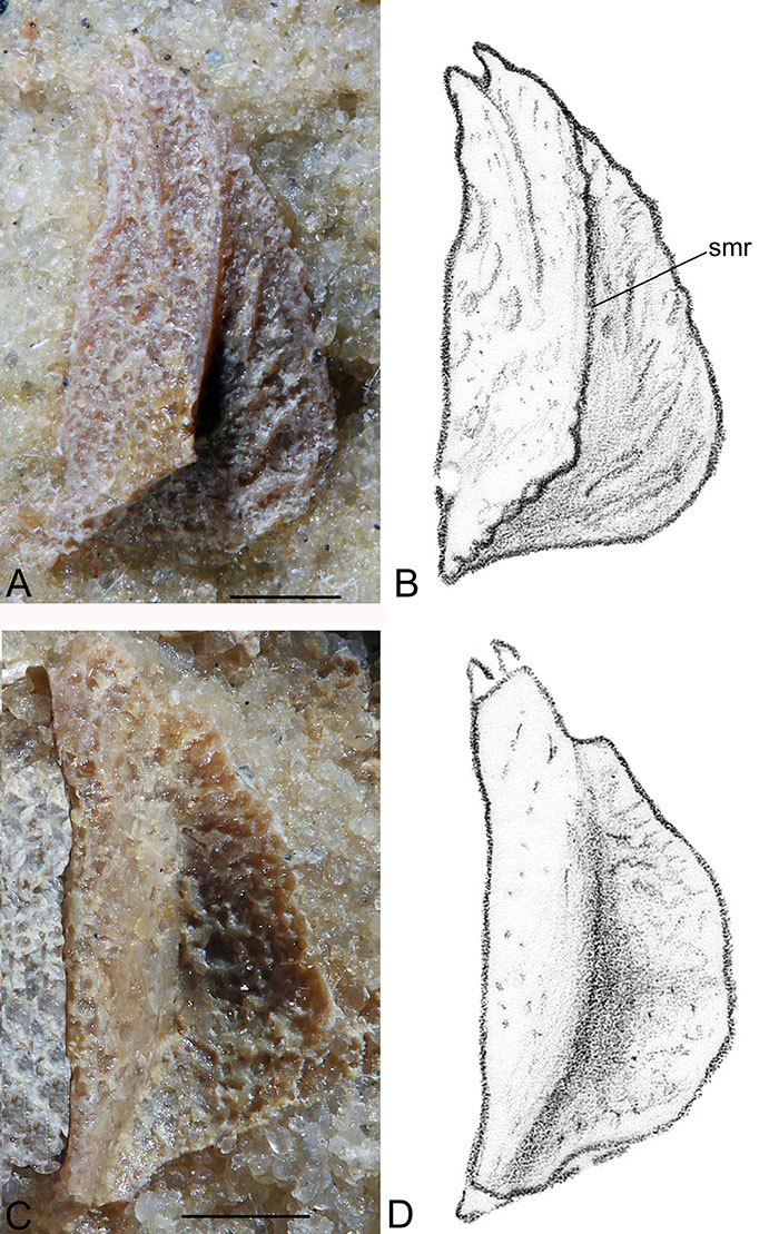
FIGURE 4. Microbrachius. Reconstruction of headshields: A, Microbrachius longi sp. nov.; B, Microbrachius dicki, modified from Hemmings 1978, fig. 36. Abbreviations: L, lateral plate; Nu, nuchal plate; Pmg, postmarginal plate; Pn, paranuchal plate; Pp, postpineal plate; Prm, premedian plate; Sm, submarginal plate.
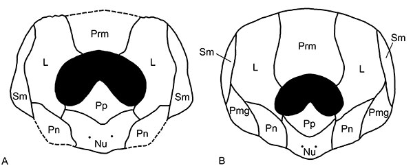
FIGURE 5. Anterior median dorsal plates (AMD) of Microbrachius from the Middle Devonian of Laurussia: A-F, Microbrachius longi sp. nov. from Essi farm in South-East Estonia: A-B, GIT 628-1 in dorsal (external) aspect; A, Photograph of the specimen: B, Illustration of the specimen; C-D, GIT 628-2 in visceral aspect; C, Photograph of the specimen; D, Illustration of the of the specimen; E-F, GIT 628-4 in visceral aspect; E, Photograph of the specimen; F, Illustration of the specimen; G-H, Microbrachius dicki from the island of Mousa in Shetland, Scotland, photographs in dorsal (external) aspect; G, GSE 13338; H, GSE 13333. Abbreviations: ADL, anterior dorso-lateral plate; AMD, anterior median dorsal plate; MxL, mixilateral plate; PMD, posterior median dorsal plate; alr, postlevator thickening of AMD; cf.ADL, area overlapping ADL; f.retr, levator fossa of AMD; lc, lateral corner of AMD; oa.ADL, area overlapped by ADL; oa.MxL, area overlapped by MxL; oa.PMD, area overlapped by PMD; pr.p, posterior median process of AMD; pt1, anterior ventral pit of dorsal wall of trunk armour. Scale bars equal 1 mm in A, C, E, G, and 5 mm in H.
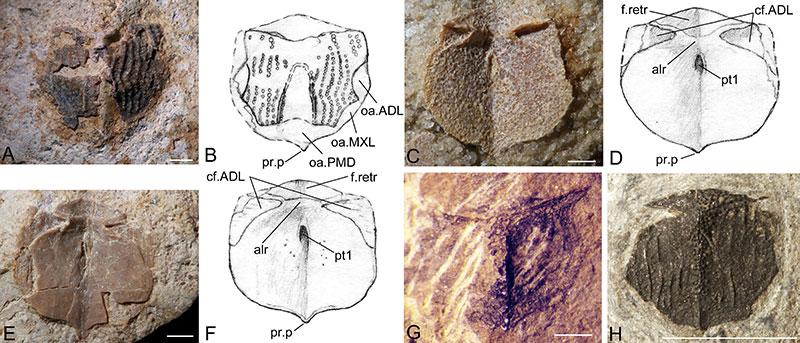
FIGURE 6. Microbrachius longi sp. nov. from Essi farm in South-East Estonia. Posterior median dorsal plates (PMD): A-B, GIT 628-3 in dorsal (external) aspect; A, photograph of the specimen; B, line drawing of the specimen; C-D, GIT 628-11 in dorsal (external) aspect; C, photograph of the specimen; D, line drawing of the specimen; E-F, GIT 628-34 in visceral aspect; E, photograph of the specimen; F, line drawing of the specimen; G-H, GIT 628-10 in visceral aspect; G, photograph of specimen; H, line drawing of specimen. Abbreviations: AMD, anterior median dorsal plate; MxL, mixilateral plate; PMD, posterior median dorsal plate; aa, anterior angle of PMD; aal, anterolateral angle of PMD; cf.AMD, area overlapping AMD; pa, posterior angle of PMD; cf.MxL, area overlapping MxL; crtp, crista transversalis interna posterior; dlg, dorsal sensory line groove; dmr, dorsal median ridge; l, lateral corner of PMD; pda, posterior dorsal angle of trunk armour; pma, posterior marginal area of PMD; pt2, posterior ventral pit of dorsal wall of trunk armour. Scale bars equal 1 mm.
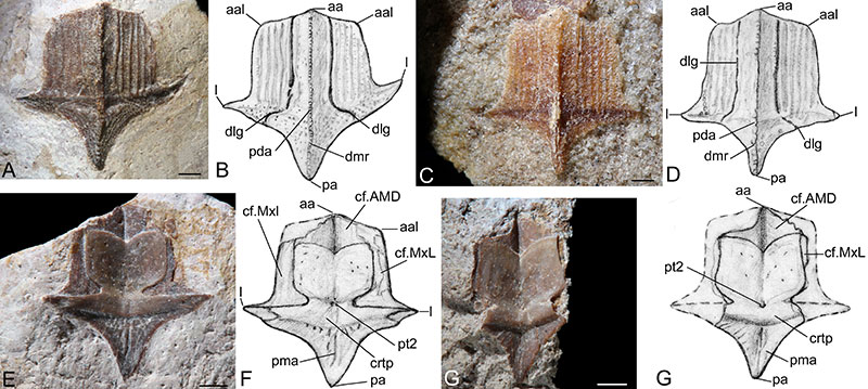
FIGURE 7. Microbrachius longi sp. nov. from Essi farm in South-East Estonia. Detail of GIT 628-3 a posterior median dorsal plate (PMD): A, detail of the dorsal median ridge; B, detail of longitudinal ridges. Scale bars equal 1 mm.
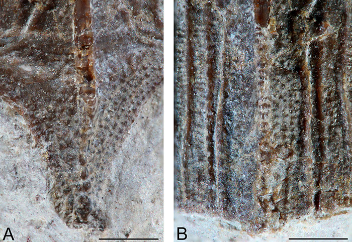
FIGURE 8. Microbrachius longi sp. nov. from Essi farm in South-East Estonia. Anterior dorsolateral plates (ADL): A-B, GIT 100-11, a right ADL in external aspect; A, photograph of specimen showing bite marks; B, line drawing of specimen with bite marks removed; C-D, GIT 628-14, a left ADL in external aspect; C, photograph of specimen; D, line drawing of specimen; E-F, GIT 6285-13, a left ADL in visceral aspect; E, photograph of specimen; F, line drawing of specimen. Abbreviations: AMD, anterior median dorsal plate; AVL, anterior ventrolateral plate; MxL, mixilateral plate; cf.AMD, area overlapping AMD; cf.MxL, area overlapping MxL; cit, crista transversalis interna anterior; dlm, dorsal lamina; dlr, dorsolateral ridge of the trunk armour; f.art, articular fossa; lcg, main lateral line groove; lateral lamina; oa.AMD, area overlapped by AMD; oa.AVL, area overlapped by AVL; pro, processus obstans. Scale bars equal 1 mm.

FIGURE 9. Microbrachius longi sp. nov. from Essi farm in South-East Estonia. Left Mixilateral plates (MxL): A, GIT 628-16; B, GIT 628-64; C, restoration of MxL plate. Abbreviations: ADL, anterior dorsolateral plate; AMD, anterior median dorsal plate; PMD, posterior median dorsal plate; PVL, posterior ventrolateral plate; cf.AMD, area that overlaps AMD, cr,tp, crista transversalis interna posterior; dlm, doral lamina; dlr, dorso-lateral ridge; lcg, main lateral line groove; llm, lateral lamina; oa.ADL, area overlapped by ADL; oa.PMD, area overlapped by PMD; oa.PVL, area overlapped by PVL; pr.dm, median dorsal process; pr.tv, posteroventral process. Scale bars equal 1 mm.
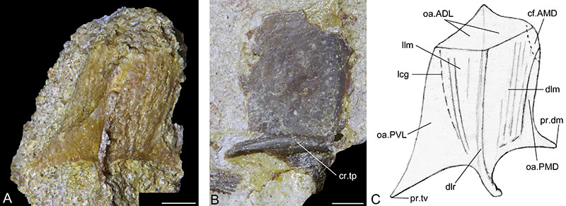
FIGURE 10. Microbrachius longi sp. nov. from Essi farm in South-East Estonia. Anterior ventrolateral plates (AVL): A-B, GIT 628-18, a left AVL in external aspect; A, photograph of specimen; B, line drawing of specimens; C-E, GIT 628-22, posterior portion of a left AVL in external aspect; C, photograph of the specimen; D, line drawing of the specimen oriented ventrally; E, line drawing of the specimen oriented more laterally; F, GIT 628-20, a left AVL in visceral aspect; G, GIT 628-23, an anterior portion of a right AVL in visceral aspect; H, GIT 628-19, a left AVL in visceral aspect; I, reconstruction of a left AVL. Abbreviations: ADL, anterior dorsolateral plate; AVL, anterior ventrolateral plate; PVL, posterior ventrolateral plate; c.al, anterolateral angle of subcephalic division of ventral lamina; c.rc, rostrocaudal canal; cf.ADL, area overlapping ADL; cf.PVL, area overlapping PVL; cit, crista transversalis anterior; f.art.d, dorsal half of the fossa articularis pectoralis; f.art.v, ventral half of the fossa articularis pectoralis; f.ax, foramen axillare; fp, funnel pit; llm, lateral lamina; m.lim, margo limitans; oa.AVL, area overlapped by AVL; pbr, processus brachialis; pdc, posterodorsal angle; pe, pars pedalis; vcl, posterior ventrolateral angle; vlm, ventral lamina; vlr, ventrolateral ridge of trunk armour. Scale bars equal 1 mm.
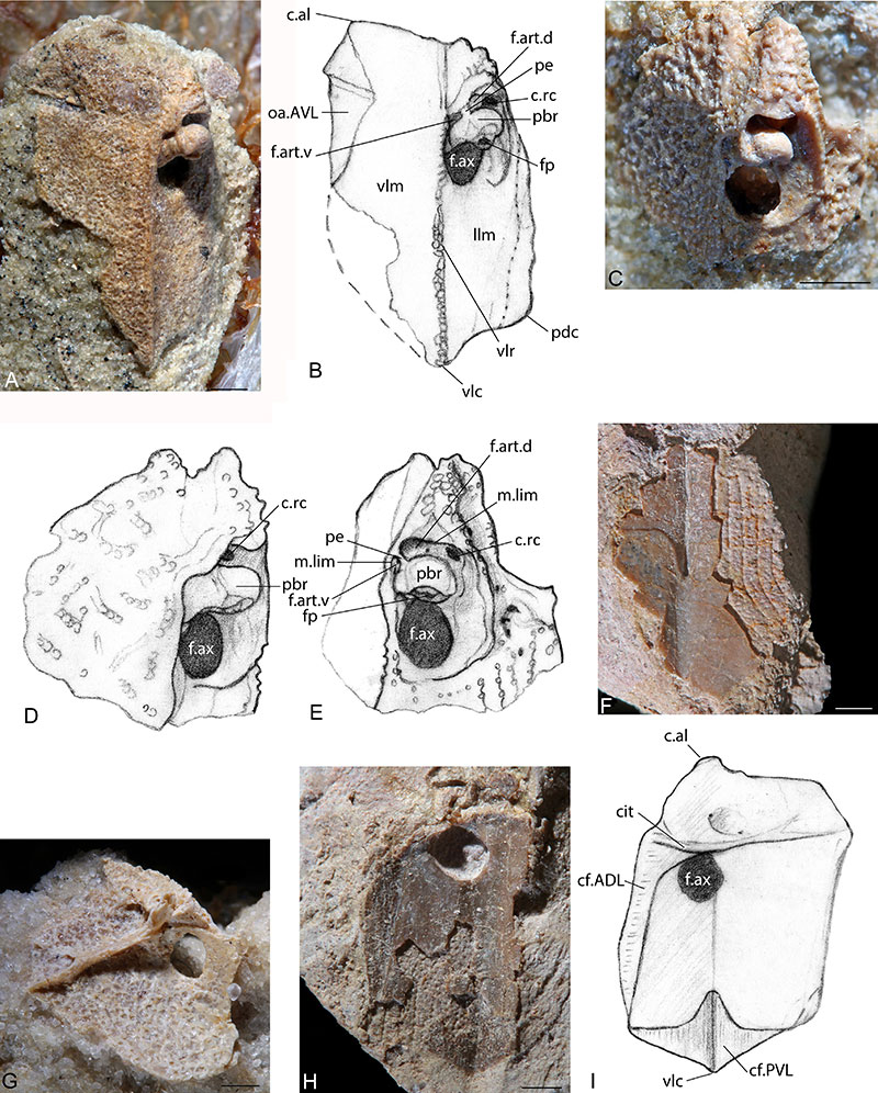
FIGURE 11. Microbrachius longi sp. nov. from Essi farm in South-East Estonia. Posterior ventrolateral plates (PVL): A-B, GIT 628-17, a left PVL in visceral aspect; A, photograph of the specimens; B, line drawing of the specimen; C-D, GIT 628-25, a left PVL in visceral aspect; C, photograph of the specimens; D, line drawing of the specimen; E, reconstruction of a left PVL in visceral aspect; F, GIT 628-24, a right PVL with attach clasper in visceral aspect. Abbreviations: MxL, mixilateral plate; cf,MxL, arear overlapping the mixilateral plate; cf.PVL, the area overlapping the posterior ventrolateral plate; cr.sp, crista transversalis interna posterior; tvr, transverse lateral ridge. Scale bars equal 1 mm.
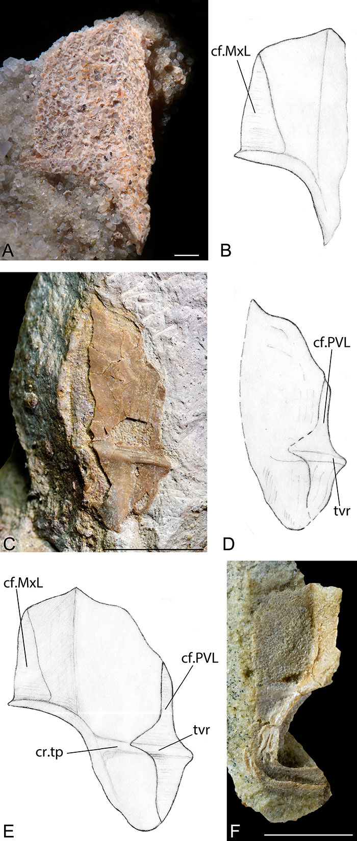
FIGURE 12. Microbrachius longi sp. nov. from Essi farm in South-East Estonia. Pectoral appendages: A-B, GIT 628-27, a right proximal appendage; A, photograph of the specimen; B, line drawing of the specimen; C-D, GIT 628-28, a right proximal appendage; C, photograph of the specimen; D, line drawing of the specimen; E, GIT 628-29, a distal appendage. Abbreviations: Cd1, dorsal central plate 1. Cv2, ventral central plate 2. Scale bars equal 1 mm.
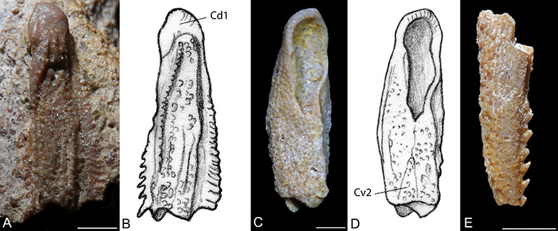
FIGURE 13. NMS G.2025.9.1.1 Microbrachius dicki from Weems Castle, South Ronaldsay, Orkney, Scotland showing the possible tail. Scale bar equals 10 mm.