FIGURE 1. Holotype of Lucidusmacula lihanjieae gen. et sp. nov. (LYU-BL2003). A, the dorsal view. B, the ventral view. C, line drawing of the dorsal view. D, line drawing of the ventral view. Scale bars: 2 mm (A-D).
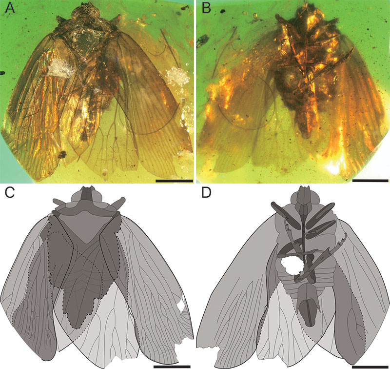
FIGURE 2. Detailed photographs of the head, pronotum and mesonotum of Lucidusmacula lihanjieae gen. et sp. nov. (LYU-BL2003). A, head and pronotum in dorsal view. B, head in ventral view. C, head and pronotum in ventral view. D, detached antenna. E, mesonotum in dorsal view. F, left tegulae. Scale bars: 0.5 mm (A, B, D); 0.2 mm (F); 0.1 mm (C, E).
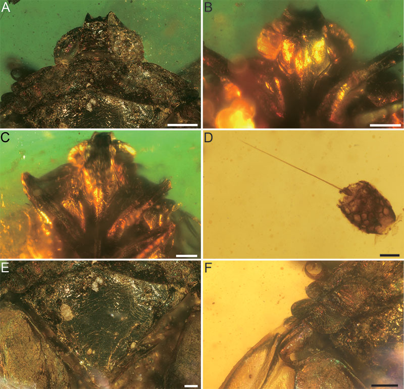
FIGURE 3. Detailed photographs and line drawing of the tegmen of Lucidusmacula lihanjieae gen. et sp. nov. (LYU-BL2003). A, the right tegmen in dorsal view. B, line drawing of the right tegmen in dorsal view. C, a distinct brighter area between ScP and RA. D, the basal cell of the left tegmen. E, the apical part of the right tegmen in dorsal view. F, claval apex of the left tegmen. Scale bars: 1.0 mm (A, B); 0.5 mm (E, F); 0.2 mm (C, D).
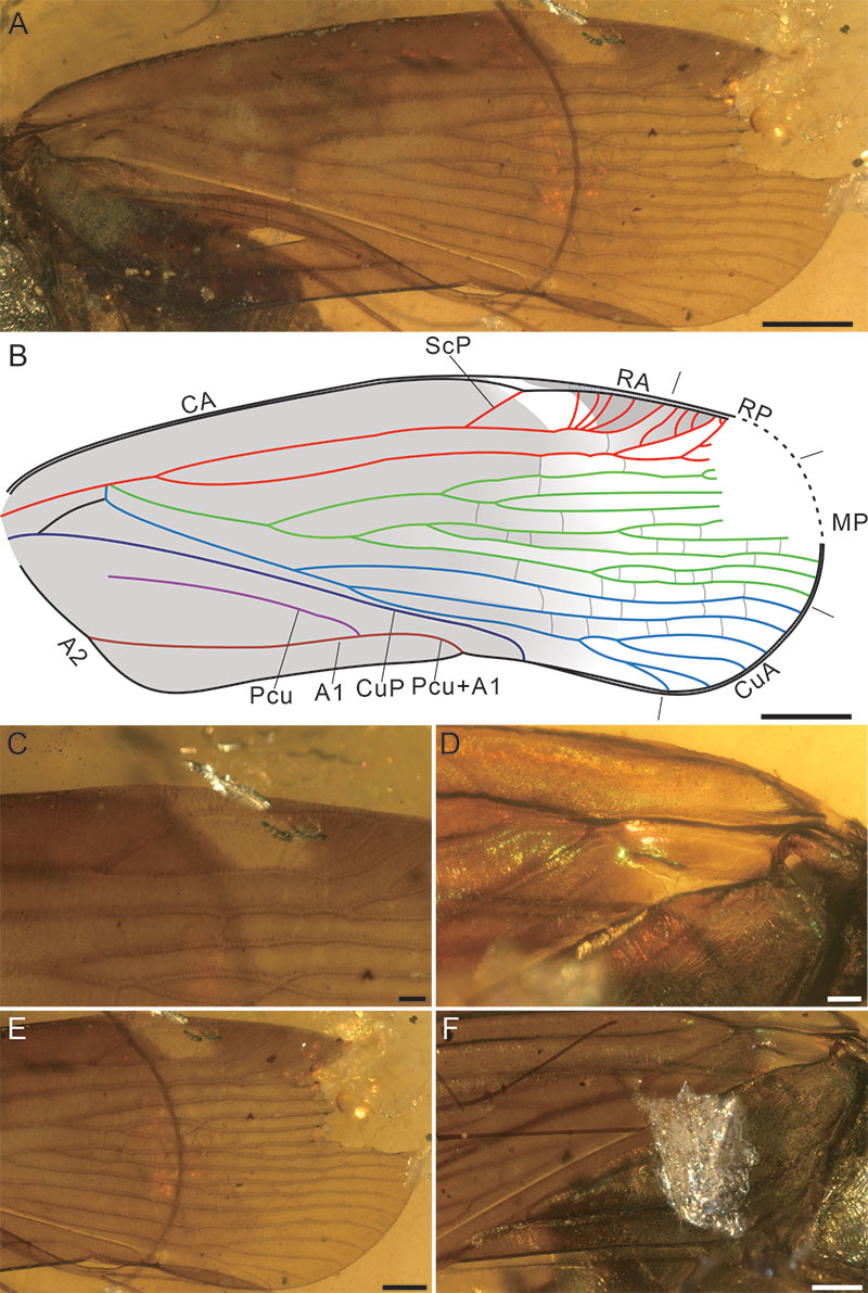
FIGURE 4. Detailed photographs and line drawing of the hind wing of Lucidusmacula lihanjieae gen. et sp. nov. (LYU-BL2003). A, the right hind wing in dorsal view. B, line drawing of the right hind wing in dorsal view. C, wing-coupling lobe (WCL) of hind wing. D, the basal part of the right hind wing in dorsal view. Scale bars: 1.0 mm (A, B), 0.5 mm (D), 0.2 mm (C).
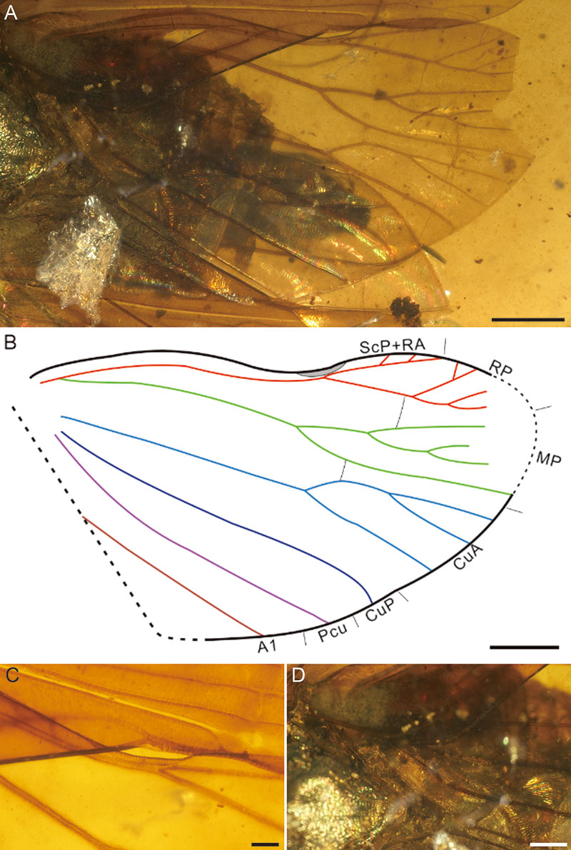
FIGURE 5. Detailed photographs of legs of Lucidusmacula lihanjieae gen. et sp. nov. (LYU-BL2003) A, the left fore leg. B, the left mid leg. C, mesotibia of the right mid leg. D, the right hind leg. E, metatibia of the right hind leg, note 3 lateral spines (arrowed). F, the apical teeth of the right metatibia. G, the apical teeth of the right basimetatarsomere, note numerous subapical platellae (arrowed). H, the apical teeth of the right midmetatarsomere. I, the claw of the right leg. Scale bars: 0.5 mm (B, D, E), 0.2 mm (A, C), 0.1 mm (F-I).
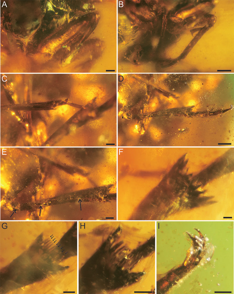
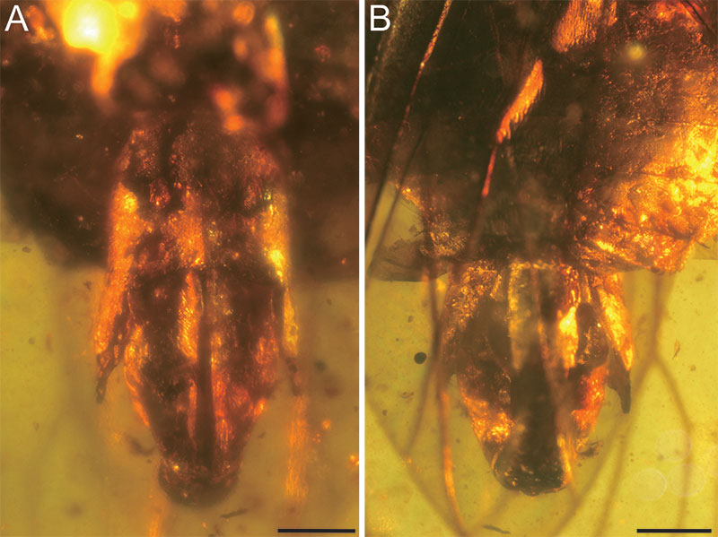
FIGURE 6. Terminalia of Lucidusmacula lihanjieae gen. et sp. nov. (LYU-BL2003). A, the ventral view. B, the dorsal view. Scale bars: 0.5 mm (A, B).