FIGURE 1. Stratigraphic and palaeogeographic distribution of piraniids, including Pirania and three new genera erected in this paper. 1, stratigraphic distribution of piraniids; 2, distribution of piraniids during the Middle and Late Ordovician (Katian, 455 Ma); 3, distribution of piraniids during the Early Ordovician (Floian, 478 Ma); 4, distribution of piraniids during the middle Cambrian (515 Ma). Base maps produced using BugPlates (Torsvik 2009).
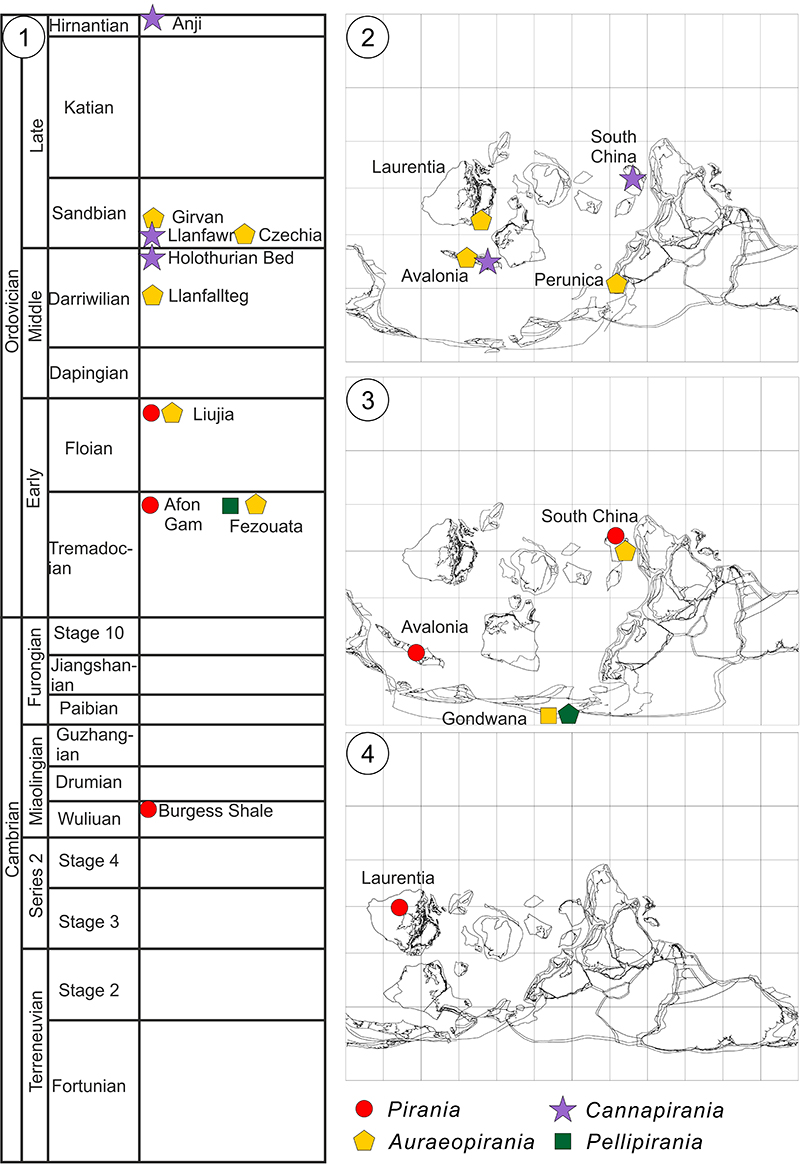
FIGURE 2. Pirania? ericia sp. nov., from the Early Ordovician (Tremadocian) Afon Gam Biota, North Wales. 1, 4: holotype NMW2015.34G.26; 1, overall view of branched specimen, and 4, detail of truncated basal stalk (counterpart). 2-3, 5-6, paratype NMW2015.34G.31; 2, overall view of unbranched sponge; 3, detail of well-preserved lateral prostalial spicule with wide base; 5, detail of flared apex showing finer spicules between prostalia; 6, detail of basal tuft of radiating fine and coarse spicules. Scale bars equal 1 mm.
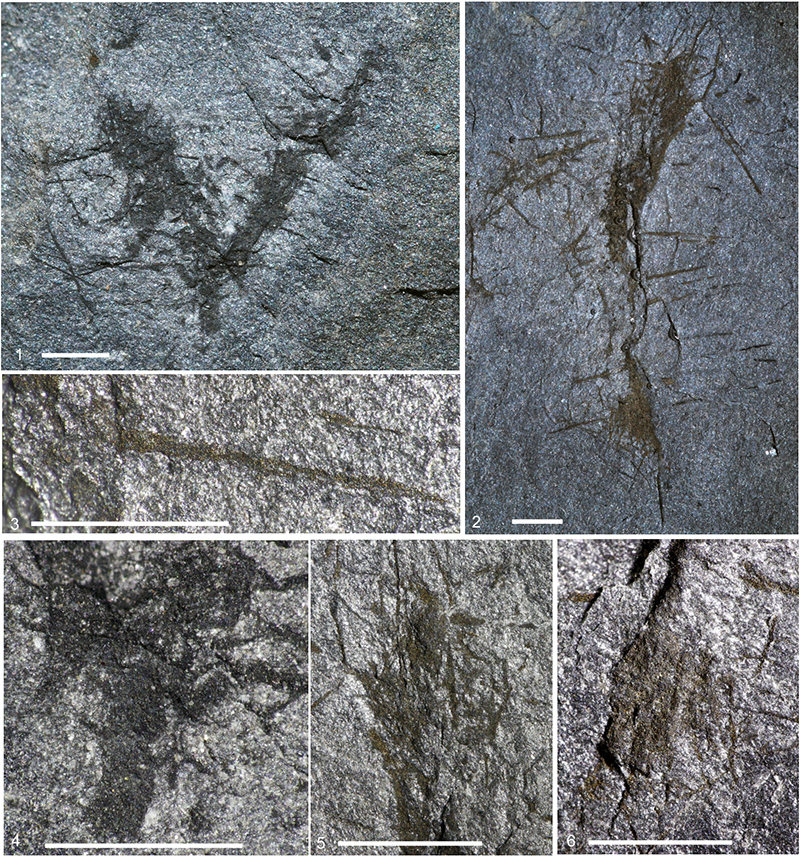
FIGURE 3. Pirania? peregrinata sp. nov. from the Floian Liujia section, Zhejiang, China; NIGP171183. 1, overall view of specimen photographed under water to highlight coarser pyritised spicules; 2, lower part photographed dry with low-angle light, showing array of fine spicules; boxed area magnified in 3, showing lack of basal spicule expansion. Scale bars equal 1, 5 mm; 2-3, 1 mm.
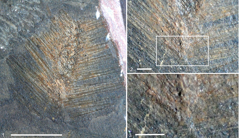
FIGURE 4. Auraeopirania pinwyddeni gen. et sp. nov., from the Middle Ordovician (Didymograptus artus Biozone) Llanfallteg Biota, Rhyd-y-Brown, South Wales; holotype NMW00.17G.113. 1, overall view of specimen; 2, magnification of upper part showing body wall and skeletal organisation; 3, detail of central part of sponge showing coarse prostalia without obvious fine monaxons; 4, lower region of sponge, with radiating basal spicule array diverging from narrowed basal part of body (arrowed). Scale bars equal 1 mm.
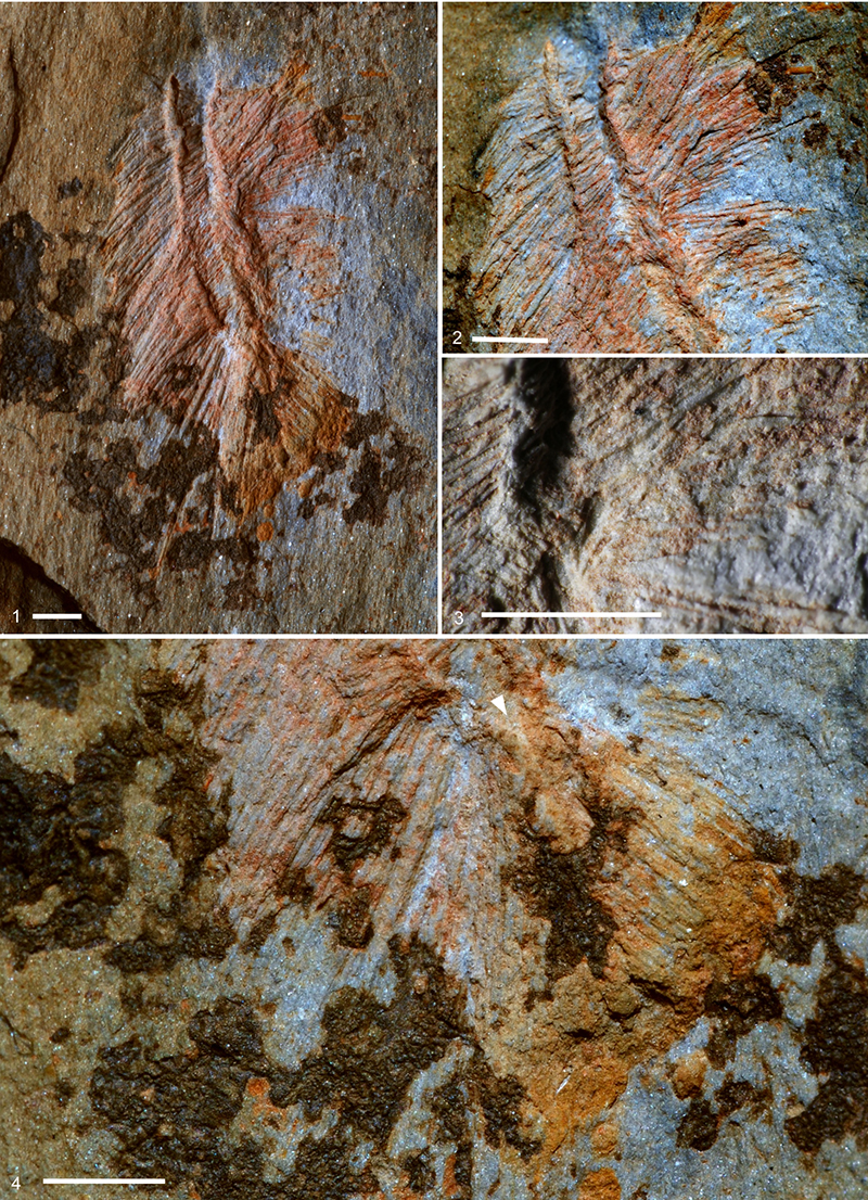
FIGURE 5. Auraeopirania sciurucauda gen. et sp. nov.; 1-4, holotype NIGP171184; 1, overall view, photographed dry; 2, the same, under water; 3, magnification of central region showing prostalia array; 4, magnification of basal region with radiating spicule tuft. 5-6, paratype NIGP171185, showing apical region; 5, overall view, photographed dry (contrast strongly enhanced); 6, detail of apex photographed under water to highlight marginal prostalia. Scale bars 5 mm.

FIGURE 6. Auraeopirania pykitia gen. et sp. nov., from the Upper Ordovician of Dalfask, Scotland. 1, 3-6, holotype GLAHM 163227; 1, overall view of specimen distorted apically where in contact with Diacanthaspis trippi specimen; 3, magnification of upper part with contrast between lateral prostalia (left) and robust marginalia (upper right); 4, magnification of basal region, including possible branch or bud at lower right (plumose spicule array, arrowed), second possible branch along lower margin of slab, and area magnified in 6 (box); 5, isolated partial branch adjacent to the holotype; 6, detail of body wall in cross section, with dense array of pyritised lateral prostalia; 2, paratype GLAHM 163228/1, overall view of weathered specimen; 7, paratype GLAHM 163228/2, overall view of pyritised specimen with coarser apical marginalia (arrowed) amongst dense lateral prostalia. Scale bars equal 1 mm.
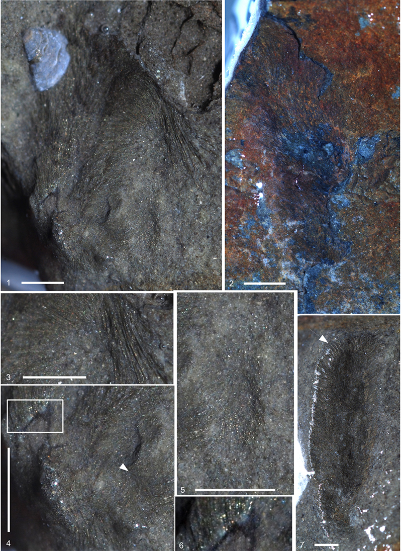
FIGURE 7. Cannapirania canna gen. et sp. nov., from the late Darriwilian (H. teretiusculus Biozone) Holothurian Bed fauna of Bach-y-Graig, Llandrindod, Wales. 1, 4, 5: holotype NMW2019.25G.1; 1, overall view of specimen with spicules largely replaced by iron oxides; 4, detail of apical region; 5, magnification of upper left lateral margin, showing rounded and occasionally expanded (tylotic; arrowed) prostalial bases. 2, 3, 6: paratype NMW2019.25G.2.; 2, overall view of specimen with spicules largely preserved as moulds with traces of siliceous minerals; 3, detail of apical end including marginalia; 6, magnification of base of marginalia, showing abrupt narrowing indicating stylose bases. Scale bars equal 1 mm.
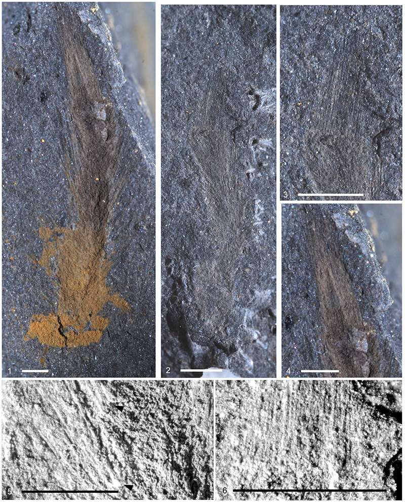
FIGURE 8. Cannapirania vermiformis gen. et sp. nov., from the Hirnantian Anji Biota of Zhejiang, China, holotype NIGP171186. 1, Overall view of branched specimen; 2, detail showing siliceous preservation of the largest spicules; 3, detail showing rounded, tylotic base (arrowed) of marginal prostalia. Scale bars equal 1 mm.

FIGURE 9. Pellipirania gloria gen. et sp. nov., from the Tremadocian Fezouata Biota of Morocco. 1, holotype NMW2015.34G.27, overall view, whitened with ammonium chloride; 2, apical region of holotype, not whitened and in low-angle light; 3, detail of prostalial spicules in paratype NMW2015.34G.26; 4, detail of fine, radially directed body wall spicules in holotype. Scale bars equal 1, 2: 10 mm; 3, 4: 5 mm.
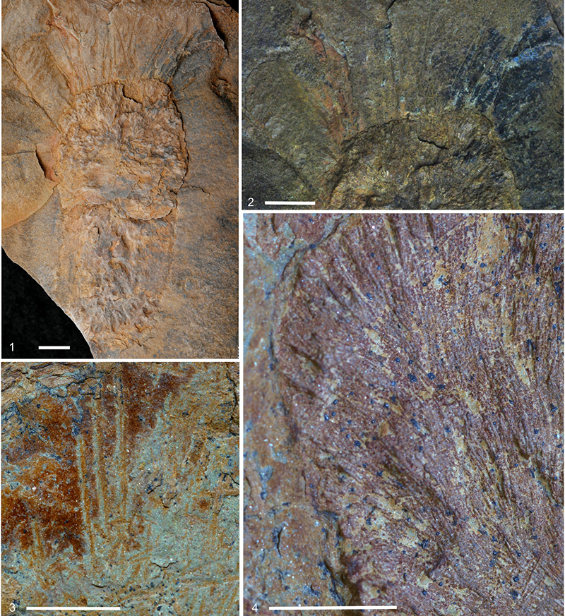
FIGURE 10. Schematic illustration of the evolution of the Piraniidae within the Protomonaxonida, with indications of what are here interpreted to be the most significant character transitions, combined with known stratigraphic ranges of taxa. Range of chancelloriids includes early appearance of distinctive sclerites, whereas other taxa are restricted to sites with exceptional preservation.

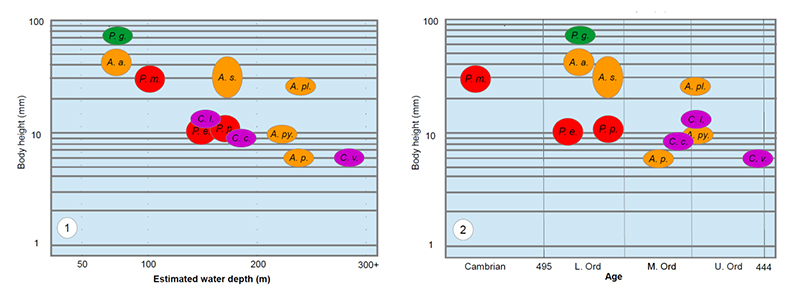
FIGURE 11. Correlation of body size of known piraniids against: 1, estimated water depth; 2, stratigraphic age of the respective deposits. Taxa: P. m.: Pirania muricata; P. e.: Pirania? ericia; P. p.: Pirania? peregrinata; A. a.: Auraeopirania auraeum; A. s.: Auraeopirania sciurucauda; A. pl.: Auraeopirania? plasi; A. py.: Auraeopirania pykitia; A. p.: Auraeopirania pinwyddeni; C. l.: Cannapirania llanfawrensis; C. c.: Cannapirania canna; C. v.: Cannapirania vermiformis; P. g.: Pellipirania gloria.