FIGURE 1. The (A, C) spatial and (B, D) temporal distribution of the various species of Tanystropheus based on the taxonomic assignments prior to this study (A-B) and following the findings of this study (C-D). Map modified from Smith et al. (1994).
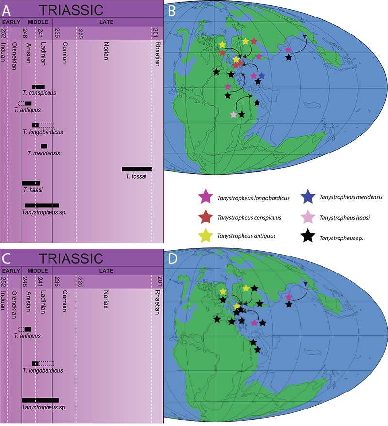
FIGURE 2. Photographs of (A) PIMUZ T 3901, the former holotype of Tanystropheus meridensis; (B) a detail of the skull of PIMUZ T 3901 in left lateral view; (C) a detail of the fifth cervical vertebra of PIMUZ T 3901 in left lateral view; and (D) a detail of a partially broken cervical vertebra of PIMUZ T 2484 referred to the small morphotype of T. longobardicus in right lateroventral view. The arrows are pointing anteriorly. Abbreviations: la, lamina.
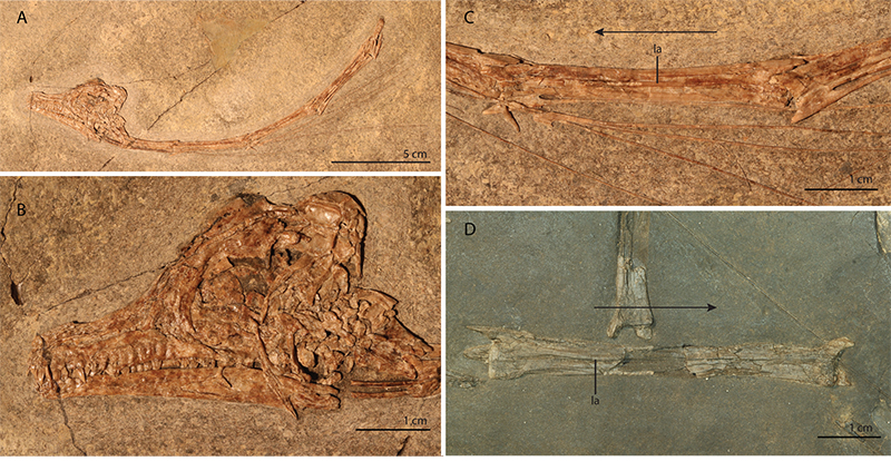
FIGURE 3. Graphs visualizing the data presented in Table 4 scaled against the relative skull size estimated of each specimen; (A) the ratio of the length of the prenarial process of the premaxilla divided by the length of the premaxillary body (=premaxillary tooth row); (B) the ratio of the length of the postnarial process of the premaxilla divided by the length of the premaxillary body; (C) the ratio of the height of the dentary keel divided by the length of the tooth row of the dentary; (D) the premaxillae of PIMUZ T 2484 indicating the measurements taken for establishing the relative length of the prenarial and postnarial processes of the premaxilla; (E) the left dentary of PIMUZ T 2819 indicating the measurements taken for establishing the relative height of the dentary keel. The black squares represent specimens assigned to the small morphotype and the grey squares represent specimens assigned to the large morphotype. Abbreviations: de, dentary; pmx, premaxilla.
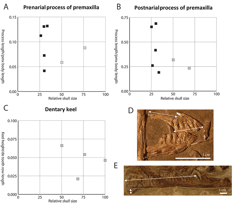
FIGURE 4. Elements exhibiting morphological variation in various specimens of Tanystropheus from Monte San Giorgio; (A) various elements of PIMUZ T 2484; (B) the parietal of PIMUZ T 2819 in dorsal view; (C) various elements of PIMUZ T 2482; (D) a pterygoid of PIMUZ T 2787; (E) the partial braincase of PIMUZ T 2819; (F) the skull of MSNM BES SC 1018 in left lateroventral view; (G) various elements of PIMUZ T 2787. Abbreviations: de, dentary; mx, maxilla; pa, parietal; pal, palatine; pmx, premaxilla; pop, paroccipital process; pt, pterygoid; vo, vomer.
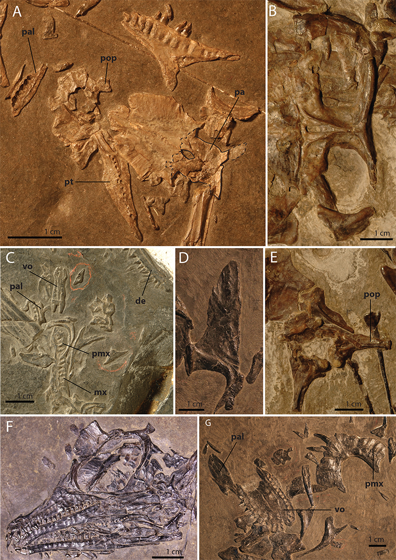
FIGURE 5. The isolated cervical vertebra U-MO BT 740, assigned as the lectotype of Tanystropheus conspicuus by Wild (1973) in (A) left lateral view; (B) right lateral view; (C) dorsal view; (D) ventral view; (E) anterior view; (F) posterior view; and (G) oblique posterodorsal view of the posterior end. The numbers refer to the following morphological characters of the cervical vertebrae (not inferring absence or presence, which is discussed in the text): (1) presence or absence of a deep horizontal groove directly dorsal to the centrum at the posterior end of the vertebrae; (2) presence or absence of thickened margins of the articulation facets of the postzygapophyses; (3) presence or absence of a straight posterior margin of the postzygapophyseal trough; (4) degree of posterior extension of the postzygapophyseal trough; (5) presence or absence of a long pointed posterior process of the neural spine that overlies the postzygapophyseal trough. The arrows are pointing anteriorly.
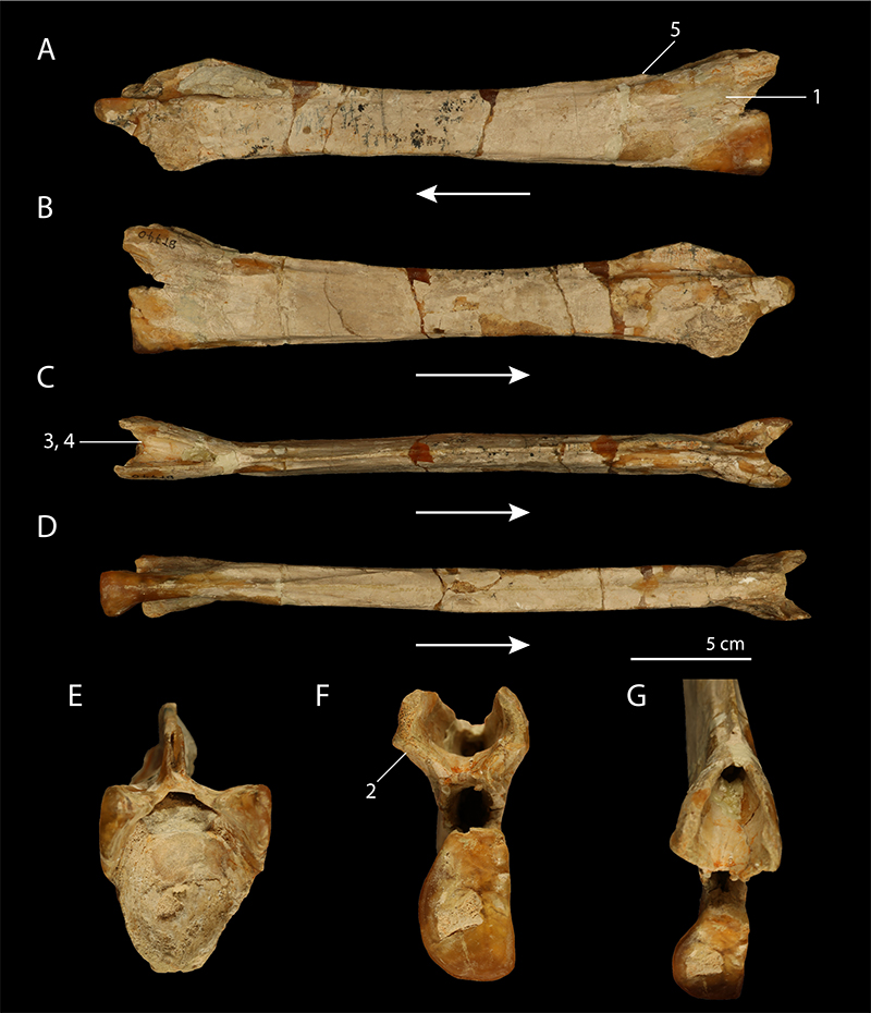
FIGURE 6. Isolated cervical vertebrae of Tanystropheus conspicuus from the Upper Muschelkalk of Bindlacher Berg housed in the U-MO; (A) U-MO BT 732 in right lateral view; (B) U-MO BT 733 in right lateral view; (C) U-MO BT 736 in right lateral view; (D) U-MO BT 737 in left lateral view; (E) U-MO BT 739 in left lateral view; (F) the preserved anterior and posterior ends of U-MO BT 738 in left lateral view; (G) the preserved posterior end of U-MO BT 734 in left lateral view. The arrows are directed anteriorly.
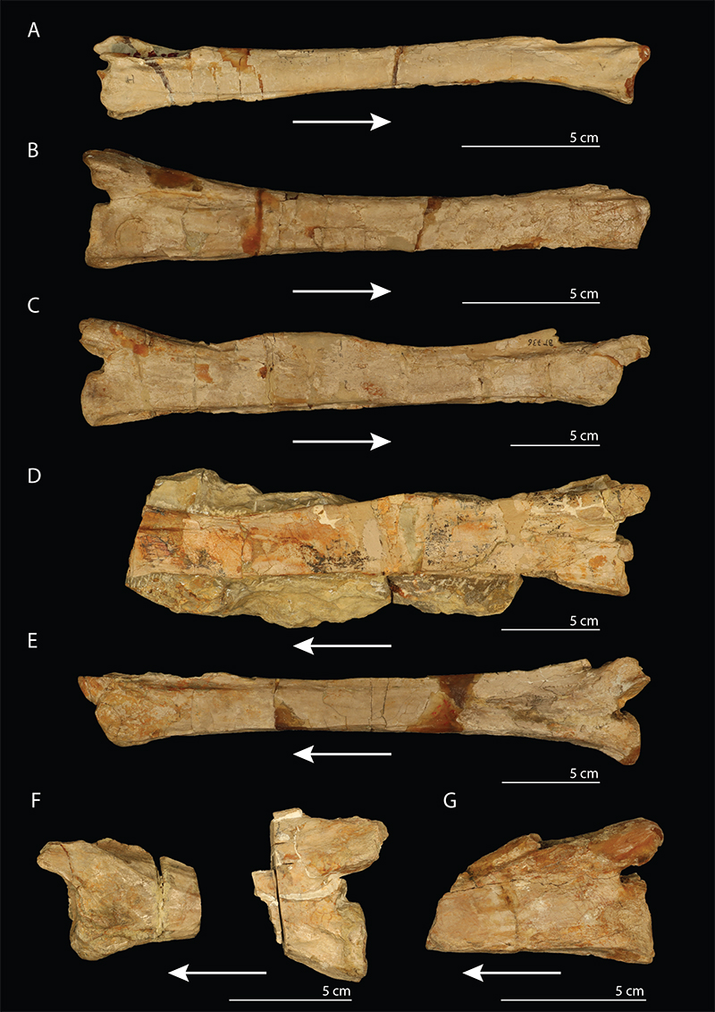
FIGURE 7. The cast of the holotype of Tanystropheus haasi (PIMUZ A III 726, cast of HUJ-Pal. TR 1), the posterior part of a mid-cervical vertebra; in (A) left lateral; (B) right lateral; (C) dorsal; (D) ventral; and (E) posterior view. The arrows are pointing anteriorly.
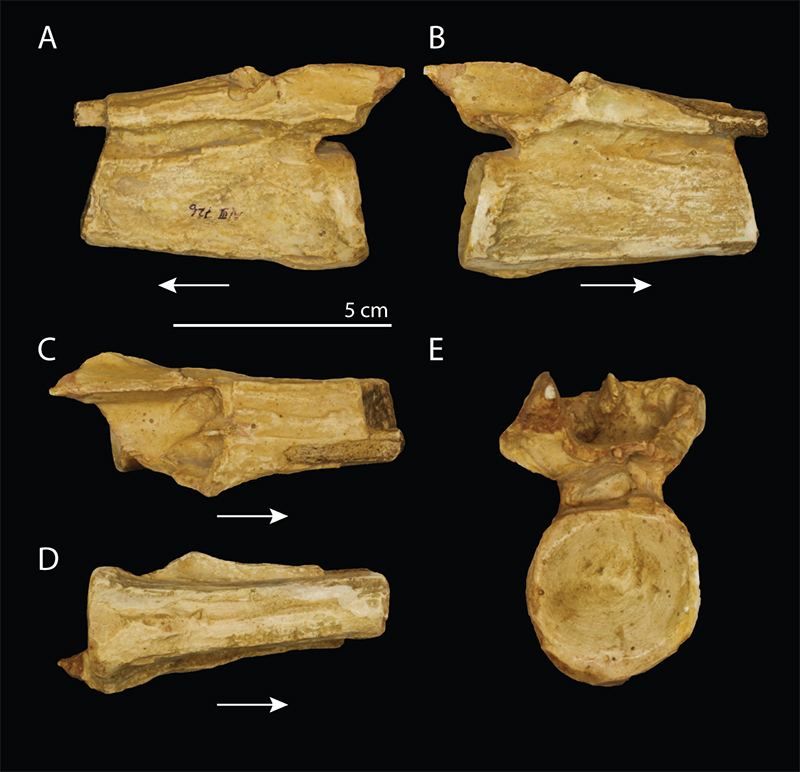
FIGURE 8. MCSNB 4035, the holotype and only known specimen of Sclerostropheus fossai; (A) the complete specimen; (B) a close up of a single vertebra including its corresponding cervical rib. The black arrows indicate the anterior direction and the white arrow indicates the apparent bifurcation of the cervical rib. Abbreviations: apr, anterior process of the cervical rib; la, lamina; ot, ossified tendons.
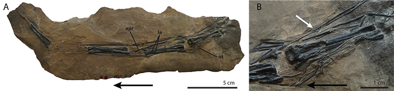
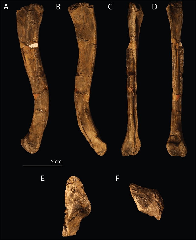
FIGURE 9. The isolated right femur, PIMUZ A/III 771, from the S-charl Formation of Piz Ravigliel, referred to Tanystropheus sp. in (A) lateral; (B) medial; (C) anterior or dorsal; (D) posterior or ventral; (E) proximal; and (F) distal view.