FIGURE 1. Photo of specimen LF 2809, a new large ctenochasmatid pterosaur. Scale bar is 10 cm.
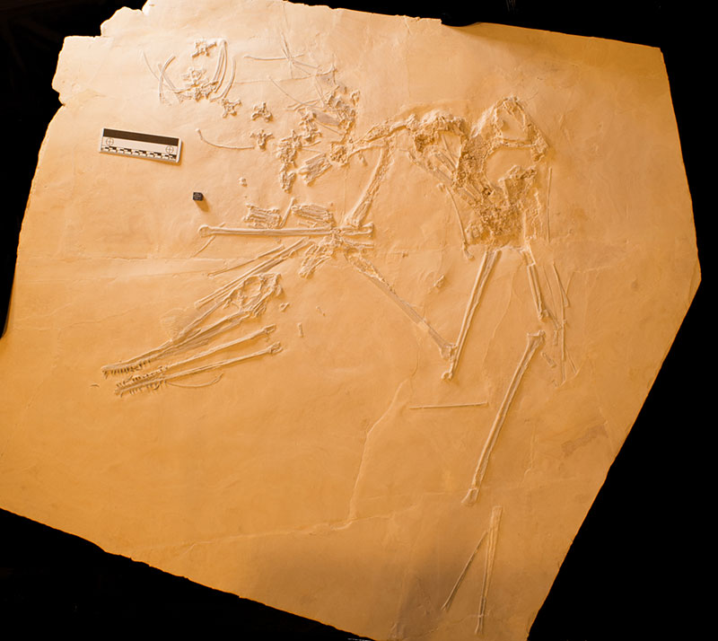
FIGURE 2. Photo of specimen LF 2809, a new large ctenochasmatid pterosaur under a mixture of UV A, B and C lighting. Scale bar is 10 cm.
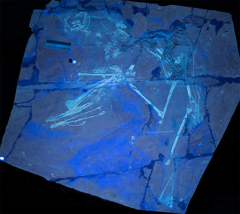
FIGURE 3. Tracing of specimen with labels. Abbreviations for this and subsequent figures are as follows: ax, axis; ca, carpal; cv, cervical vertebra; dr, dorsal ribs; dv, dorsal vertebra; fe, femur; fi, fibula; g, gastralia; hu, humerus; il, ilium; mc1-3, metacarpals 1-3; mph, manual phalanges; mn, mandible; mt I-V, metatarsals; ppb, prepubis; pph, pedal phalanges; pt, pteroid; r-u; radius and ulna; sc, scapulocoracoid; sk skull; sv, sacral vertebrae; ta, tarsal; ti, tibia; u, ungual; wmc, wing metacarpal; wpx 1-4, wing phalanges. Scale bar is 100 mm.
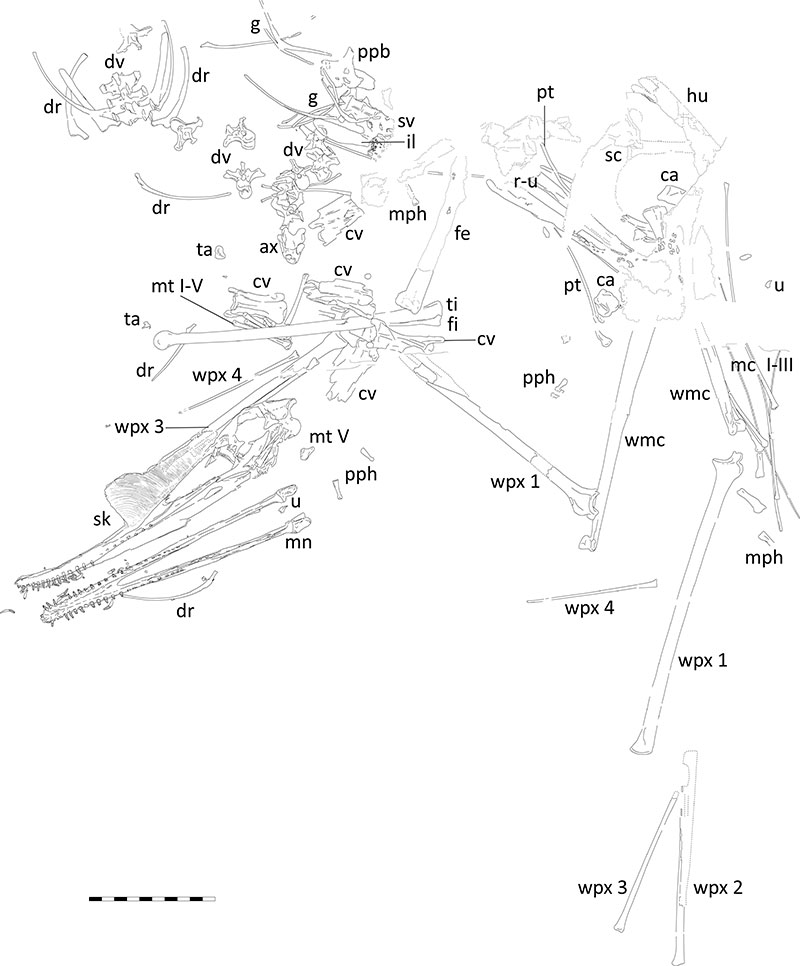
FIGURE 4. Close up of skull of Petrodactyle in A, natural light and B, UV light (mixture of UV A, B and C). Scale bar is 10 cm.
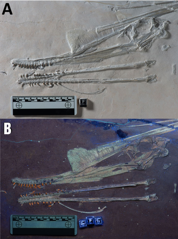
FIGURE 5. Tracing of the skull of Petrodactyle. Many sutures are uncertain and these labels are for general guidance. Abbreviations are as follows: an, angular; ar, articular; fpc, frontoparietal crest; j, jugal; lc, lacrimal; m, maxilla; n, nasal; naof, nasoantorbital fenestra; o, orbit; p, palate; po, postorbital; ppf, postpalatine fenestra; pm, premaxilla; pmc, premaxillary crest; q, quadrate; qj, quadratojugal; sa, surangular; t, tooth. Scale bar is 100 mm.
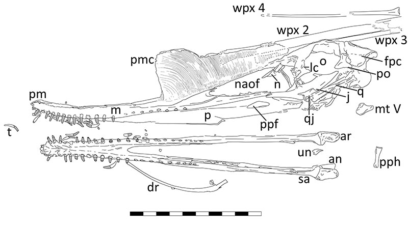
FIGURE 6. Close ups of key parts of the skull of Petrodactyle. A, detail of the upwards curved rostrum and anterior dentition under natural light (scale bar with 1 mm division). B, detail of the premaxillary crest under natural light showing the striations, the differing texture on the anteriodorsal part of this (scale bar in cm with mm divisions). C, photo of the notch at the anterior base of the crest (arrow), posterior alveoli and part of the palate under UV light showing (scale bar is 10 mm).
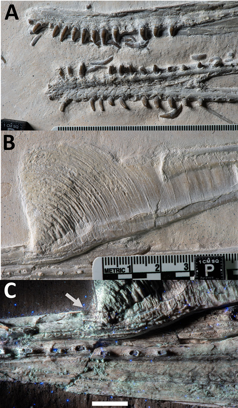
FIGURE 7. Close up of middle cervical vertebrae of Petrodactyle. A, in left lateral view, and B, in ventral view. Scale bar is 10 mm.
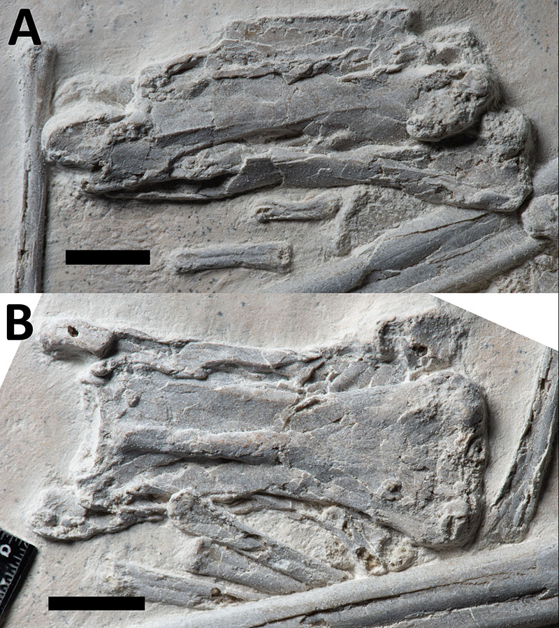
FIGURE 8. Close up of dorsal vertebrae of Petrodactyle. A, single mid dorsal seen in posterioventral view (scale bar is 10 mm), and B, anterior dorsal vertebral series seen in dorsal view with articulated anterior dorsal ribs, and two disarticulated dorsal vertebrae seen in anterior view (scale bar with 1 mm divisions).
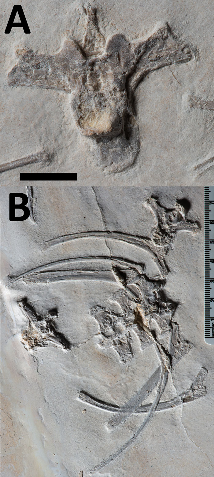
FIGURE 9. Close up of the pelvic region of Petrodactyle in A, natural light and B, a mixture of UV AB and C light. Abbreviations are as in Figure 3. Scale bar for both is 20 mm.
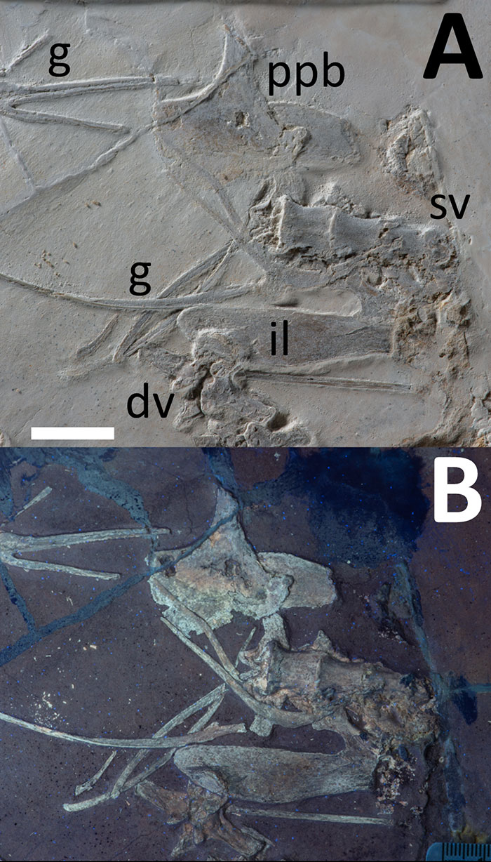
FIGURE 10. Close up of the poorly preserved area of the Petrodactyle holotype that contains the pectoral girdles, seen under UV C light to highlight the bone fragments. Abbreviations as in Figure 3. Scale bar is 50 mm.
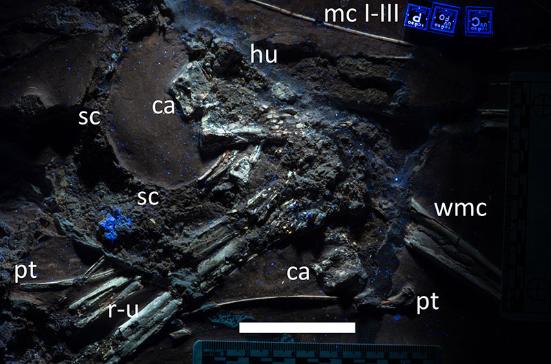
FIGURE 11. Close up of manual elements of Petrodactyle. In a mixture of UV A, B and C light. Abbreviations as in Figure 3. Scale bar is 100 mm.
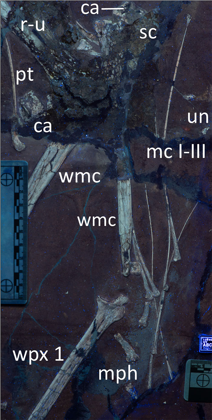
FIGURE 12. ‘Nopsca chart’ showing major proportions of the skeleton comparing Petrodactyle to other large Solnhofen region ctenochasmatids (data in Table 1). Petrodactyle shows a similar pattern to other animals, though with a proportionally larger head and a long tibia.
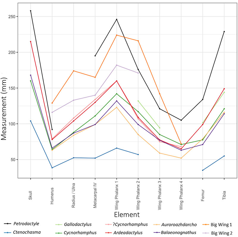
FIGURE 13. Comparison drawing of restored skulls of assorted Solnhofen ctenochasmatids (not to scale). A, Petrodactyle; B, Cycnorhamphus (following Bennett, 2013); C, Pterodactylus (following Wellnhofer, 1970), D, Ardeadactylus; E, Germanodactylus (following Unwin, 2003).
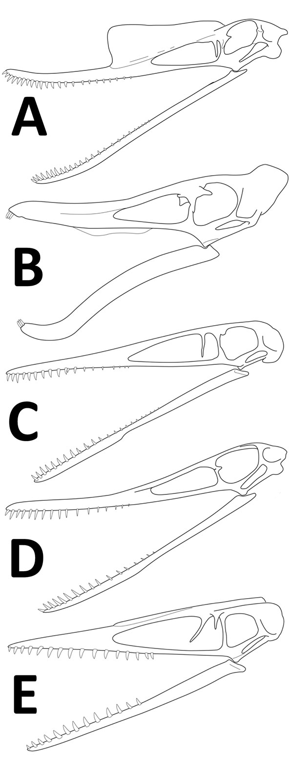
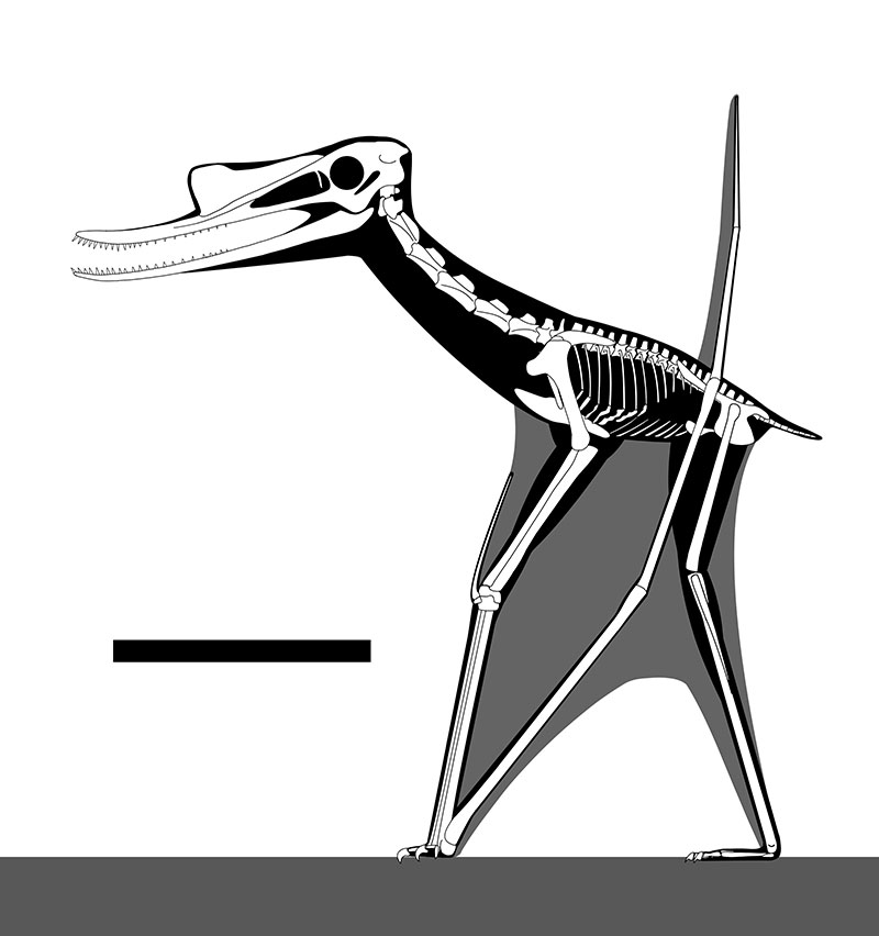
FIGURE 14. Reconstruction of the complete skeleton of Petrodactyle wellnhoferi in a standing pose. Scale bar is 200 mm. Missing parts based on Cycnorhamphus following Witton (2013).