FIGURE 1. Thalamoporella sp. 1; PMC EDM-Collection J.H.B.119a. A-B, sample 19206. A, fragment general view (500 μm). B, close-up of autozooids (200 μm). C-E, sample 19084. C, fragment general view (500 μm). D, close-up of an opesia with preserved adoral tubercle (100 μm). E, close-up of autozooids and putative vicarious avicularium (200 μm).
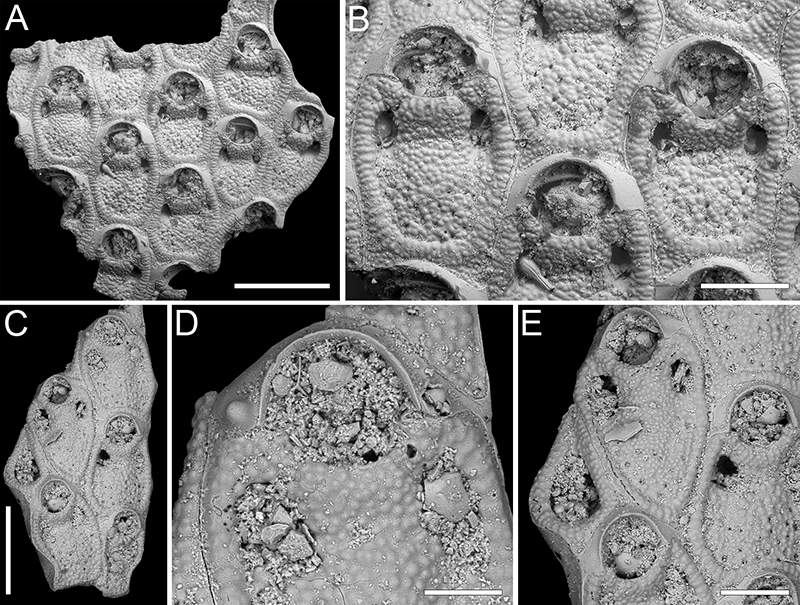
FIGURE 2. Steginoporella sp. 1; PMC EDM-Collection J.H.B.120a, sample 19210. A, group of zooids (1 mm). B-C, close-up of A- and B-zooids (500 μm). D, close-up of the median process (250 μm).
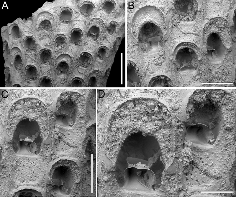
FIGURE 3. Steginoporella sp. 2; PMC EDM-Collection J.H.B.121a, sample 19210. A, E, portion of two cylindrical branch fragments showing groups of zooids (1 mm). B, close-up of two A-zooids (500 μm). C, close-up of a B-zooid (500 μm). D, F, close-up of polypide tubes in two A-zooids (200 μm).
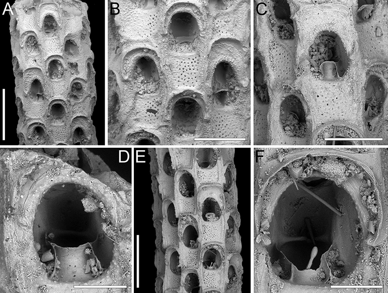
FIGURE 4. Copidozoum rhoae Yang, Seo and Gordon, 2018; PMC EDM-Collection J.H.B.122a. A-B, sample 19033. A, colony fragment general view (200 μm). B, close-up of two ovicellate zooids and subvicarious avicularium (200 μm). C, sample 19017, close-up of two autozooids (200 μm). D, sample 19021, close-up of two ovicellate zooids and subvicarious avicularium (200 μm).
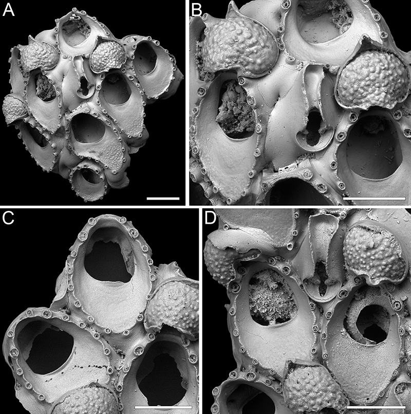
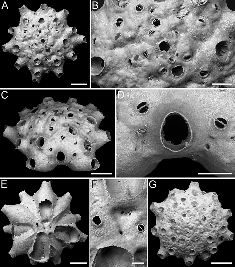
FIGURE 5. Ammatophoroides angeloi gen. et sp. nov. Di Martino, Rosso and Taylor. A-B, holotype PMC. B44. 29.7.2024a, sample 19027. A, general view of the colony fragment (1 mm). B, close-up of three zooids, two ovicellate (500 μm). C-D, paratype PMC. B44. 29.7.2024b1, sample 19016. C, general view of the colony fragment (500 μm). D, close-up of three zooids, one with signs of regeneration and one ovicellate (250 μm). E-F, paratype PMC. B44. 29.7.2024b2, sample 19002. E, general view of the colony fragment (200 μm). F, close-up of an ovicellate zooid (200 μm). G, paratype PMC. B44. 29.7.2024b3, sample 19018, close-up of lateral walls with uniporous septula (200 μm).
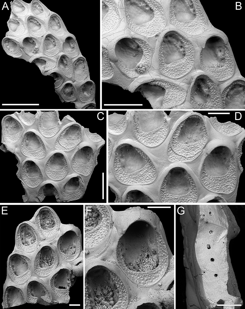
FIGURE 6. Antropora typica (Canu and Bassler, 1928); PMC EDM-Collection J.H.B.123a. A-C, sample 19072. A, colony fragment general view (200 μm). B, close-up of two autozooids, one with multiple opesial rims owing to intramural budding, and several interzooidal avicularia (200 μm). C, close-up of autozooids and interzooidal avicularia (200 μm). D-E, sample 19230, close-up of two ovicellate zooids and interzooidal avicularia (150 μm).
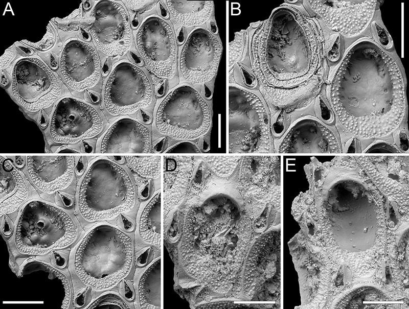
FIGURE 7. Chaperia robusta sp. nov. Di Martino, Rosso and Taylor. A-D, holotype PMC. B45. 29.7.2024a, sample 19081. A, colony general view (1 mm). B, close-up of ancestrula, first two budded autozooids, and an additional subsequent one, all with closure plates (500 μm). C, close-up of four autozooids (500 μm). D, close-up of an autozooid showing 13 spines and the thick lateral walls with pore-chamber windows (200 μm). Distal is to the left. E-F, paratype PMC. B45. 29.7.2024b, sample 19059. E, colony fragment general view (400 μm). F, close-up of two autozooids (200 μm).
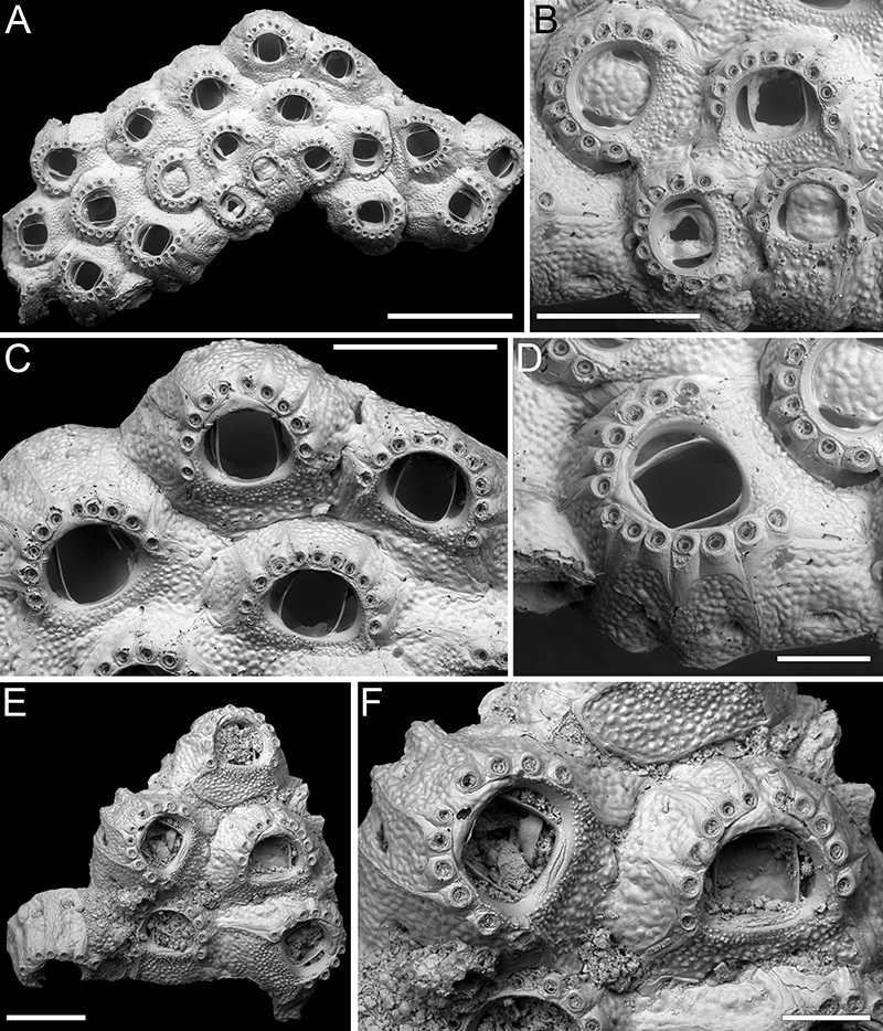
FIGURE 8. Foveolaria retiformis Harmer, 1926; PMC EDM-Collection J.H.B.124a. A-C, sample 19072. A, branch fragment general view (1 mm). B, group of autozooids with adventitious avicularia (500 μm). C, close-up of avicularium (100 μm). D-E, sample 19232, close-up of ovicells (D 500 μm, E 250 μm). F, sample 19227, branch dorsal side (500 μm).
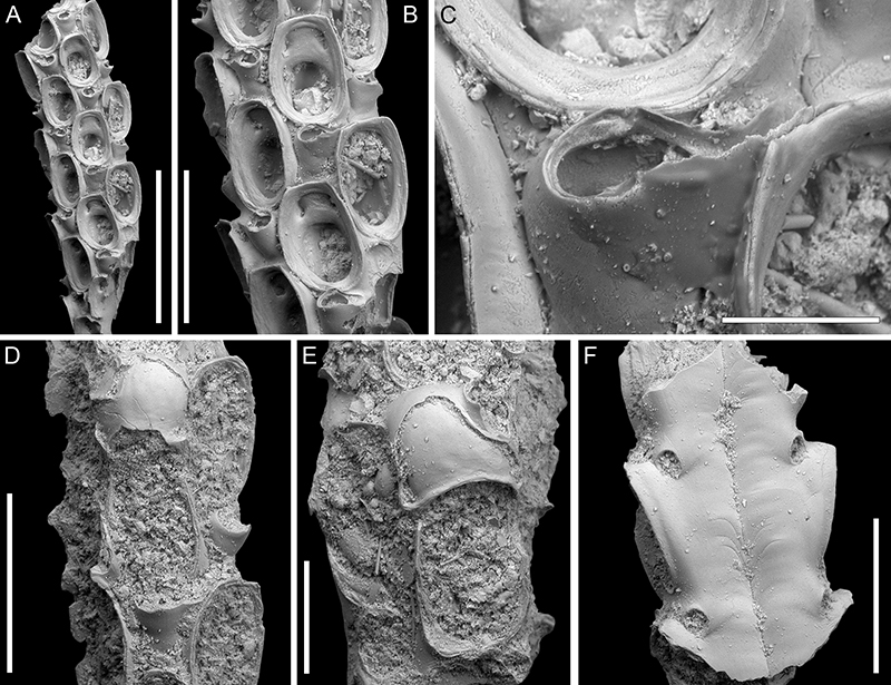
FIGURE 9. Amastigia okinawaensis sp. nov. Di Martino, Rosso and Taylor. A-B, E-F, holotype PMC. B46. 29.7.2024a, sample 19035. A, partial view of a branch fragment including several zooids (200 μm). B, close-up of autozooids and adventitious avicularia (100 μm). C-D, PMC. B46. 29.7.2024b, sample 19065, close-ups of ovicellate zooids (200 μm). E-F, dorsal view with vibracula (200 μm).
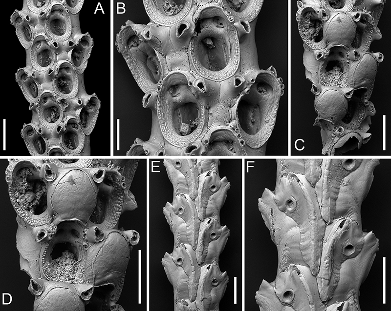
FIGURE 10. Canda foliifera Harmer, 1926; PMC EDM-Collection J.H.B.125a. A-C, F, sample 19071. A, lateral view of the branch fragment (500 μm). B, close-up of autozooids at branch bifurcation (200 μm). C, close-up of two autozooids and associated vibracula (200 μm). D-E, sample 19200. D, frontal view of the branch fragment (500 μm). E, close-up of an autozooid (300 μm). The arrow indicates the attachment base of the scutum. F, dorsal side with vibracula (200 μm).
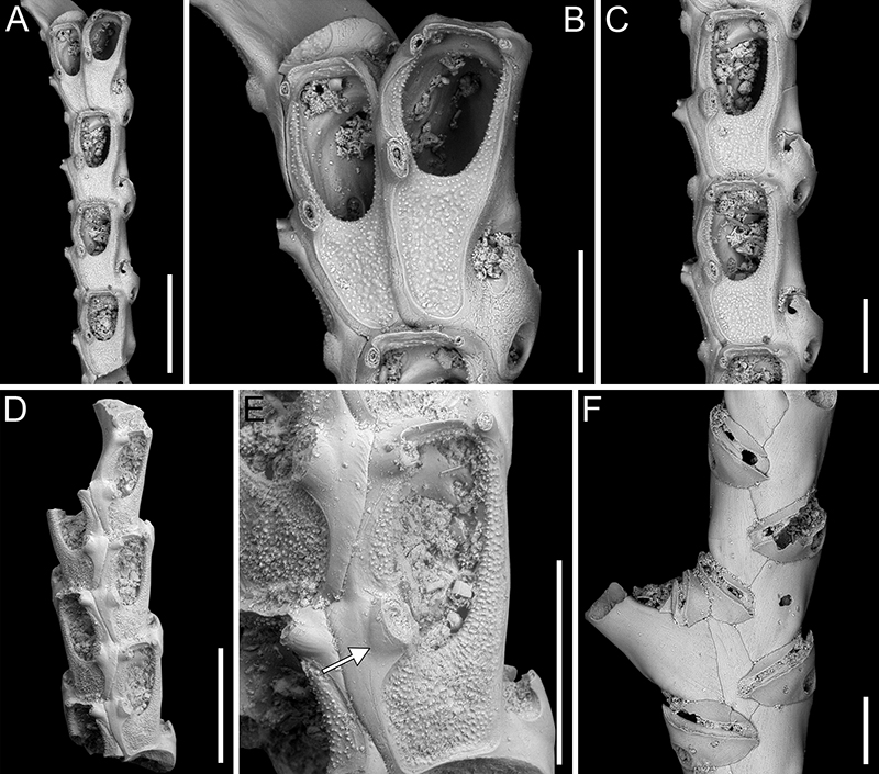
FIGURE 11. Canda scutata Harmer, 1926; PMC EDM-Collection J.H.B.126a. A-C, sample 19200. A, general view of a fertile branch fragment (1 mm). B, close-up of zooids, the one on top with signs of regeneration, those at the bottom ovicellate (250 μm). C, close-up of ovicellate zooids (250 μm). D, sample 19029, close-up of two autozooids and associated vibracula in frontal view (200 μm). The arrow indicates the attachment base of the scutum. E-F, sample 19227, dorsal side with vibracula (E 500 μm, F 250 μm).
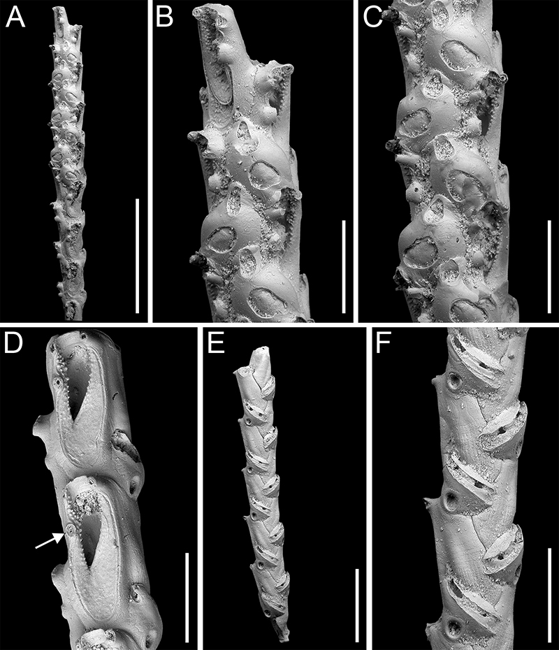
FIGURE 12. Canda lunata sp. nov. Di Martino, Rosso and Taylor. A-E, holotype PMC. B47. 29.7.2024a, sample 19029. A, lateral view of the branch fragment (200 μm). B, close-up of two autozooids, one with signs of regeneration (200 μm). C, close-up of an autozooid and interzooidal avicularium (100 μm). D-E, dorsal side with vibracula (D 200 μm, E 100 μm). F-G, paratype PMC. B47. 29.7.2024b, sample 19227. F, lateral view of a branch fragment with ovicellate zooids (500 μm). G, close-up of an ovicellate zooid (100 μm).
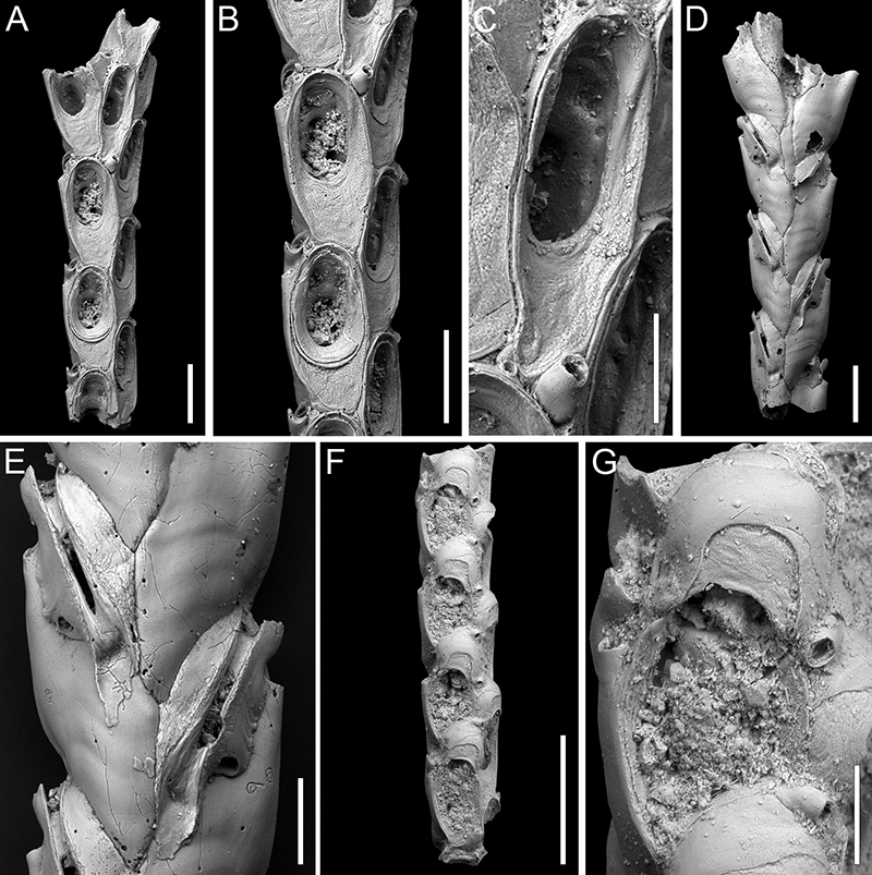
FIGURE 13. Pomocellaria spatulata sp. nov. Di Martino, Rosso and Taylor. Holotype PMC. B48. 29.7.2024a, sample 19032. A, general view of the internode (500 μm). B, close-up of the giant spatulate avicularia and the single frontal avicularium observed (200 μm). C, close-up of an autozooid (200 μm). D, close-up of two ovicellate zooids, one with preserved hooked rostrum of the lateral avicularium (200 μm). F, general view of the dorsal side (500 μm). G, close-up of vibracula (200 μm).
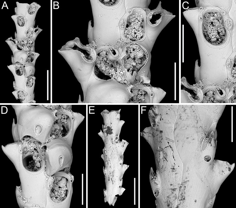
FIGURE 14. Scrupocaberea contraria sp. nov. Di Martino, Rosso and Taylor. A-B, F-G, paratype PMC. B49. 29.7.2024b1, sample 19065. A, general view of the internode (200 μm). B, close-up of autozooids (100 μm). C-E, holotype PMC. B49. 29.7.2024a, sample 19032. C, general view of the internode (500 μm). D, group of ovicellate zooids (200 μm). E, close-up of an ovicellate zooid with giant frontal avicularium (200 μm). F-G, dorsal side with vibracula (F 400 μm, G 200 μm).
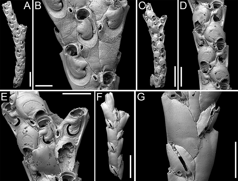
FIGURE 15. Scrupocaberea maderensis (Busk, 1860); PMC EDM-Collection J.H.B.127a. A-C, sample 19019. A, general view of an internode (600 μm). B, close-up of three autozooids (100 μm). C, close-up of an autozooid, with frontal avicularium, at branch bifurcation (100 μm). D-E, sample 19016. D, group of ovicellate zooids (200 μm). E, close-up of an ovicellate zooid with denticulated distal margin of opesia (100 μm). F-G, sample 19147. F, close-up of opesia and lateral avicularium (100 μm). G, close-up of lateral avicularium (50 μm). H-I, sample 19019, dorsal side with vibracula (H 1 mm, I 200 μm).
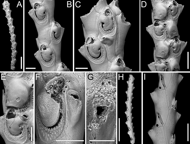
FIGURE 16. Poricellaria ratoniensis (Waters, 1887); PMC EDM-Collection J.H.B.128a. A-B, sample 19085. A, branch fragment general view (500 μm). B, close-up of an autozooid with adventitious avicularium in lateral view (100 μm). C, sample 19048, close-up of an autozooid with adventitious avicularium in frontal view (100 μm). D, sample 19085, close-up of an autozooid showing the slit-like opesiule (100 μm).
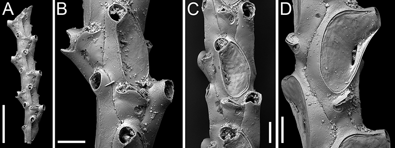
FIGURE 17. Cellaria levigata sp. nov. Di Martino, Rosso and Taylor. A-C, holotype PMC. B50. 29.7.2024a, sample 19200. A, inflated fertile internode general view (1 mm). B, close-up of the inflated portion of the internode with fertile zooids (150 μm). C, close-up of autozooids (200 μm). D-F, paratype PMC. B50. 29.7.2024b1, sample 19227. D, inflated fertile internode general view (500 μm). E, close-up of an autozooid (100 μm). F, close-up of a maternal zooid (100 μm). The arrows indicate the two U-shaped indentations of the endotoichal ovicell. G-J, paratype PMC. B50. 29.7.2024b2, sample 19230. G, inflated fertile internode with vicarious avicularia (500 μm). H, close-up of the inflated fertile portion of the internode (200 μm). I, close-up of an autozooid and an avicularium (100 μm). J, close-up of two maternal zooids (100 μm).
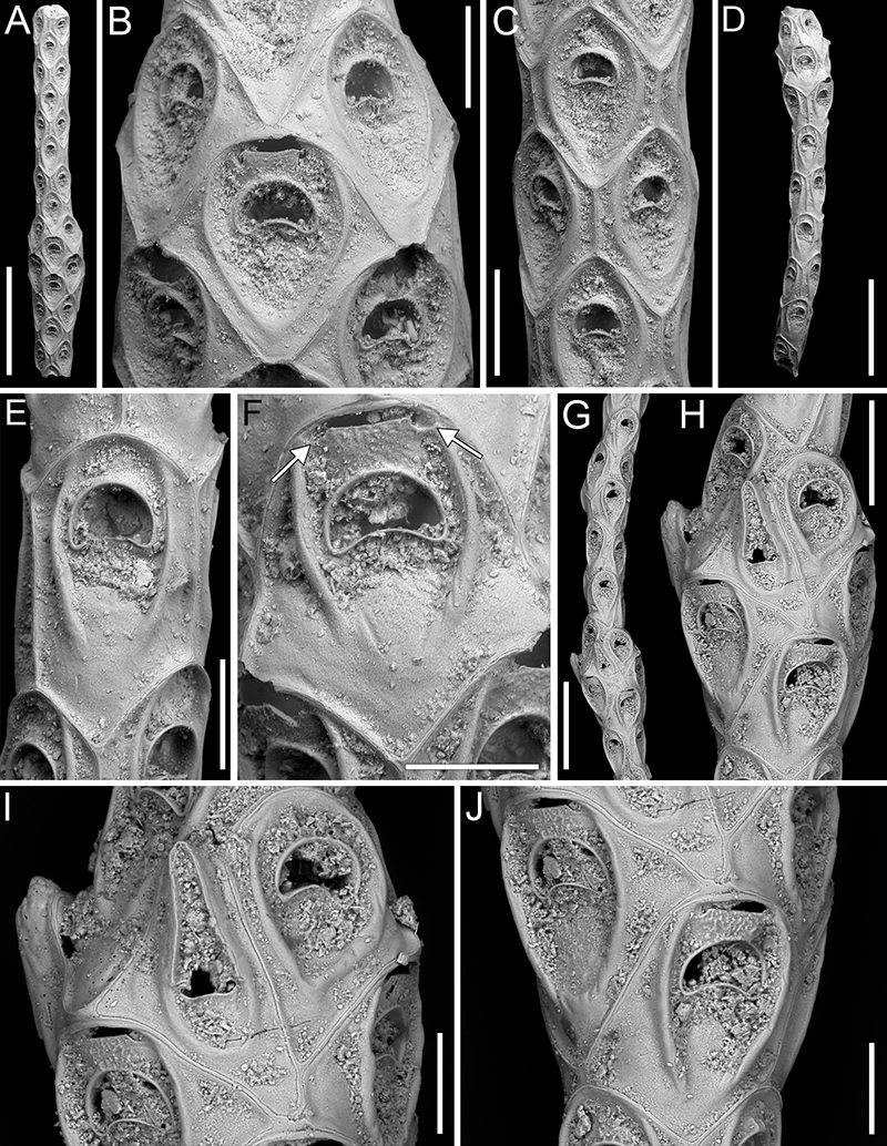
FIGURE 18. Cribrilaria harmeri Ristedt, 1985; PMC EDM-Collection J.H.B.129a, A-B, sample 19069. A, colony fragment general view (500 μm). B, close-up of autozooids and interzooidal avicularia (200 μm). C-E, sample 19019. C, close-up of an autozooid and an ovicellate zooid (ooecium type B produced by the distal kenozooid) with adventitious avicularia (200 μm). D, close-up of an autozooid and an ovicellate zooid (ooecium type A produced by the distal autozooid) with adventitious avicularia (100 μm). E, group of autozooids and a kenozooid (200 μm). F, sample 19072, close-up of a kenozooidal ooecium, paired adventitious avicularia, and an interzooidal avicularium (100 μm). G-I, sample 19113. G, colony fragment general view (500 μm). H, group of zooids, some ovicellate, with ooecia formed either by distal kenozooids or distal autozooids, and paired adventitious avicularia, kenozooids and interzooidal avicularia (200 μm). I, close-up of an ooecium formed by the distal autozooid and adventitious avicularia budded from the lateral gymnocyst (100 μm).
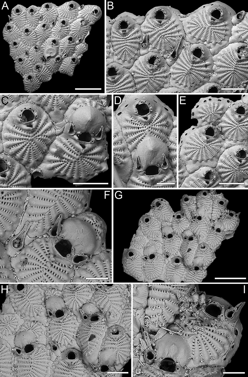
FIGURE 19. Glabrilaria biavicularia (Kataoka, 1961); PMC EDM-Collection J.H.B.130a, sample 19025. A, colony fragment general view (200 μm). B, close-up of autozooids with avicularia and a kenozooid (100 μm).
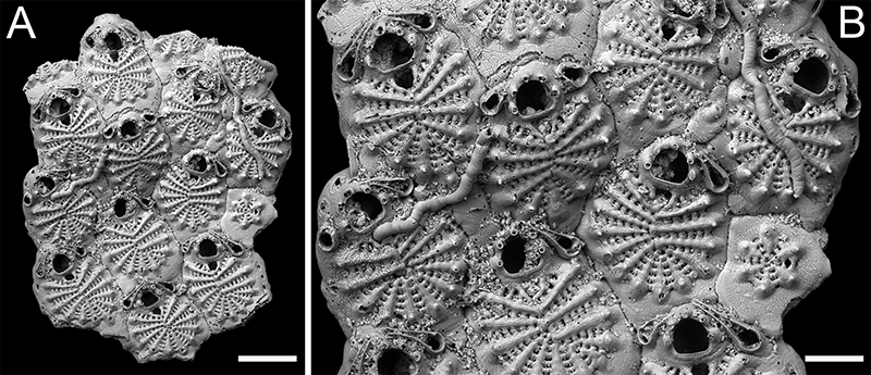
FIGURE 20. Glabrilaria ossuosa sp. nov. Di Martino, Rosso and Taylor. A-D, holotype PMC. B51. 29.7.2024a, sample 19071. A, colony fragment general view, with arrows indicating two kenozooids (400 μm). B, group of zooids, mostly ovicellate (200 μm). C, group of autozooids (200 μm). D, close-up of an orifice with closure plate (60 μm). E-F, PMC. B51. 29.7.2024b, sample 19053. E, close-up of zooids with semi-erect, latero-oral avicularia showing the needle-like rostrum (200 μm). F, close-up of autozooids at colony edge with visible pore-chamber windows. The zooid on the left, with dimorphic orifice and only four spines, is likely to be forming an ovicell (100 μm).
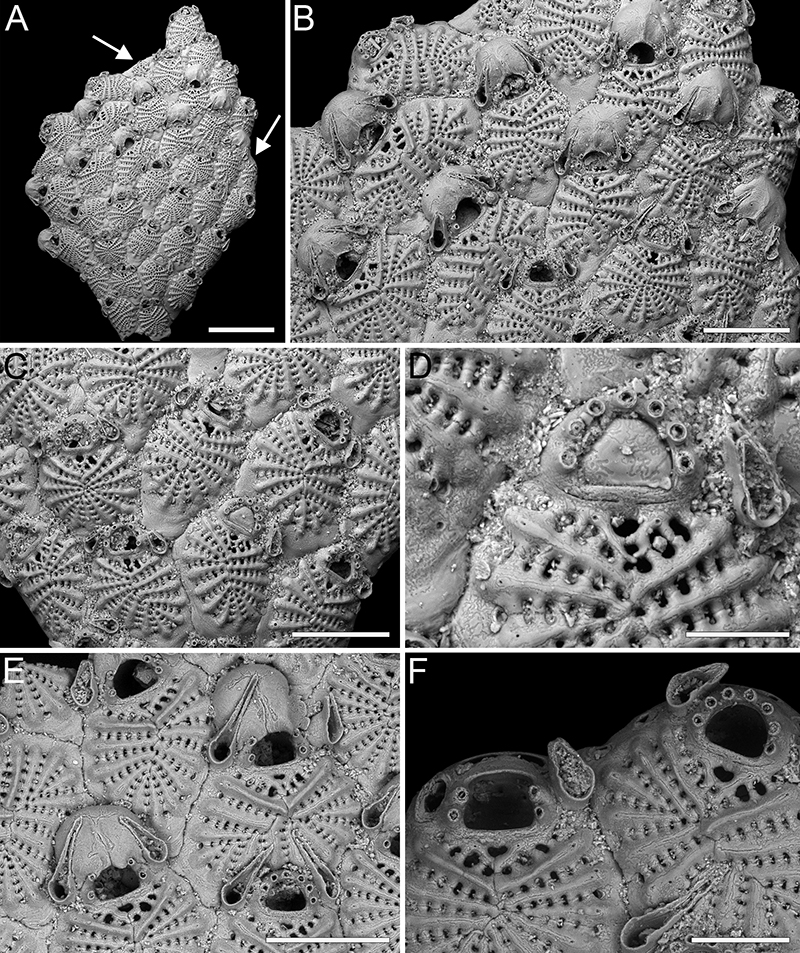
FIGURE 21. Auricellina biserialis gen. et sp. nov. Di Martino, Rosso and Taylor, holotype PMC. B52. 29.7.2024a, sample 19132. A, colony fragment general view (200 μm). B, close-up of orifice showing median suture and drop-shaped cryptocystal pore area flanking it (100 μm). C, close-up of an autozooid and an ovicell (100 μm). D, close-up of an ovicell with slit-like, arched fenestra and quadrangular ooecial opening (100 μm). E, close-up of an ovicell with two separate fenestrae, one small and circular, the other slit-like and obliquely placed, and longitudinal median carena (100 μm). F, dorsal side general view (200 μm). G, close-up of the dorsal side showing the general placement of pore areas (100 μm). H-I, close-up of the cryptocystal pore areas (50 μm).
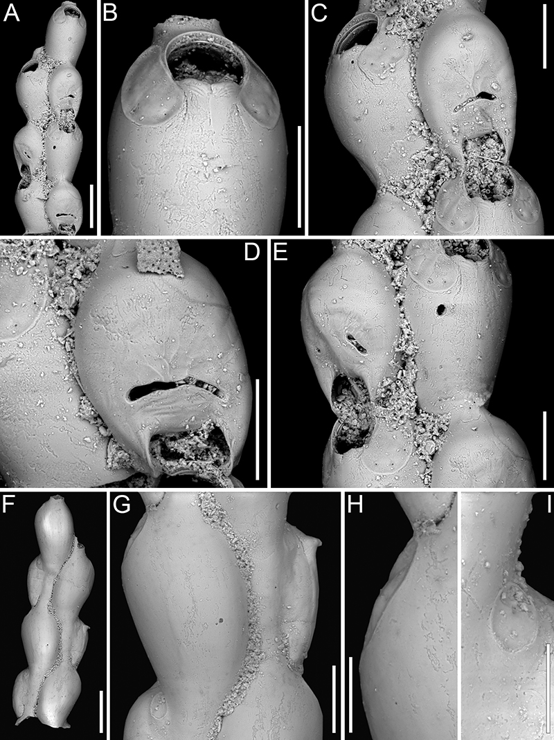
FIGURE 22. Exechonella coronida sp. nov. Di Martino, Rosso and Taylor. A-C, holotype PMC. B53. 29.7.2024a, sample 19010. A, colony fragment general view with self-overgrowth (1 mm). B, group of autozooids, some with single or paired curved avicularia (500 μm). C, close-up of autozooids with avicularia (200 μm). D, paratype PMC. B53. 29.7.2024b1, sample 19007, close-up of autozooids with preserved collar-like peristome and avicularia (400 μm).
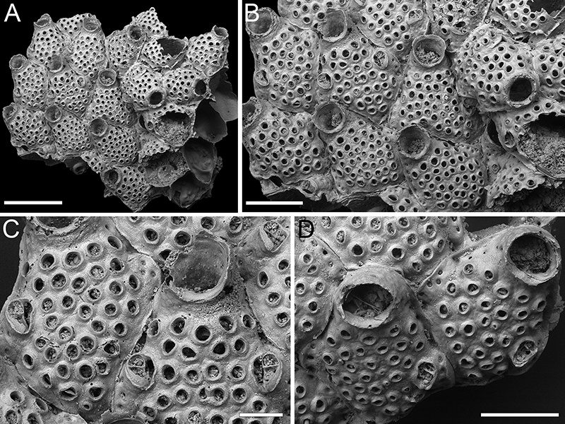
FIGURE 23. Exechonella juliae sp. nov. Di Martino, Rosso and Taylor. A-B, paratype PMC. B54. 29.7.2024b1, sample 19059. A, colony fragment general view (1 mm). B, close-up of autozooid (500 μm). C, paratype PMC. B54. 29.7.2024b2, sample 19015, close-up of an autozooid with a frontal projection (500 μm). D-E, holotype PMC. B54. 29.7.2024a, sample 19014. D, close-up of an autozooid with frontal projection and view of peristome internal surface (200 μm). E, close-up of the primary orifice (100 μm); F, paratype PMC. B54. 29.7.2024b3, sample 19042, group of zooids showing the flared, jagged rim of the tubular peristomes (500 μm).
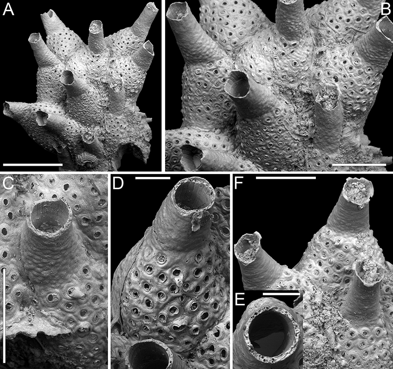
FIGURE 24. Exechonella verrucosa (Canu and Bassler, 1927); PMC EDM-Collection J.H.B.131a. A-D, sample 19074. A, tilted lateral view of colony fragment (400 μm) exposing communication pores. B, frontal view of the same fragment (500 μm). Arrows indicate the nipple-like structure typical of the avicularia of this species. C, close-up of fused foramina with frontal projection and nipple-like structure of an avicularium (100 μm). D, close-up of the primary orifice (150 μm). E-F, sample 19125. E, colony fragment general view (500 μm). F, close-up of a preserved peristome with spiny processes, the lateral one bifurcated (200 μm).
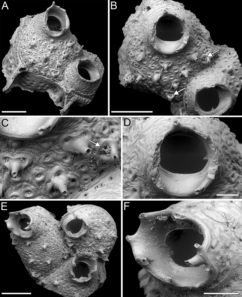
FIGURE 25. Crepidacantha longiseta Canu and Bassler, 1928; PMC EDM-Collection J.H.B.143a, sample 19119. A, colony general view (200 μm). B, D, close-up of three autozooids (200 μm). C, close-up of the distal margin of an autozooid with arrow indicating a spine base (100 μm).
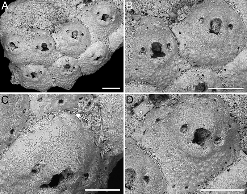
FIGURE 26. Escharoides cavernicolus sp. nov. Di Martino, Rosso and Taylor. A-D, holotype PMC. B55. 29.7.2024a, sample 19132. A, colony fragment general view (1 mm). B, close-up of an ovicellate zooid with avicularian configuration type (ii) (500 μm). C, close-up of an autozooid with avicularian configuration type (iv) (500 μm). D, close-up of two autozooids with avicularian configurations type (i) and (iii) (500 μm). E-F, paratype PMC. B55. 29.7.2024b1, sample 19105. E, colony fragment general view (500 μm); F, close-up of ancestrula (300 μm). G, paratype PMC. B55. 29.7.2024b2, sample 19125, view of the dorsal side showing conical pillars (1 mm). H, paratype PMC. B55. 29.7.2024b3, sample 19094, close-up of primary orifice showing the extension of the median denticle inside the peristome (200 μm).
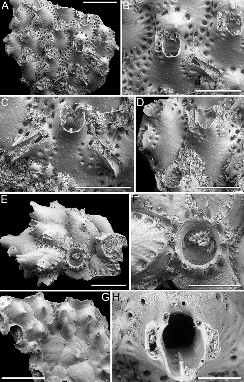
FIGURE 27. Hippopleurifera hirosei sp. nov. Di Martino, Rosso and Taylor. A-D, holotype PMC. B56. 29.7.2024a, sample 19143. A, colony fragment general view (1 mm). B, close-up of orifice with six spine bases and lateral, spoon-shaped avicularium (200 μm). C, intramurally budded autozooid with reverse polarity and frontal, pear-shaped avicularium (400 μm). D, close-up of two autozooids with eight and nine oral spine bases (400 μm). E, paratype PMC. B56. 29.7.2024b1, sample 19227, colony fragment consisting of two autozooids one with broken ovicell (500 μm); F, paratype PMC. B56. 29.7.2024b2, sample 19135, close-up of an autozooid distal half, showing hooked, downwardly directed condyles and two frontal, spoon-shaped avicularia, one with broken rostrum (300 μm). G, paratype PMC. B56. 29.7.2024b3, sample 19217, two autozooids with single frontal avicularium and frontal shield with narrow imperforate central area (1 mm).
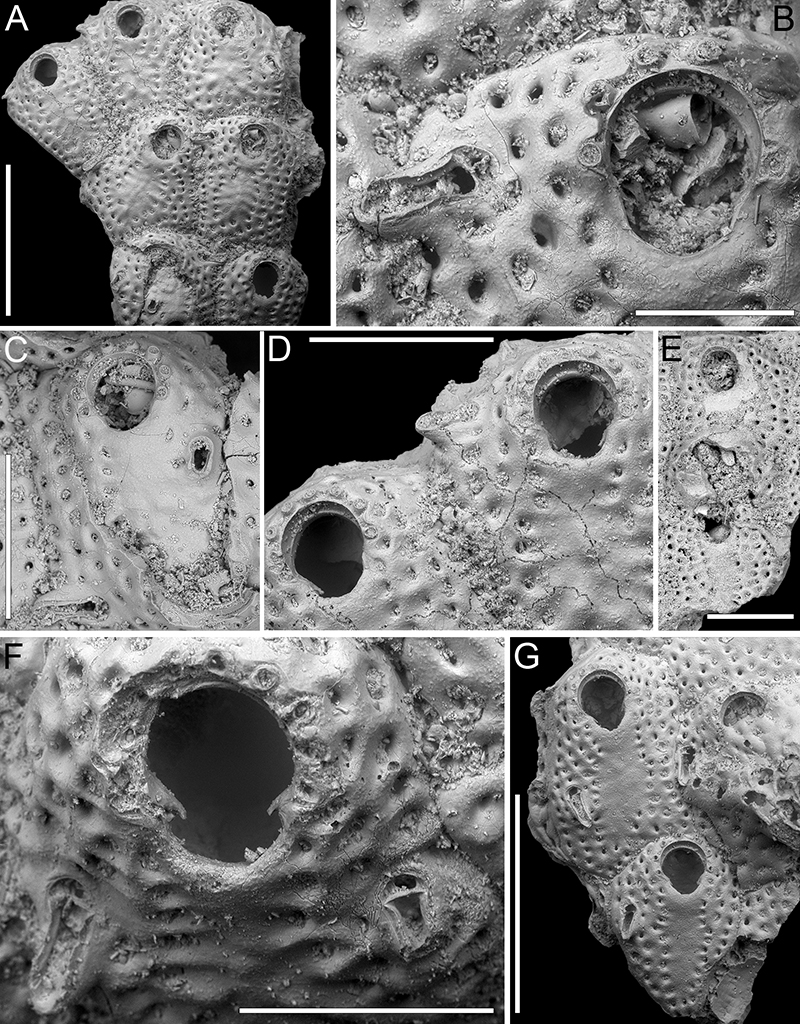
FIGURE 28. Hemismittoidea dicki sp. nov. Di Martino, Rosso and Taylor. A-C, holotype PMC. B57. 29.7.2024a, sample 19061. A, colony fragment general view (400 μm). B, close-up of three zooids, one ovicellate (200 μm). C-D, close-up of ovicell and avicularium (200 μm). Note the sharp constriction of the secondary orifice. D, paratype PMC. B57. 29.7.2024b, sample 19210, group of autozooids, some with visible lyrula (500 μm). Arrows in A and D indicate the putative spine bases.
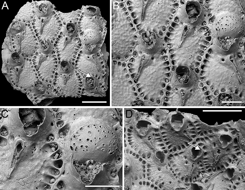
FIGURE 29. Smittoidea ligata sp. nov. Di Martino, Rosso and Taylor. A-D, holotype PMC. B58. 29.7.2024a, sample 19052. A, colony fragment general view (400 μm). B-C, close-up of ovicellate zooids with faint ligula preserved in the suboral avicularium crossbar (200 μm). D, close-up of broken orifice showing one of the slightly downwardly curved tips of the lyrula and one acute condyle, and the suture linking the peristomial pseudosinus to the avicularium (50 μm). E-F, paratype PMC. B58. 29.7.2024b1, sample 19185. E, colony fragment general view showing the frontally curved peristomes (500 μm). F, close-up of an ovicell with partially exposed endooecium (150 μm). G-H, paratype PMC. B58. 29.7.2024b2, sample 19210. G, pseudoancestrula and surrounding autozooids (500 μm). H, close-up of pseudoancestrula (100 μm). I-K, paratype PMC. B58. 29.7.2024b3, sample 19044. I, colony fragment general view (400 μm). J, close-up of pseudoancestrula (50 μm). K, close-up of orifice with eight distal spines and suboral avicularium (50 μm).
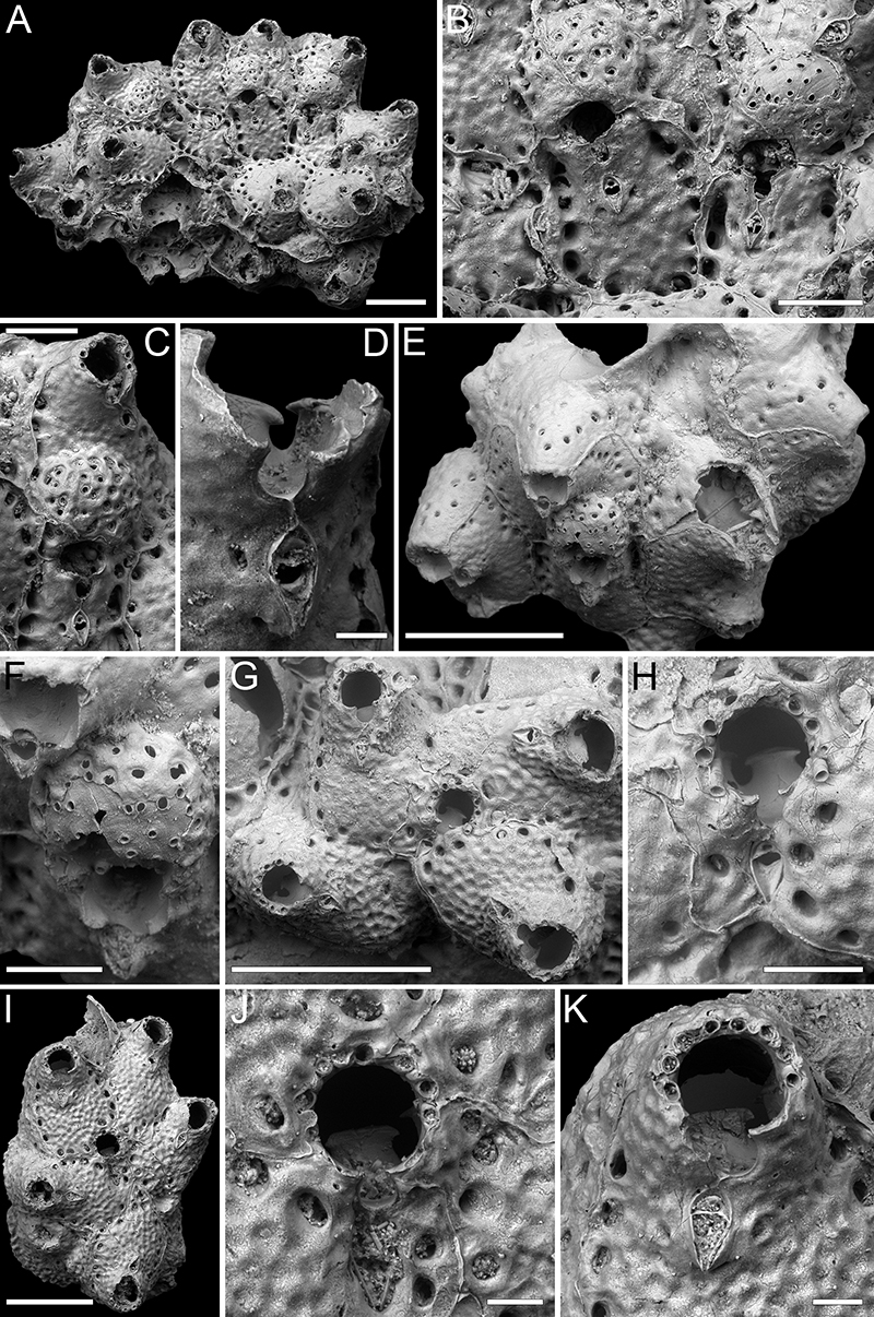
FIGURE 30. Smittoidea notoensis Hayami, 1975; PMC EDM-Collection J.H.B.132a, sample 19072. A, colony fragment general view (500 μm). B, close-up of ovicellate zooids with ligula preserved in the suboral avicularium crossbar (200 μm). C, close-up of orifice with distal spines (50 μm).
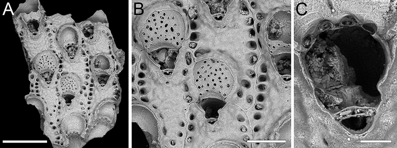
FIGURE 31. Parasmittina cf. delicatula (Busk, 1884); PMC EDM-Collection J.H.B.133a, sample 19210. A, general view of the colony (500 μm). B, close-up of the distal half of the autozooid with orifice and single, median spine and paired dimorphic avicularia (100 μm). C, close-up of autozooid with large spatulate avicularium (150 μm). D, close-up of orifice (100 μm).
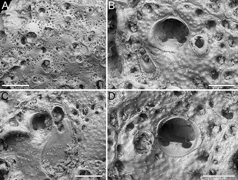
FIGURE 32. Parasmittina ligulata sp. nov. Di Martino, Rosso and Taylor, holotype PMC. B59. 29.7.2024a, sample 19178. A, general view of the colony (1 mm). B-C, groups of zooids, some ovicellate (500 μm). D, close-up of an ovicell, lyrula and avicularia (150 μm).
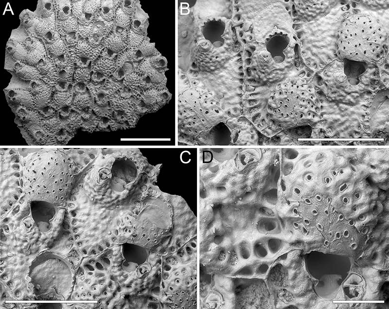
FIGURE 33. Metroperiella montferrandi (Audouin, 1826); PMC EDM-Collection J.H.B.134a, sample 19227. A, general view of the colony fragment (1 mm). B, close-up of two autozooids, one ovicellate (500 μm). C, close-up of orifice and avicularium with broken crossbar (200 μm). D, close-up of avicularium with preserved crossbar (100 μm).
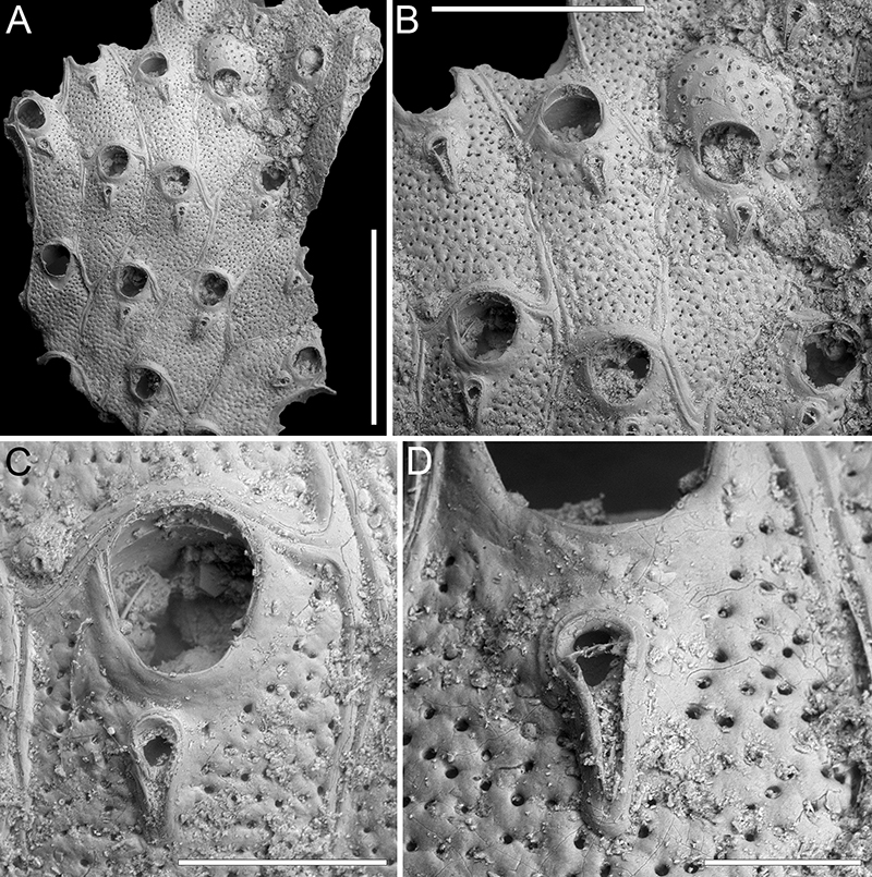
FIGURE 34. Parkermavella daidokutsuensis sp. nov. Di Martino, Rosso and Taylor. A-C, holotype PMC. B60. 29.7.2024a, sample 19113. A, general view of the colony fragment (200 μm). B, close-up of autozooids, mostly ovicellate (200 μm). C, close-up of orifice, ovicell and latero-oral avicularia, one with preserved crossbar and ligula (100 μm). D-G, paratype PMC. B60. 29.7.2024b1, sample 19135. D, general view of the colony fragment (500 μm). E, close-up of ovicell and suboral avicularium (100 μm). F, group of autozooids (200 μm). G, close-up of orifice showing spines and lateral avicularia on raised cystid (100 μm). H-J, paratype PMC. B60. 29.7.2024b2, sample 19160. H, general view of the colony fragment (200 μm). I-J, close-ups of ovicells and paired or single avicularia (100 μm).
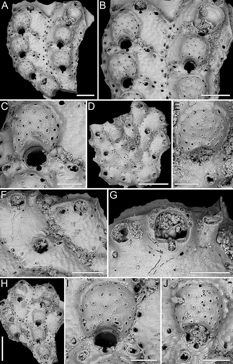
FIGURE 35. ?Bitectiporidae sp. ind.; PMC EDM-Collection J.H.B.135a. A-B, sample 19229. A, general view of the colony fragment (1 mm). B, close-up of autozooids (500 μm). C, sample 19231, close-up of orifice (100 μm).
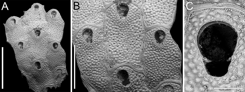
FIGURE 36. Tetraplaria stellifera sp. nov. Di Martino, Rosso and Taylor. A-C, holotype PMC. B61. 29.7.2024a, sample 19044. A, general view of the internode (200 μm). B, close-up of ovicell (100 μm). C, close-up of frontal and ooecial pseudopores (60 μm). D-G, paratype PMC. B61. 29.7.2024b, sample 19028. D, lateral view of the internode (200 μm). E, close-up of the stellate frontal shield pseudopores (100 μm). F, frontal view of an autozooid (200 μm). G, close-up of orifice (100 μm).
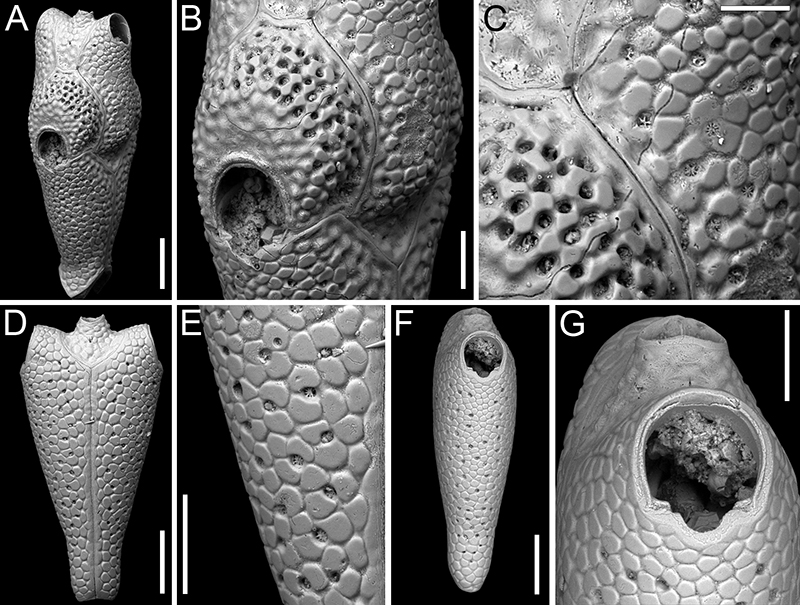
FIGURE 37. Margaretta gracilior (Ortmann, 1892); PMC EDM-Collection J.H.B.136a. A-C, sample 19200. A, general view of a fertile internode (1 mm). B, close-up of an autozooid (250 μm). C, close-up of brooding zooids with swollen peristome base (250 μm). D-F, sample 19195. D, general view of a curved internode with base of rami (1 mm). E, close-up of base of rami (500 μm). F, close-up of an autozooid with internode attachment base (500 μm).
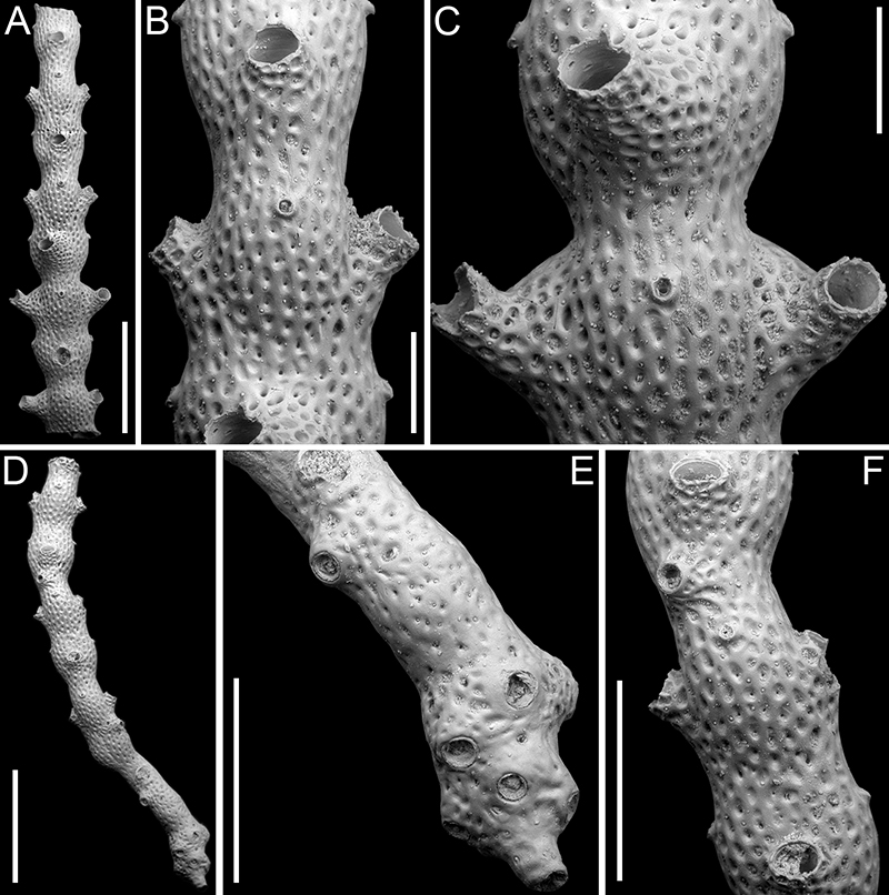
FIGURE 38. Margaretta cf. tenuis Harmer, 1957; PMC EDM-Collection J.H.B.137a. A-F, sample 19047. A, general view of a fertile internode with broken or not yet developed peristomes (1 mm). B, close-up of an autozooid (400 μm). C, close-up of brooding zooids with swollen peristome base (400 μm). D, general view of a curved internode with bipartite base of rami (500 μm). E, close-up of base of rami (200 μm). F, close-up of an autozooid (400 μm). G, sample 19185, fertile internode with long upward-curving peristomes (1 mm).
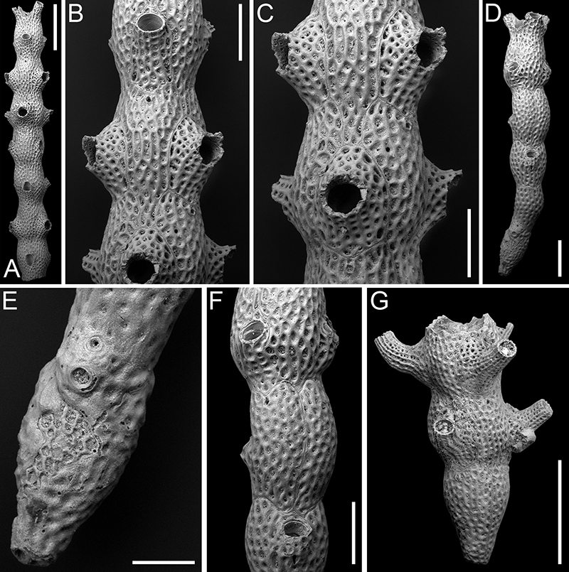
FIGURE 39. Hippopodina feegeensis (Busk, 1884); PMC EDM-Collection J.H.B.138a. A-B, sample 19185. A, unique complete autozooid available (500 μm). B, close-up of orifice and avicularia (150 μm). C, sample 19160, autozooid with broken orifice (200 μm).
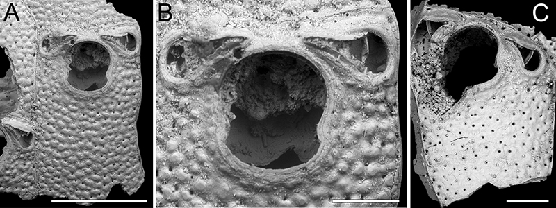
FIGURE 40. Bryopesanser inflexus sp. nov. Di Martino, Rosso and Taylor, holotype PMC. B62. 29.7.2024a, sample 19229. A, colony fragment general view (500 μm). B, close-up of orifice with peristomial proximal mucro curved upward and ooecium proximal projection curved downward (150 μm). C-D, close-ups of orifice, ovicells and avicularia (100 μm). E, close-up of the frontal shield texture with multiporous pseudopores blurred by diagenesis.
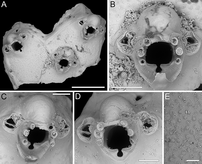
FIGURE 41. Bryopesanser minutiporosus sp. nov. Di Martino, Rosso and Taylor. A-E, holotype PMC. B63. 29.7.2024a, sample 19217. A, colony general view (1 mm). B, close-up of zooids, one ovicellate (250 μm). C, close-up of avicularium (50 μm). D, close-up of orifice showing sign of intramural budding, and avicularia (100 μm). E, group of autozooids with seven oral spine bases and pore-chamber windows (250 μm). F-H, paratype PMC. B63. 29.7.2024b, sample 19210. F, colony general view (500 μm). G, close-up of two autozooids, one with partially formed ooecium (200 μm). H, close-up of orifice and avicularia (100 μm).
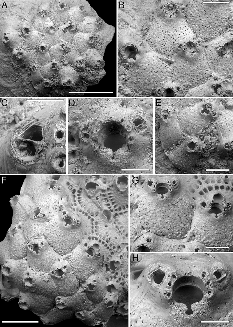
FIGURE 42. Gigantopora sp. 1; PMC EDM-Collection J.H.B.139a, sample 19140. A, general view of the colony fragment (500 μm). B, close-up of an avicularium and spiramen (100 μm). C, close-up of the tubular peristome and spiramen (100 μm).
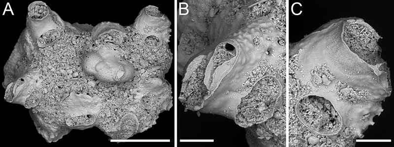
FIGURE 43. Paragigantopora echinata gen. et sp. nov. Di Martino, Rosso and Taylor, holotype PMC. B64. 29.7.2024a, sample 19140. A, general view of the cylindrical branch fragment (1 mm). B, close-up of an autozooid (250 μm). C, close-up of an ovicellate zooid (250 μm).
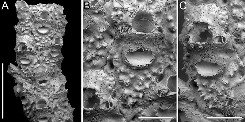
FIGURE 44. Phoceana gabrielei sp. nov. Di Martino, Rosso and Taylor. A-B, holotype PMC. B65. 29.7.2024a, sample 19140. A, tubular hollow colony general view (1 mm). B, close-up of ovicellate zooids (500 μm). C-E, paratype PMC. B65. 29.7.2024b1, sample 19185. C, close-up of ovicell (150 μm). D, colony fragment general view (1 mm). E, close-up of autozooid with subconical marginal tubercles (400 μm). F-H, paratype PMC. B65. 29.7.2024b2, sample 19125. F, colony fragment general view (500 μm). G-H, close-ups of orifice with flared, undulose peristome (150 μm).
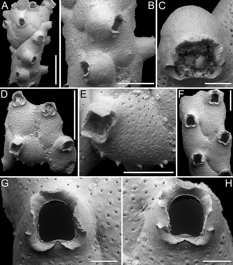
FIGURE 45. Calloporina biavicularia Kataoka, 1961; PMC EDM-Collection J.H.B.140a, sample 19093. A, colony fragment general view (500 μm). B, group of autozooids (250 μm). C-D, close-ups of orifice, spine bases, ascopore and avicularia (100 μm).
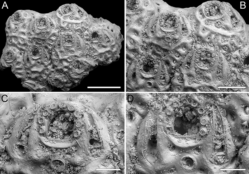
FIGURE 46. Mucropetraliella cf. robusta (Canu and Bassler, 1929). A-E, PMC EDM-Collection J.H.B.141a, sample 19083. A, colony fragment general view (1 mm). B, close-up of autozooids (200 μm). C, close-up of ovicell and suboral complex (150 μm). D-E, close-up of orifice showing paired distolateral spines, variability of median denticle and condyles (100 μm). F, sample 19135, close-up of an autozooid with latero-frontal avicularium (100 μm). G, sample 19193, close-up of the trifurcate suboral mucro (200 μm). H, sample 19229, general view of the colony fragment (500 μm). I-J, sample 19178, close-ups of the enlarged latero-oral avicularium (200 μm).
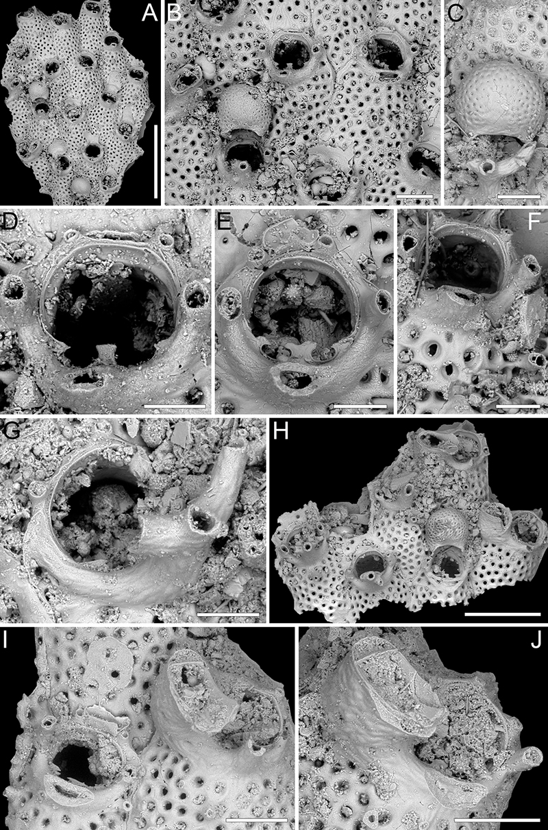
FIGURE 47. Mucropetraliella cf. tubulifera (Canu and Bassler, 1929); PMC EDM-Collection J.H.B.142a. A-E, sample 19035. A, colony fragment general view (400 μm). B, close-up of orifice (120 μm). C-D, close-ups of orifices showing at least four and five spines and tri- to quadri-furcated suboral complex with additional conical processes (100 μm). E, close-up of two autozooids with frontal conical processes (200 μm). F, sample 19095, bush-like suboral complexes of a group of autozooids in tilted view (500 μm).
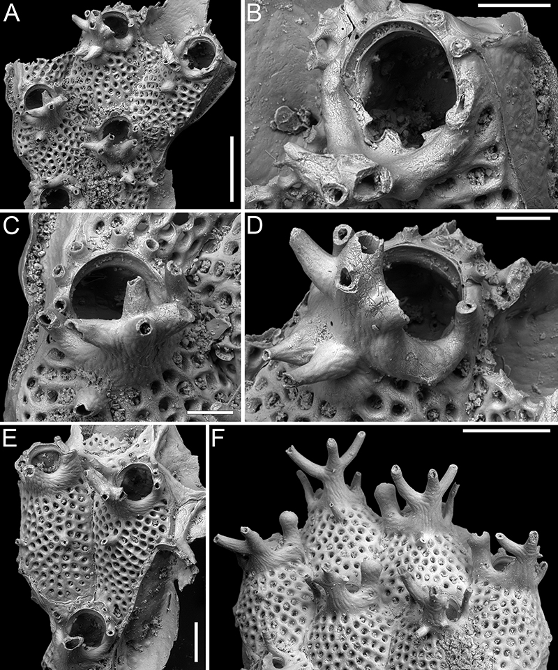
FIGURE 48. Arthropoma iuncta sp. nov. Di Martino, Rosso and Taylor. A-C, holotype PMC. B67. 29.7.2024a, sample 19041. A, colony fragment general view (1 mm). B, close-up of two ovicellate zooids (200 μm). C, close-up of the orifice (50 μm). D-F, paratype PMC. B67. 29.7.2024b1, sample 19178; paratype PMC. B67. 29.7.2024b2, sample 19085; paratype PMC. B67. 29.7.2024b3, sample 19092; close-ups of vicarious avicularia (D, F 200 μm; E 250 μm). Arrows in D and F indicate distal spines. G-H, paratype PMC. B67. 29.7.2024b4, sample 19104. G, general view of part of the colony showing the multilaminar growth (500 μm). H, close-up of an autozooid (200 μm).
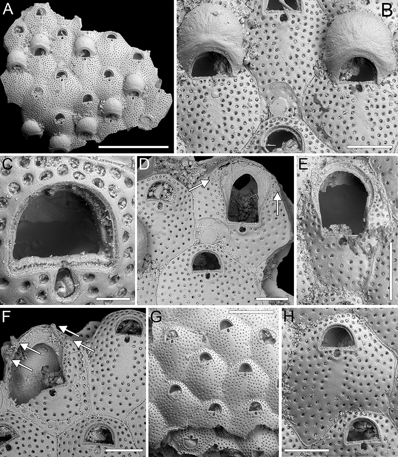
FIGURE 49. Arthropoma semipunctata sp. nov. Di Martino, Rosso and Taylor. A-D, holotype PMC. B66. 29.7.2024a, sample 19048. A, colony fragment general view (400 μm). B, close-up of an ovicell (200 μm). C-D, close-up of two orifices (100 μm). E-F, paratype PMC. B66. 29.7.2024b1, sample 19025. E, colony fragment general view (400 μm). F, close-up of two autozooids with signs of regeneration (200 μm). G-H, paratype PMC. B66. 29.7.2024b2, sample 19041. G, colony fragment general view (500 μm). H, close-up of an ovicellate zooid (250 μm).
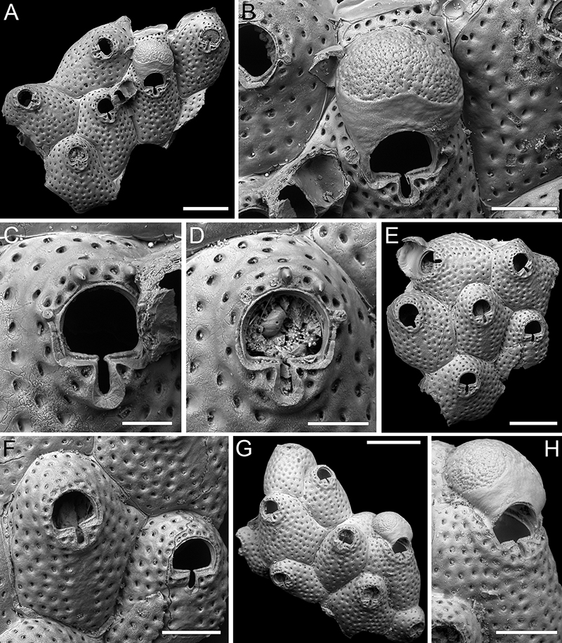
FIGURE 50. Cribellopora ? fissurata sp. nov. Di Martino, Rosso and Taylor, holotype PMC. B68. 29.7.2024a, sample 19088. A, colony general view (250 μm). B, close-up of zooids and ovicells (300 μm). C, close-up of an autozooid with preserved spine bases (150 μm).
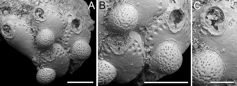
FIGURE 51. Characodoma erinaceum sp. nov. Di Martino, Rosso and Taylor. A-B, paratype PMC. B69. 29.7.2024b1, sample 19025. A, general view of a colony in erect phase of growth (500 μm). B, group of autozooids (200 μm). C-D, paratype PMC. B69. 29.7.2024b2, sample 19041, close-up of autozooids, some with avicularium and ovicell in formation (C 150 μm; D 180 μm). E, G, holotype PMC. B69. 29.7.2024a, sample 19183, close-up of autozooids, some with avicularia and complete ovicells (200 μm). F, paratype PMC. B69. 29.7.2024b3, sample 19068, close-up of orifice and partially calcified ooecium (100 μm). H, paratype PMC. B69. 29.7.2024b4, sample 19228, general view of a colony in encrusting phase of growth (1 mm).
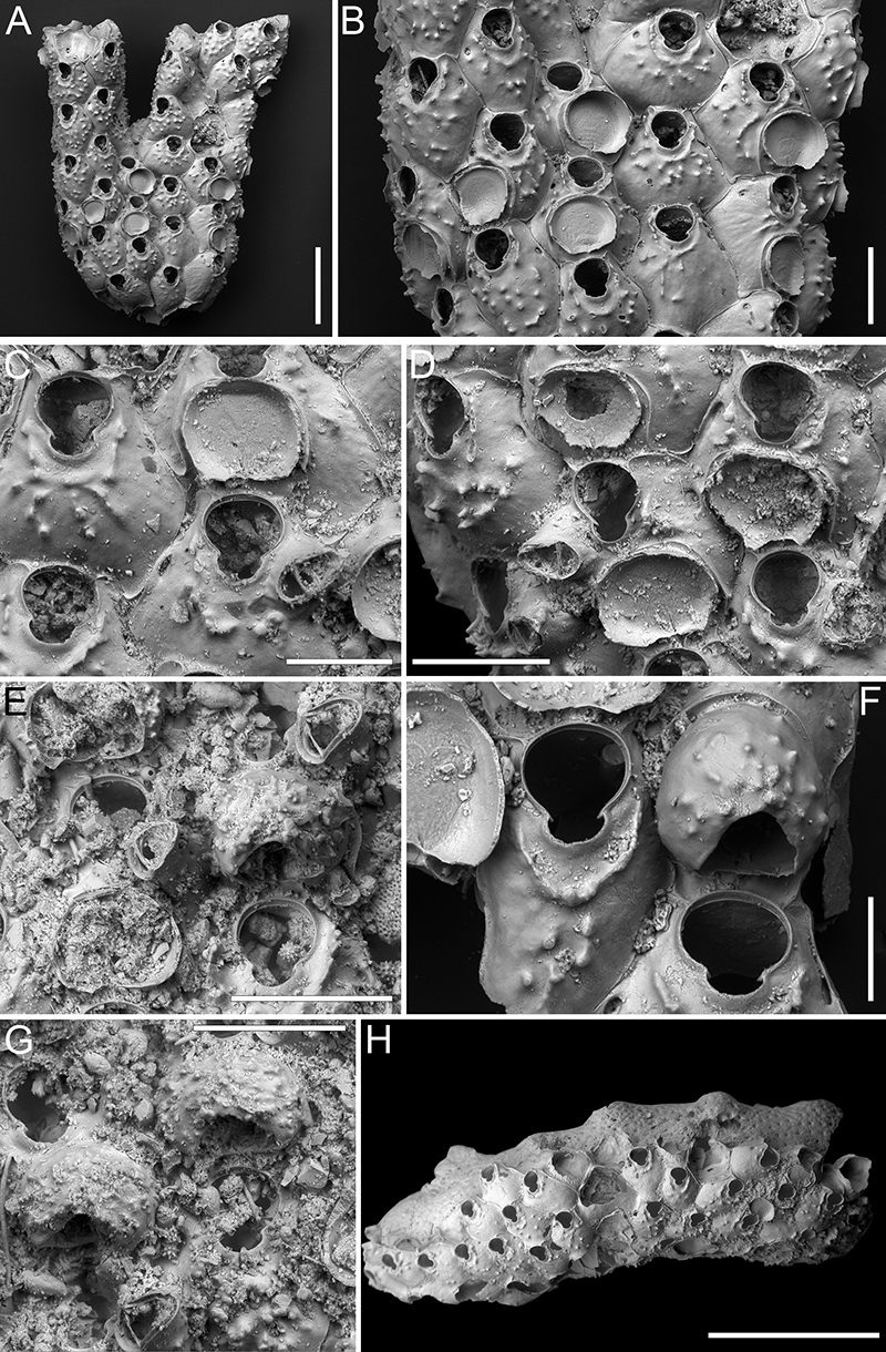
FIGURE 52. Buffonellaria conformis sp. nov. Di Martino, Rosso and Taylor. A-D, holotype PMC. B70. 29.7.2024a, sample 19174. A, colony general view (500 μm). B, close-up of two autozooids (200 μm). C, close-up of orifice and avicularia (100 μm). D, close-up of ovicells (200 μm). E-F, paratype PMC. B70. 29.7.2024b, sample 19115. E, colony general view (500 μm). F, close-up of ovicells (200 μm).
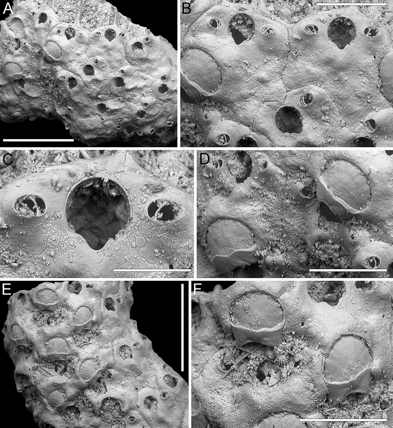
FIGURE 53. Celleporaria cf. calva Tilbrook, 2006; PMC EDM-Collection J.H.B.144a, sample 19125, colony without spines. A, colony fragment general view (500 μm). B-D, sample 19114. B, fragment general view (500 μm). C-D, close-up of orifice and adjacent avicularia (300 μm).
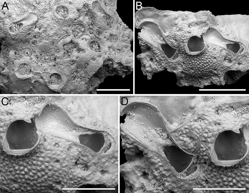
FIGURE 54. Celleporaria cf. calva Tilbrook, 2006; PMC EDM-Collection J.H.B.144a, sample 19127, colony with spines. A, colony fragment general view (1 mm). B, close-up of orifice (100 μm). C, close-up of an autozooid with three spine bases and parallel-sided avicularium of medium size (500 μm). D, close-up of an avicularium (100 μm). E, group of autozooids with four spine bases partially obliterated (500 μm). F, close-up of an avicularium and orifice with partially obliterated spine bases (500 μm).
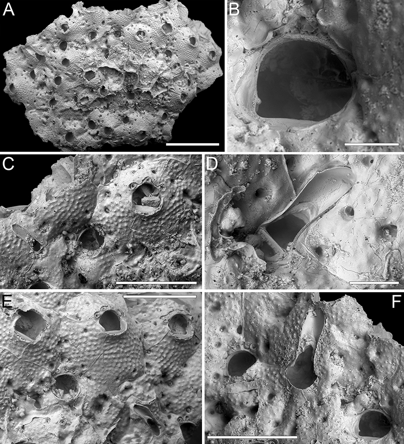
FIGURE 55. Celleporaria cf. repens Canu and Bassler, 1929; PMC EDM-Collection J.H.B.145a, sample 19127, colony with spines. A, colony fragment general view (500 μm). B, close-up of two ovicells and suboral avicularium (150 μm). C, close-up of ovicell encircling and obscuring the orifice and suboral avicularium (100 μm). D, close-up of orifices showing semircircular proximolateral condyles (150 μm). E, close-up of an autozooid with developed suboral mucro (200 μm). F, close-up of orifice with trapezoidal condyle (50 μm).
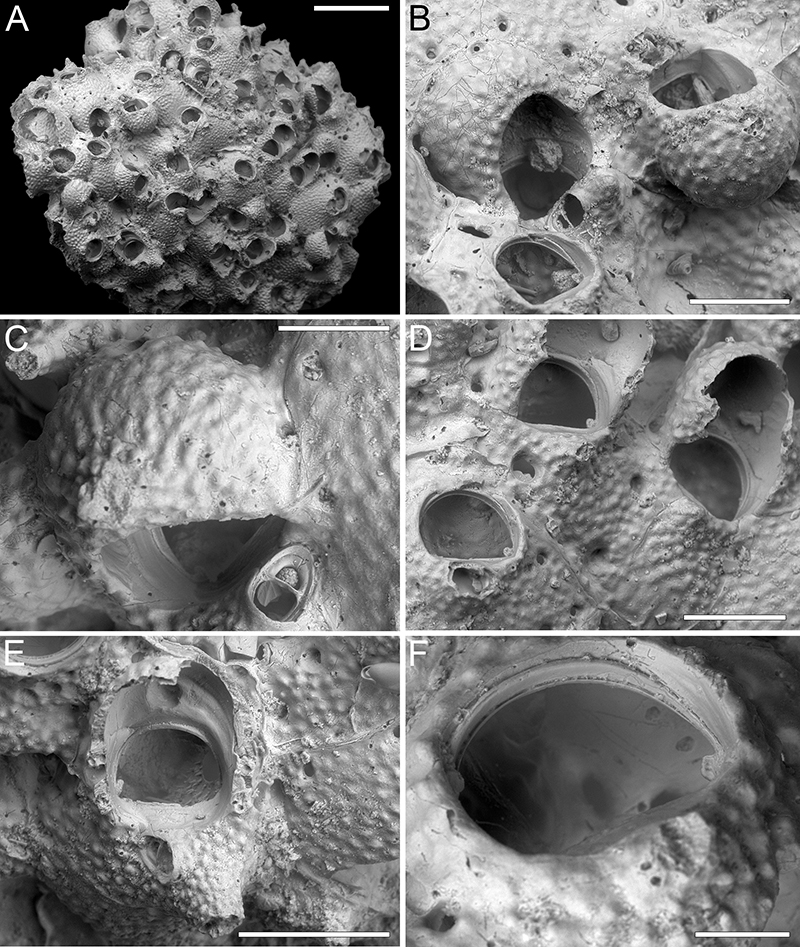
FIGURE 56. Celleporaria cf. triangula Seo, 1994; PMC EDM-Collection J.H.B.146a, sample 19057, colony with spines. A, colony fragment general view (500 μm). B, close-up of autozooids and avicularia (200 μm). Arrows indicate a spine base and the third denticle difficult to discern because partially broken.
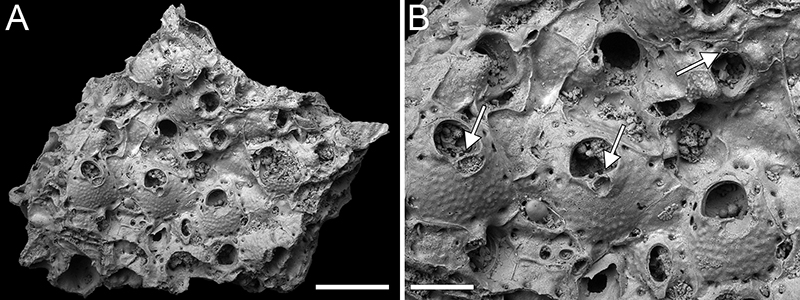
FIGURE 57. Reteporella phimostoma sp. nov. Di Martino, Rosso and Taylor, holotype PMC. B71. 29.7.2024a, sample 19061. A, colony fragment general view (500 μm). B, close-up of ovicellate autozooids (200 μm). C, close-up of the complex peristomial avicularian structure and coalescent spines (100 μm). D, close-up of non-ovicellate autozooids (60 μm). E, close-up of autozooid with secondary calcification (100 μm). F, close-up of avicularium and ovicell (50 μm). G, general view of the dorsal side (500 μm). H, close-up of dorsal vibices and avicularia (200 μm). I, close-up of dorsal avicularium (50 μm).
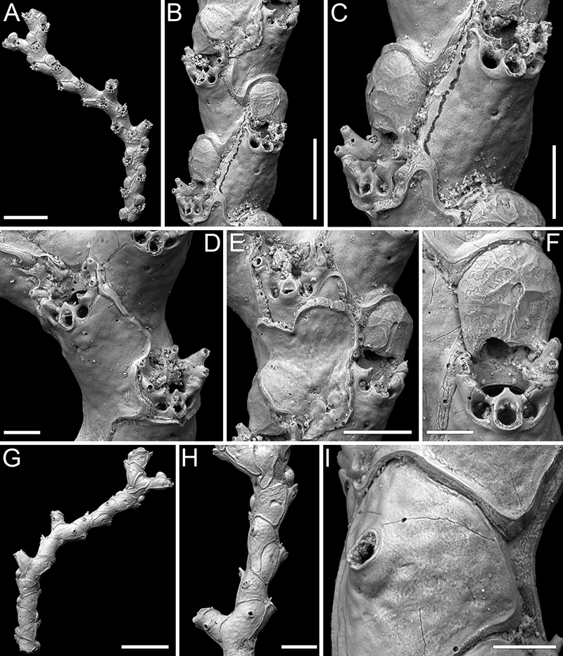
FIGURE 58. Reteporella sp. 1; PMC EDM-Collection J.H.B.147a. A-E, sample 19015. A, colony fragment general view (400 μm). B, close-up of autozooids (200 μm). C, close-up of secondary orifice and suboral avicularium (40 μm). D, group of zooids some with up to six spines and some ovicellate (200 μm). E, close-up of ovicells with the short labellum visible, and suboral avicularia (100 μm). F-K, sample 19025. F, colony fragment general view (300 μm). G, group of zooids some ovicellate (200 μm). H-J, close-ups of ovicells and avicularia (100 μm). K, general view of the dorsal side (200 μm).
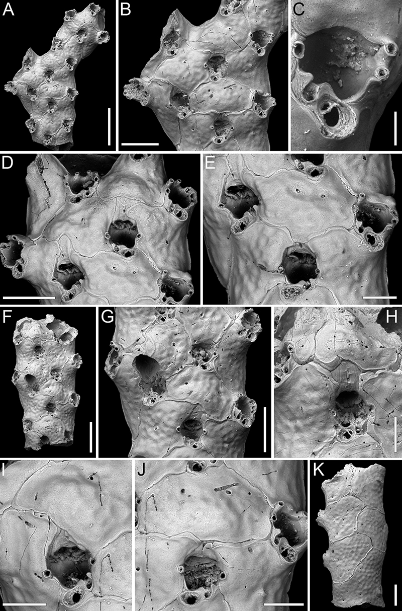
FIGURE 59. Reteporellina denticulata (Busk, 1884); PMC EDM-Collection J.H.B.148a. A-B, sample 19044. A, colony fragment general view (400 μm). B, close-up of autozooids (200 μm). C-I, sample 19021. C, colony fragment general view (400 μm). D, close-up of autozooids with suboral avicularium (200 μm). E, close-up of ovicell with labellum and suboral avicularium (40 μm). F, close-up of avicularium (40 μm). G, general view of fragment dorsal side (400 μm). H-I, close-up of the dorsal side with avicularia and vibices (H 400 μm, I 40 μm).
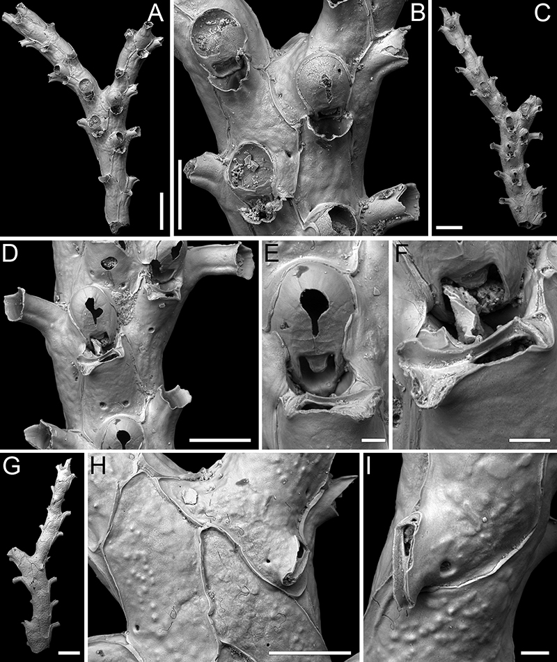
FIGURE 60. Rhynchozoon lunifrons Dick and Grischenko, 2017; PMC EDM-Collection J.H.B.149a. A-C, sample 19033. A, colony with frontally budded second layer (400 μm). B, close-up of the pseudoancestrular area, referred to as the first type in the text (200 μm). C, close-up of autozooids at the margin (100 μm). D, sample 19015, close-up of orifice with arrow indicating one preserved condyle (40 μm). E, sample 19031, close-up of ovicellate zooids (200 μm). F, sample 19041, pseudoancestrular area, referred to as the second type in the text (200 μm). G-I, sample 19036. G, general view of colony fragment with numerous ovicells and two frontal avicularia arrowed (300 μm). H, close-up of a suboral avicularium (60 μm). I, close-up of a frontal avicularium, an ovicell, and bifurcated lateral mucrones (100 μm).
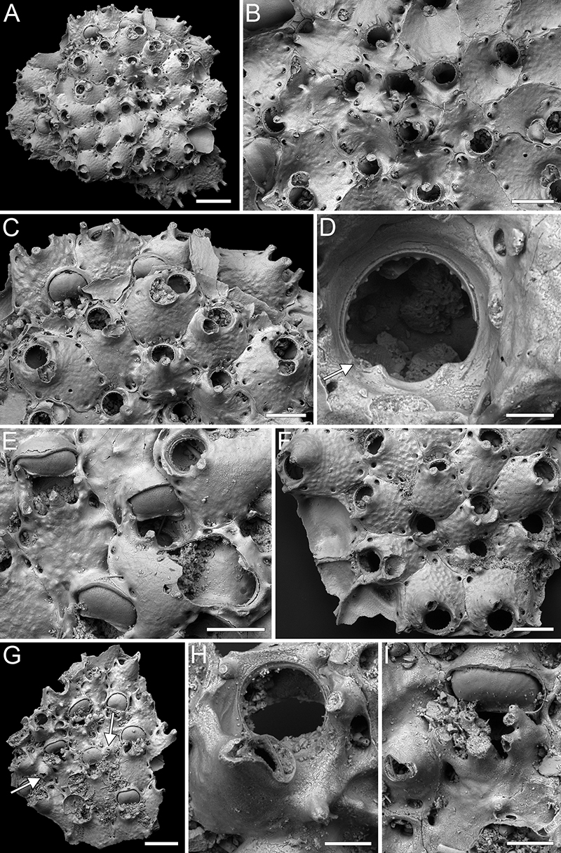
FIGURE 61. Stephanollona pachycondylata sp. nov. Di Martino, Rosso and Taylor. A-E, holotype PMC. B72. 29.7.2024a, sample 19185. A, colony fragment general view (1 mm). B, close-up of orifice (100 μm). C, close-up of a large avicularium (100 μm). D, close-up of orifice with small lateral avicularium (100 μm). E, close-up of ovicell (100 μm). F-G, paratype PMC. B72. 29.7.2024b, sample 19140. F, colony fragment general view (250 μm). G, close-up of orifice and large avicularium (100 μm).
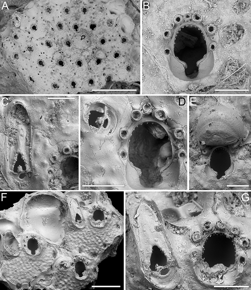
FIGURE 62. Triphyllozoon angustum sp. nov. Di Martino, Rosso and Taylor. A-D, G-I, holotype PMC. B73. 29.7.2024a, sample 19113. A, frontal view of the reticulate fragment (1 mm). B, close-up of an ooecium covered with secondary calcification and secondary orifice with lateral oral spine bases, peristomial avicularium, and sparse frontal avicularia (200 μm). C-D, ovicellate zooids (300 μm). E-F, paratype PMC. B73. 29.7.2024b1, sample 19055. E, close-up of an ovicellate zooid with two types of frontal avicularia and ovicell in lateral view (200 μm). F, close-up of ooecium and secondary orifice with peristomial avicularium and deep, drop-shaped pseudosinus (100 μm). G, dorsal view of the reticulate fragment (1 mm). H-I, close-ups of dorsal avicularia (H 500 μm, I 300 μm).
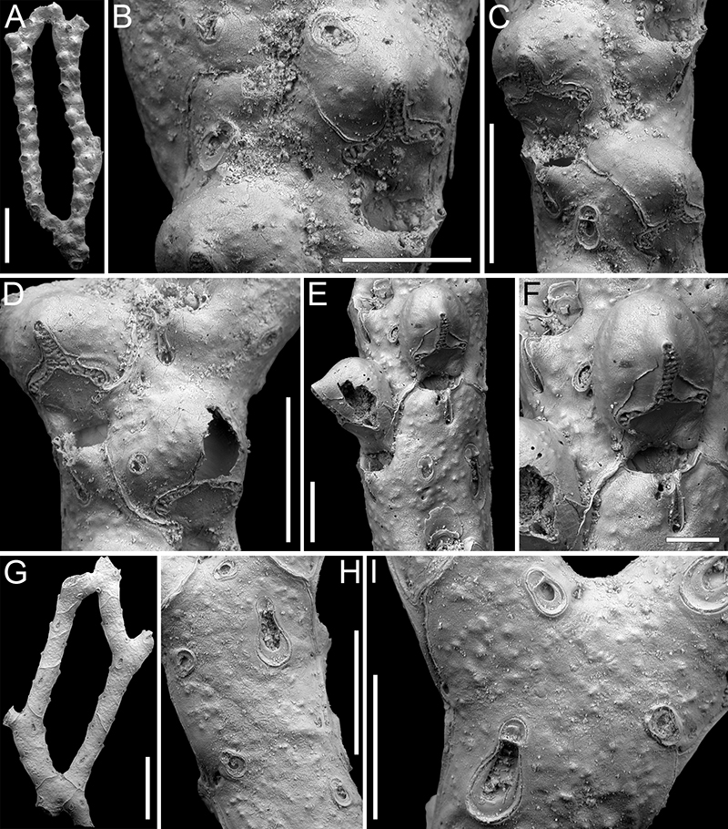
FIGURE 63. Conescharellina brevirostris Silén, 1947; PMC EDM-Collection J.H.B.150a. A-B, sample 19053. A, colony general view (400 μm). B, close-up of two orifices with peristomes and associated distal avicularia (100 μm). C-D, sample 19095. C, ovicellate colony general view (500 μm). D, close-up of ovicell (100 μm). E-F, sample 19081. E, general view of the antapical surface (500 μm). F, close-up of basal avicularia (100 μm).
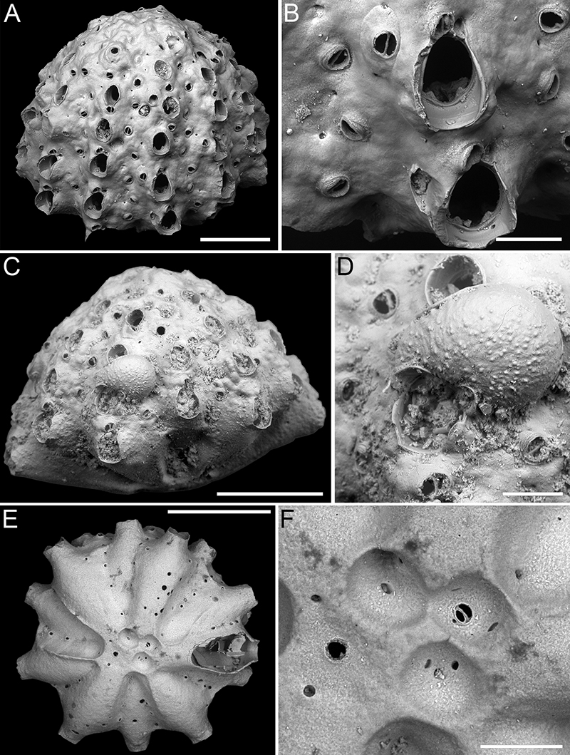
FIGURE 64. Conescharellina sp. 1; PMC EDM-Collection J.H.B.151a. A-F, sample 19063. A, general view of the colony apical surface (200 μm). B, close-up of the tubercular apical area with avicularia and kenozooidal openings (100 μm). C, colony lateral view (200 μm). D, close-up of orifice and interzooidal avicularia (100 μm). E, general view of the antapical surface (200 μm). F, close-up of basal avicularia (40 μm). G, sample 19206, general view of the colony apical surface (250 μm).