FIGURE 1. Geological map of the sponge locality in the San Juan province Argentina.
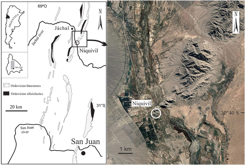
FIGURE 2. Stratigraphic column of the Niquivil section showing the location of the sponge level in the reef mounds interval. Field photographs showing a single reef mound and scheme, and a close-up of the same structure showing orchoclad “lithistids” demosponges in transversal and longitudinal views.
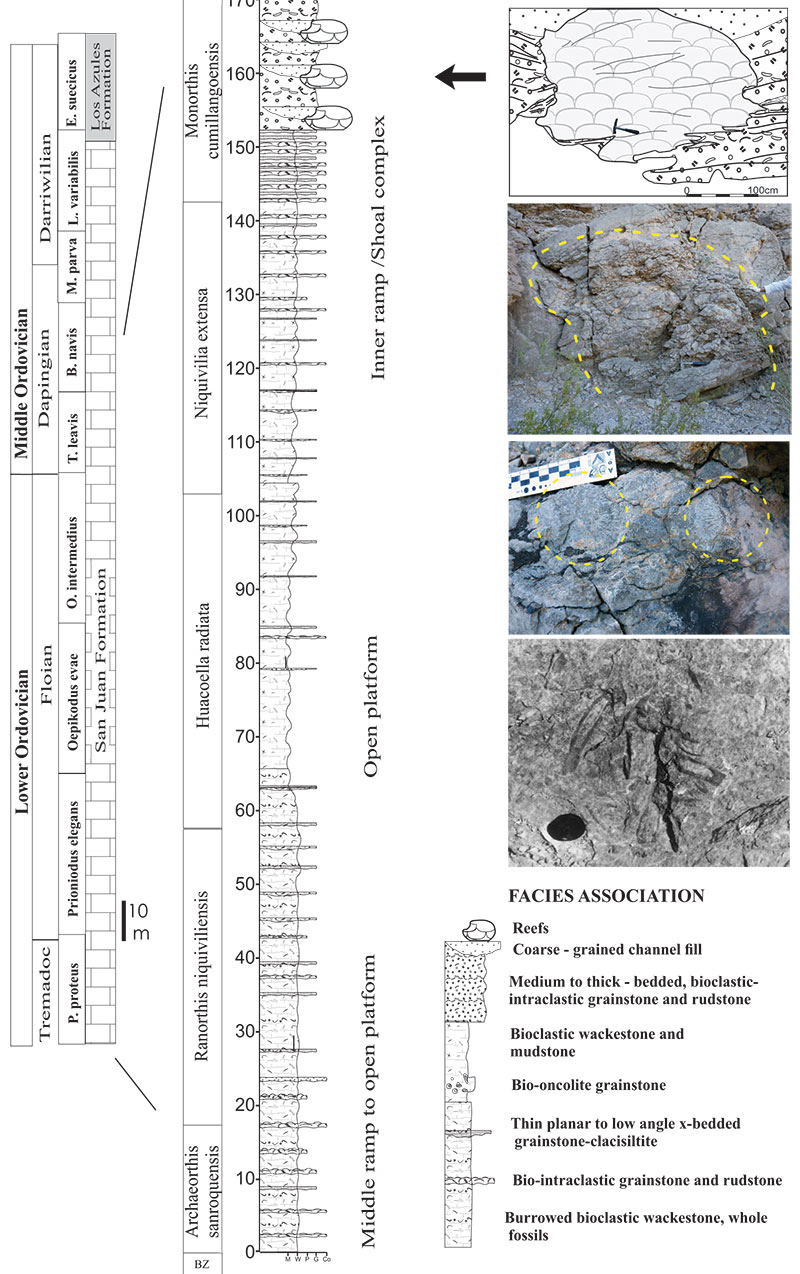
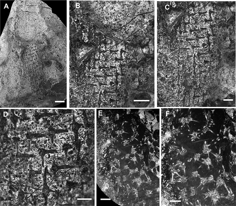
FIGURE 3. Niquivilispongia asteria gen. et sp. nov. A. General view of an almost complete specimen (thin section CEGH-UNC 27595) showing a laminar to encrusting morphology of the sponge interrupted by vertical dissolution gaps, scale bar equals 1 cm. B. General view of the specimen in thin section (Holotype CEGH-UNC 27593), showing a mound shaped sponge form at the top and a laminar to encrusting form at the base, scale bar equals 1 cm. C. Close-up of 3B showing the mound shaped form at the top, irregular arrangement of heteractinid spicules and below the base the microproblematicum Allonema, scale bar equals 5 mm. D. Detailed view of figure 3A showing a partially dissolved sponge structure with isolated hexaradiate heteractinid spicules, scale bar equals 5 mm. E. Close-up of figure 3C with hexaradiate heteractinids and views of heteractinids showing slightly curved rays, scale bar equals 1 mm. F. Detailed view of the previous thin section figure 3E, showing heteractinids with the view of four curved rays, scale bar equals 1 mm. G. Close-up of figure 3E showing heteractinids in different views with six rays in a plane and four curved rays in a plane. Arrows show two divergent ray bases in the center of an apparent “four rayed hexactin”, and interposed rays bisecting the space between two others, scale bar equals 1 mm. H. Transversal view of at the base of the sponge (CEGH-UNC 27594) showing modified undulate monaxon-like spicules or modified rays of heteractinid-based spicules, scale bar equals 1 mm. I. Transversal view of the same thin section showing the transition of the main sponge spicules and the modified spicules (hypertrophied heteractinid rays and possible monaxons) of the root tuft structure, scale bar equals 1 mm. J. The same transversal view showing an adjacent extended part of the root tuft structure and the transition of the main sponge spicules, scale bar equals 1 mm.
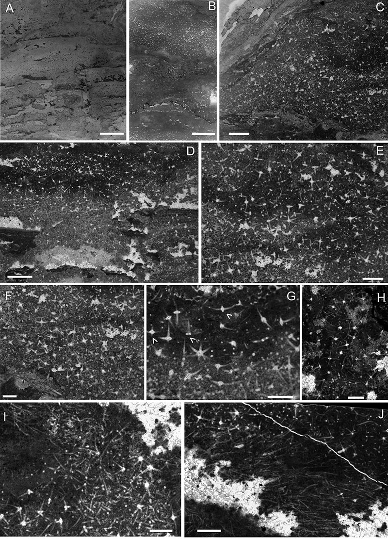
FIGURE 4. Niquivilispongia asteria gen. et sp. nov. A. Detailed view of a thin section (Holotype CEGH-UNC 27593) with heteractinid spicules with filled or hollow axial regions preserved as calcareous blocky or drusy cement, scale bar equals 0.5 mm (500 µm). B. Another view of the same thin section mainly showing hexaradiate heteractinids, scale bar equals 0.5 mm (500 µm). C. Detailed view of the long undulate rods of monaxons or expanded hypertrophied spicule rays of the root tuft structure (CEGH-UNC 27594) scale bar equals 0.2 mm (200 µm). D. Close-up of heteractinid spicules (Holotype CEGH-UNC 27593), including “four rayed” visual sections, with spar filled cement and a blocky differential cement in the center or a hollow core in others, scale bar equals 0.5 mm (500 µm). E. Detailed view of a four rayed heteractin with a core filling by a differential blocky spar cement, scale bar equals 0.2 mm (200 µm). F. Detailed view of an heteractinid showing orthogonal rays to the six rays in a plane, and a view of a partial section of an heteractinid with interposed ray bisecting the space between two others, scale bar equals 0.2 mm (200 µm). G. Close-up of an heteractinid with apparently six rays in one plane and a vertical ray, scale bar 0.2 mm (200 µm). H. A close-up of a curve rayed heteractinid with a big crystal spar cement in the axial core, scale bar equals 0.2 mm (200 µm). I. Four rayed view of a heteractinid with spar axial infilling, scale bar equals 0.2 mm (200 µm). J. A detailed view of figure 5D showing a hollow core, scale bar equals 0.2 mm (200 µm). K. A detailed view of figure 4A showing another hollow core in the axial infilling, scale bar equals 0.2 mm (200 µm).
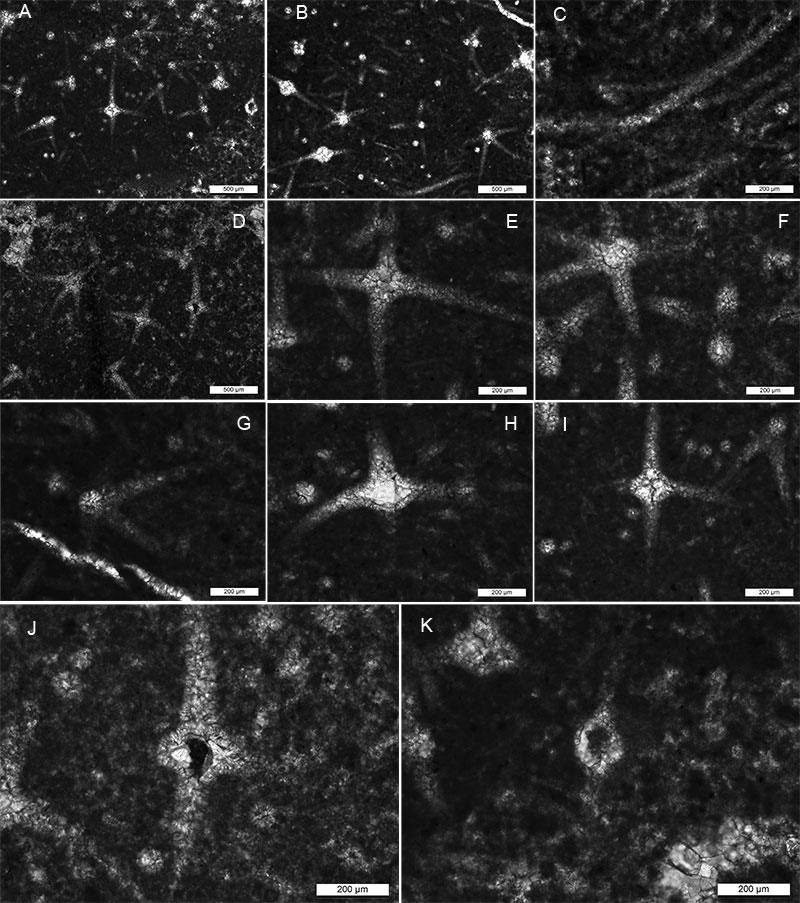
FIGURE 5. External views and thin sections of ? Mattaspongia sp. and Astylospongiidae indet. A. General view of the fragmented sponge ? Mattaspongia sp. (CEGH-UNC 27596), showing an apparent cylindrical or tubular body and a faintly marked oscular margin, scale bar equals 5 mm. B. Detailed of the upper part of the sponge fragment showing detailed of the apparent oscular margin and dermal hexactins, scale bar equals 2 mm. C. General view of the main body spicules composed of coarse, predominantly aligned hexactin-type forms, mostly of the same size, scale bar equals 1 mm. D. Close-up of the main body spicules showing alternated vertically aligned rays and small monaxons in between, scale bar equals 1 mm. E. Thin section of a fragmented Astylospongiidae indet. (CEGH-UNC 27597), showing sphaeroclone spicules forming isodictyal net with triangular interspaces in all directions, scale bar equals 1 mm. F. Close-up of the sphaeroclone anaxial desmas with five to six ray-like arms extended from one side of a globular centrum formed by the fusion of the zygome endings of the rays, scale bar equals 1 mm.