FIGURE 1. Reflection of elements. A. Selection of the section found complete in the skull of a Pascualgnathus polanskii (MLP 65-VI-18-1) with Metashape. B. Generation of the model of the isolated section with Metashape and mirroring in Blender. C. Alignment and fusion of both models in Blender to restore the skull.
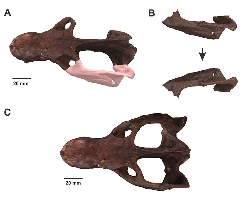
FIGURE 2. Superimposition. A. Mandible of Massetognathus pascuali PVL 4613, specimen A to be restored. B. Mandibles of Massetognathus pascuali PVL 3901 used as specimen B. C. Isolated coronoid process of specimen B. D. Restored mandibles of Massetognathus pascuali PVL 4613.
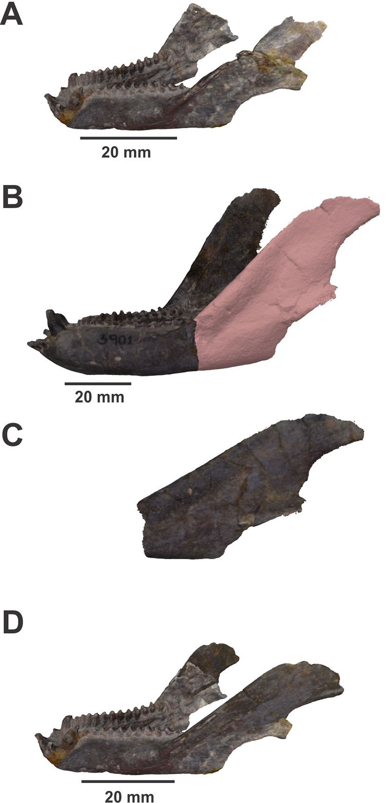
FIGURE 3. Retrodeformation. A. Mandible in anterior view of Massetognathus pascuali (PVL 4726) shear deformed. B. Retrodeformed model of the mandible in the Landmark program.
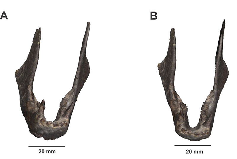
FIGURE 4. Dismantling of elements for restoration separately. A. Pascualgnathus polanskii (PVL 4416) with a high deformation grade. B. Disarticulated and independently modeled skull and mandible. C. Skull and mandible restored using the steps explained in the text (mirroring of elements).
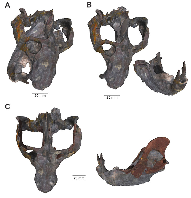
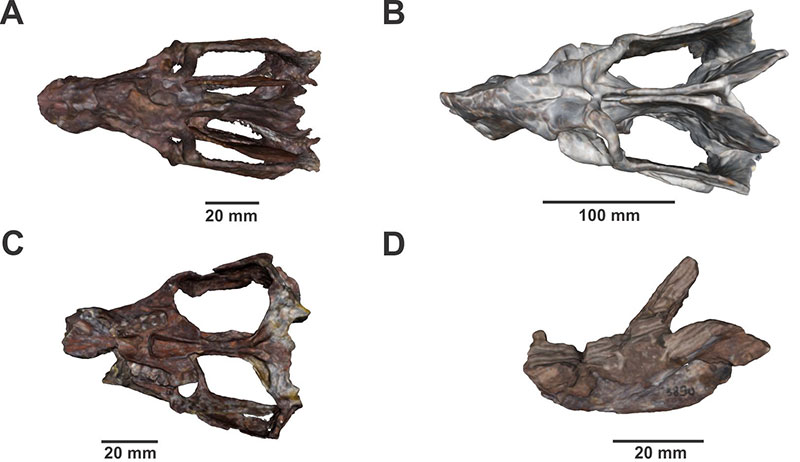
FIGURE 5. Cynognathian specimens which could not be fully restored (A, B) and specimens that were left out from the restoration process due to the level of damage could not be resolved with any of the restoration techniques described (C, D). A. Dorsal view of skull and mandibles of Andescynodon mendozensis PVL 3092 showing a slightly mediolaterally compress. B. Dorsal view of the skull of Exaeretodon PVL 2056b showing pronounced mediolaterally compression. C. Ventral view of skull of Andescynodon mendozensis PVL 3892 in which it can be appreciated the missing anterior part of the snout. D. Dorsal view of incompletely prepared mandibles of Andescynodon mendozensis PVL 3890.