FIGURE 1. Location map of Scontrone and Gargano. From Mazza (2013a), modified.
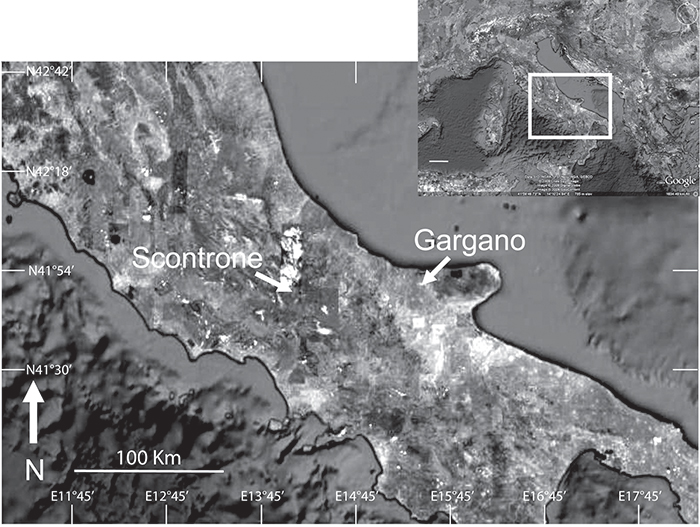
FIGURE 2. Schematic stratigraphy of the early Tortonian site of Scontrone (from Patacca et al., 2008a, modified). Succession showing the stratigraphic position of the bonebeds within the Scontrone Member of the Lithothamnium Limestone Formation. RC Rudist-bearing Calcarenite; SC Scontrone Member of the Lithothamnium Limestone Formation; SCa-SCd facies units of the Scontrone Member recording major shifts in the depositional setting and biotic associations (SCa = costal bar deposits; SCb = tidal creek deposits; SCc = marsh deposits; SCd = lagoon deposits). 1-Calcareous marls; 2-Bioclastic calcarenites; 3-Bioclastic calcarenites with oversized well-rounded lithoclast lags; 4-Lithoclastic calcirudites; 5-Major disconformity; 6-Low-angle cross-bedding; 7-Trough cross-bedding; 8-Root traces; 9-Oyster shell lags; 10-Hydrobiids; 11-Bonebeds; 12-Rudists. For further details see Patacca et al. (2008a).
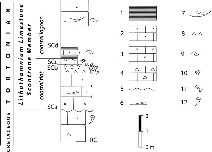
FIGURE 3. Guide to dental morphology terms used in this paper, drawn from Engesser (1980), Gould (1995), Lopatin (2006) by Masini and Fanfani (2013), and slightly modified here. 1, Upper molar. 2, Lower molar; (a) paralophid (paracristid) = paraconid crest + preprotocristid sensu Lopatin (2006); (b) postcristid (hypolophid) = postentocristid + posthypocristid sensu Lopatin (2006).
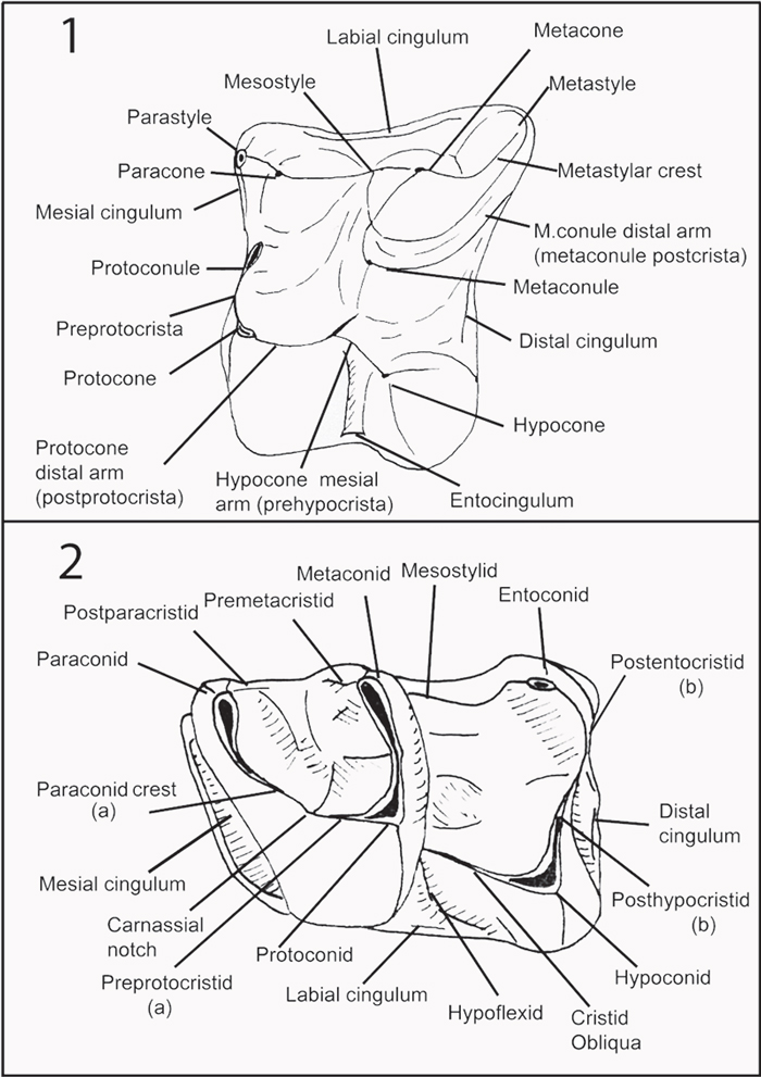
FIGURE 4. 1, Left M1 (F9-018), undetermined Deinogalerix sp. from fissure F9, oblique, occluso-distal view. 2, same as 1 with sketch showing contact of the distal arm of the metaconule with the uninterrupted distal cingulum. 3, left m1 (paratype SCT 347), Deinogalerix samniticus sp. nov., occlusal view. 4, same specimen as 3, lingual view. Arrows in 3 and 4 showing mesiolingual bulge. 5, left p4 (holotype SCT 246), Deinogalerix samniticus sp. nov., occlusal view. 6, same as 5, with sketch showing roundish, delimited lingually, trigonid valley. 7, right P3-P4 (paratype SCT 19), Deinogalerix samniticus sp. nov., occlusal view. 8, same as 7 with sketch showing the weak separation of protocone and hypocone. Hyc = Hypocone, Mecl = Metaconule, Mec = Metacone, Mecd = Metaconid, Pac = Paracone, Pacd = Paraconid, Prc = Protocone, Prcd = Protoconid. Figures not to scale.
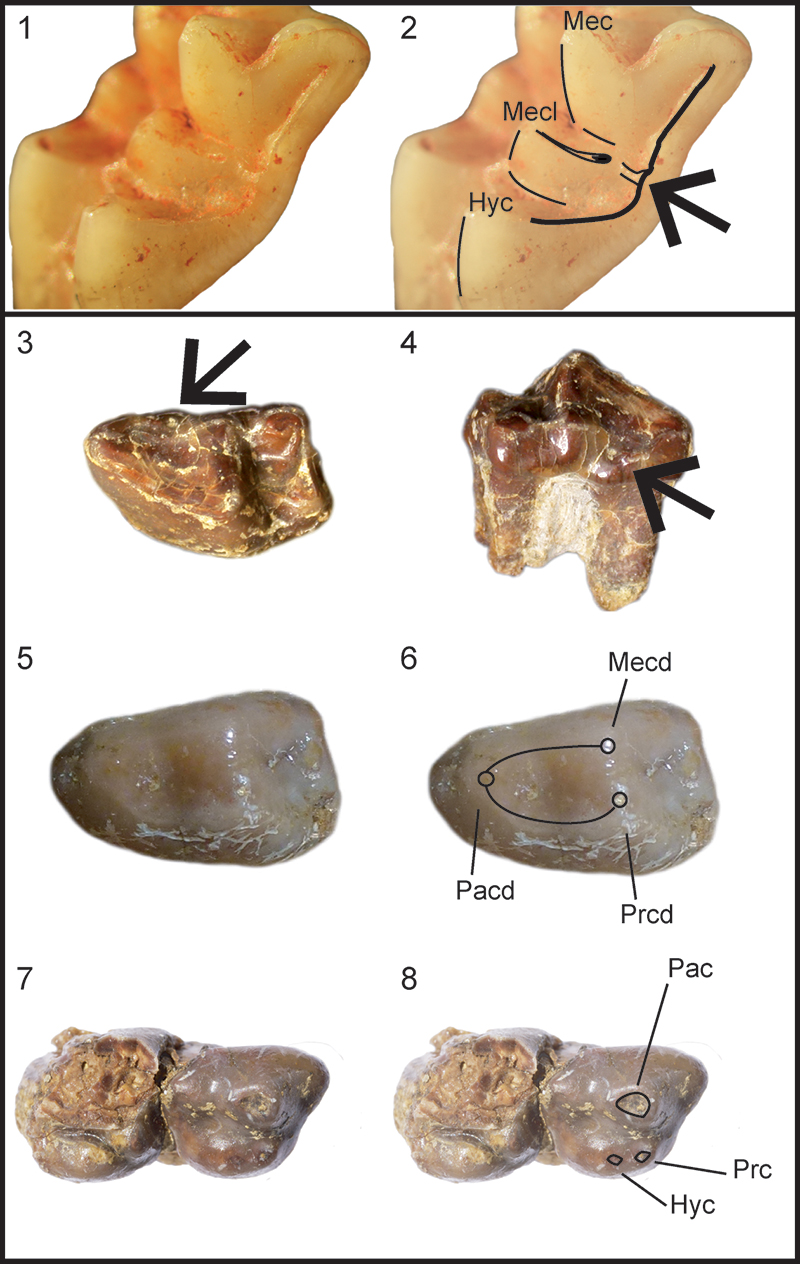
FIGURE 5. Deinogalerix samniticus sp. nov., from Scontrone. 1-2, Holotype, fragment of left hemimandible, with p3, p4, alveoli of p2, broken m1, m2, and m3 partially embedded in the rock (SCT 246). 1, labial view; 2, occlusal view. 3-5, Paratype, isolated left m1 (SCT 347). 3, occlusal view; 4, labial view; 5, lingual view. 6-7, Paratype, fragmental right maxillary with complete P3 and fragmented P4 (SCT 19). 6, occlusal view; 7, lingual view. 8, Paratype, fragmental right maxillary, with M3 and postero-lingual portion of M2 (SCT 232).
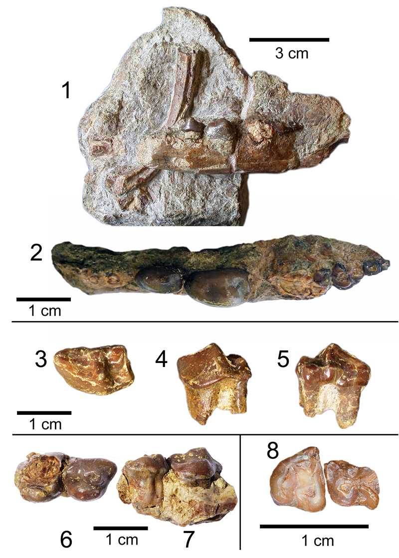
FIGURE 6.1-3, Holotype SCT 246 during its preparation from the rock matrix by vibrotool. The figure shows the premolars still partially encased in the rock matrix.
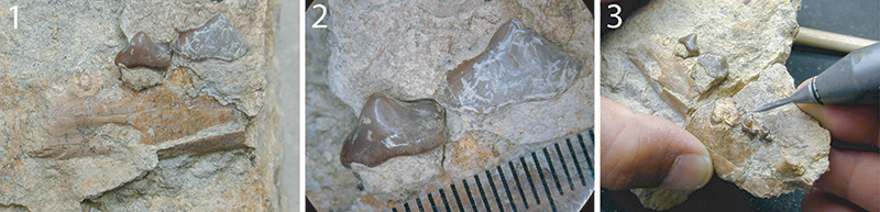
FIGURE 7. Comparisons of p3-m3 lengths in the different species of Deinogalerix, as well as in Parasorex, Galerix, Schizogalerix, and Apulogalerix (measurements and abbreviations in Table 3). 1, The graph shows that in Apulogalerix, Parasorex and other continental galericines molars grow progressively larger towards m1, which is the largest tooth of the row. The fourth lower premolar is larger than p3, which, in contrast, is the smallest of the toothrow (it is somewhat smaller than m3). Compared to the mainland counterparts, Deinogalerix bears a very large m1 (due to the increase of the trigonid length) and larger premolars, that grow progressively smaller moving rostrally, but less than in the continental genera (p3 intermediate in size between m1 and m2). The Scontrone specimen shows the size increase from m3 to m1 typical of Deinogalerix. In contrast, it bears a very large p4, similar in size to m1 and in the dimensional ranges of D. koenigswaldi, and a very reduced p3, more similar to those of the smaller and more primitive species of Deinogalerix from Gargano. 2, Ratio diagram comparing the p3-m3 lengths (see Table 4) in Deinogalerix samniticus sp. nov. with other species of Deinogalerix, using Parasorex socialis from La Grive as the standard (horizontal line). All the Deinogalerix specimens show a similar trend where the proportions of m2 and m3 are parallel to the standard, whereas, starting from m1, the teeth proportionally increase in size respect to the standard. It is noteworthy that this trend reaches its maximum expression in D. koenigswaldi. Compared to the other species of Deinogalerix, the curve of D. samniticus sp. nov.,shows a steeper rise toward p4, and it is the only one that drops toward p3.
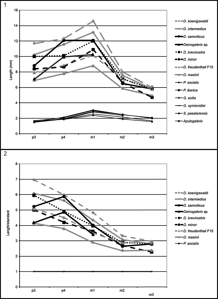
FIGURE 8. Scatter plot of Lp4 vs Lp3 (values in Table 5) for the species of Deinogalerix. The Deinogalerix specimens from Gargano align roughly in a linear trend. D. samniticus sp. nov. and Deinogalerix sp. in contrast, display an anomalously short p3. It is worth noting that also the smaller and more primitive species, e.g., D. freudenthali from F15 and D. masinii from M013, have fairly short p3s. Measurements in mm.
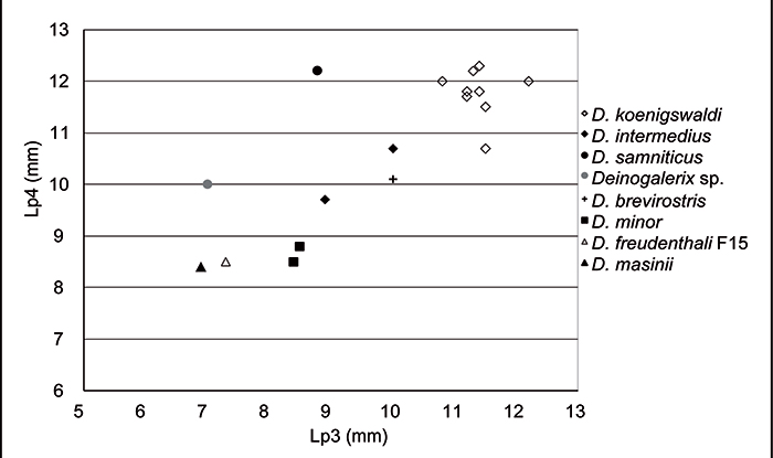
FIGURE 9. Left mandible (SCT 243), Deinogalerix sp. 1, lateral view; 2, dorsal view.
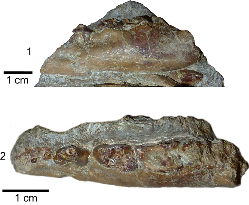
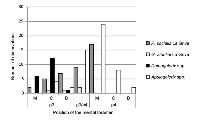
FIGURE 10. Histogram of the positions of the mental foramen in Deinogalerix and in other galericines. M = foramen located under the mesial root of p3 or p4, D = under the distal root of p3 or p4, C ("Central") = between the roots of p3 or p4, I ("Intermediate") = Foramen located between p3 and p4.