FIGURE 1. Geographical and stratigraphical occurrence of MPEF-PV 8166. 1 location of Cañadón Vaca, Chubut, Argentina; 2 Paleogene time table and South American Land Mammal Ages (SALMAs) after (Woodburne et al. 2014a,b); 3 skeletal restoration of Thomashuxleya modified from Simpson (1936); 4 artistic reconstruction of Thomashuxleya externa (by Stjepan Lukac).
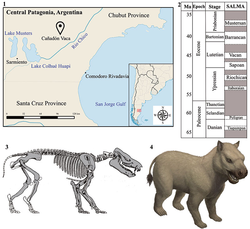
FIGURE 2. Skull of T. externa (MPEF-PV 8166). 1 ventral view; 2 lateral view; 3 occiput in caudal view; 4 detail of upper dentition in occlusal view. Abbreviations are prot=protocone (part of the protoloph), par=paracone (part of the ectoloph), parst=parastyle, met=metacone (part of the ectoloph), metst=metastyle, meph=metaloph, pr.l.f.=primary lingual fold, m.cin=mesial cingulum, l.cin=lingual cingulum.
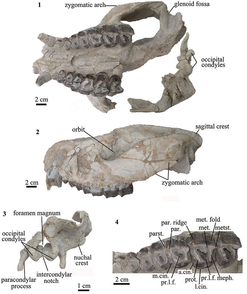
FIGURE 3. Mandible of T. externa (MPEF-PV 8166) in 1 dorsal and 2 lateral views; 3 lower dentition in occlusal view. Abbreviations are tri=trigonid, tal=talonid, prod=protoconid, metd=metaconid, entd=entoconid, hypd=hypoconulid.
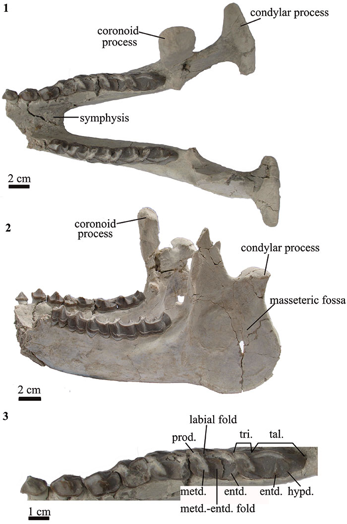
FIGURE 4. Scapulae and humeri of T. externa (MPEF-PV 8166). 1 right and 2 left scapulae in proximal view; 3 left scapula in dorsolateral view; right and left humeri in 4 right and left humeri in anterior (top) and distal (bottom) views; 5 right and left humeri in posterior view.

FIGURE 5. Forelimb of T. externa (MPEF-PV 8166). 1 right and left ulnae in frontal view; 2 left ulna in medial view; right radius in 3 frontal, 4 lateral, and 5 posterior views.
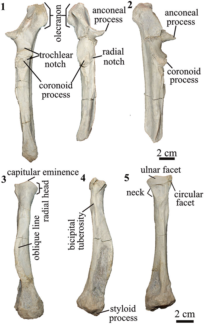
FIGURE 6. Hindlimb of T. externa (MPEF-PV 8166). right pelvis in 1 dorsal and 2 lateral views; 3 right femur in anterior view; 4 right tibia in proximal (top), anterior (middle) and distal (bottom) views; left astragalus in 5 dorsal and 6 plantar views; 7 right navicular in proximal view; 8 navicular and entocuneiform in plantar views.
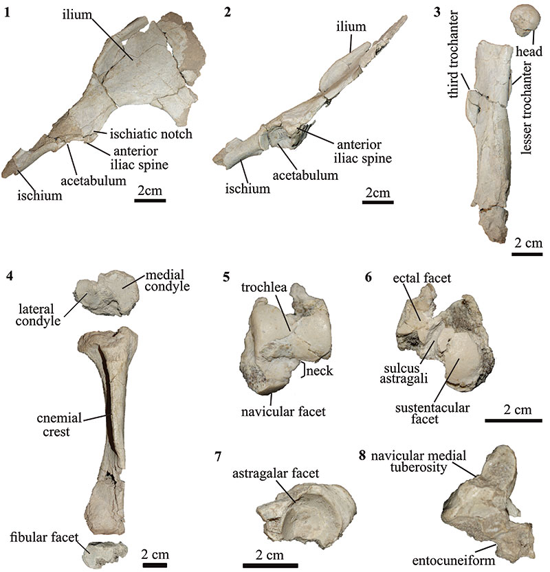
FIGURE 7. Bone histology and microstructure of T. externa (MPEF-PV 8166). 1 Cross section of the midshaft of the right femur; 2 same as 1 after conversion to a binary image (black represents bone and white the cavities); 3 bone histology under linear polarized light, with black arrows pointing the lines of arrested growth (LAGs). OCL= Outer circumferential layer. 4 Bone histology under cross polarized light.
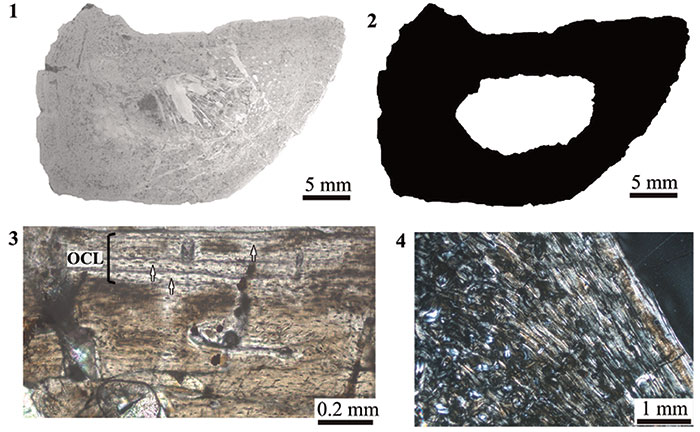
FIGURE 8. Strict consensus of 281 trees, 420 steps in length showing the phylogenetic relationships of Thomashuxleya within Notoungulata based on the morphological dataset of Deraco and García-López (2015). Numbers indicate bootstrap values above 50.
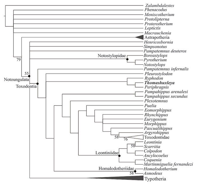
FIGURE 9. Strict consensus of 510 trees, 122374 steps in length from unconstrained parsimony analysis of combined proteomic and morphological data.
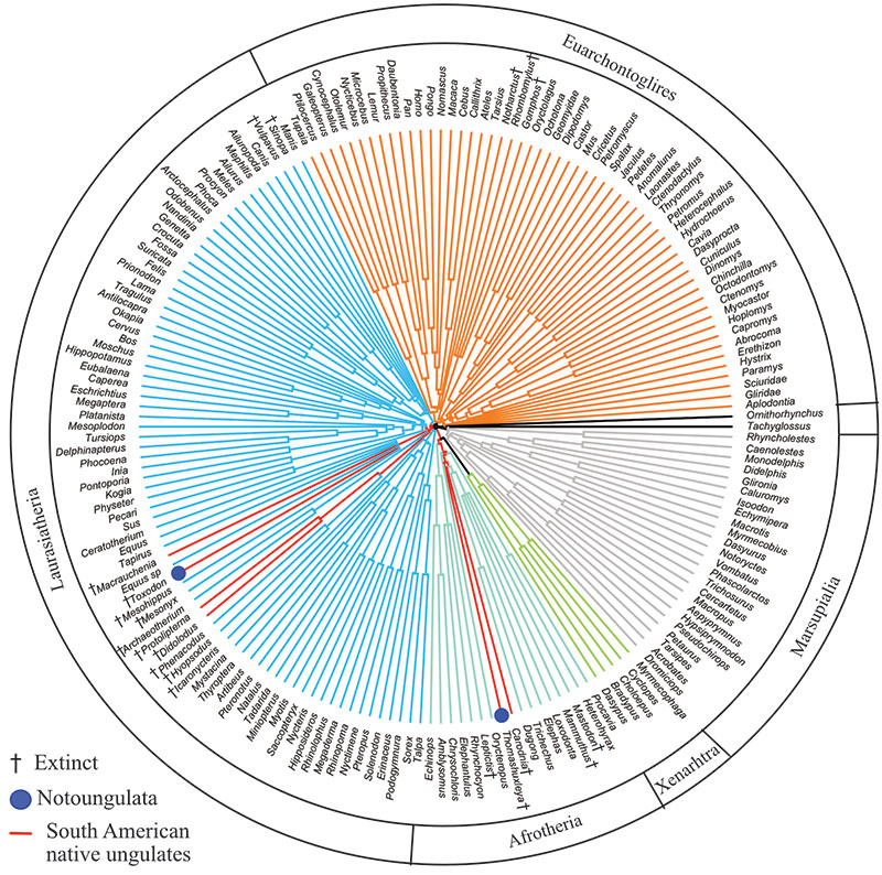
FIGURE 10. Strict consensus of 620 trees, 122391 steps in length from parsimony analysis of combined proteomic and morphological data constraining monophyly of each of two clades (but not both together): Notoungulata (i.e., Thomashuxleya and Toxodon) and Litopterna (i.e., Protolipterna and Macrauchenia).
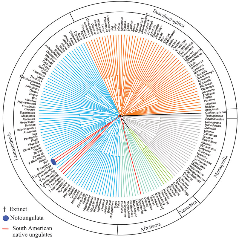
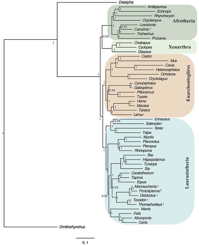
FIGURE 11. Optimal Bayesian tree (i.e., 50% majority rule of post-burn-in trees) of combined proteomic and morphological data. Numbers represent Bayesian posterior probabilities; daggers indicate fossil taxa.
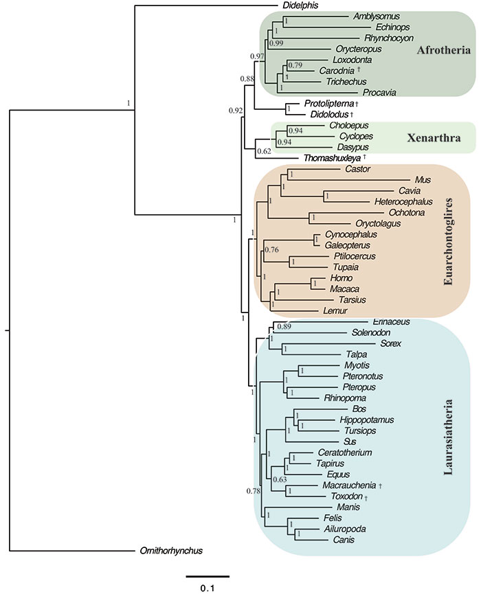
FIGURE 12. Optimal Bayesian tree (i.e., 50% majority rule of post-burn-in trees) of combined proteomic and morphological analysis constraining monophyly of each of two clades (but not both together): Notoungulata (i.e., Thomashuxleya and Toxodon) and Litopterna (i.e., Protolipterna and Macrauchenia). Numbers represent Bayesian posterior probabilities; daggers indicate fossil taxa.