FIGURE 1. Fused cervical vertebrae 12 and 13 in juvenile Muraenosaurus sp. (NWM 19.96.G17) in 1. ventral 2. left lateral 3. right lateral and 4. dorsal view. 5. Ventral view of vertebral series (numbered), showing abnormal position of ventral foramina in vertebrae 10 and 11, and symmetrical positioning in 14, 15. 6. Reconstruction of Muraenosaurus (from Andrews, 1910) showing position of vertebral fusion. Abbreviations: for, foramina subcentralia; naf, neural arch facets. Scale bar equals 4 cm.
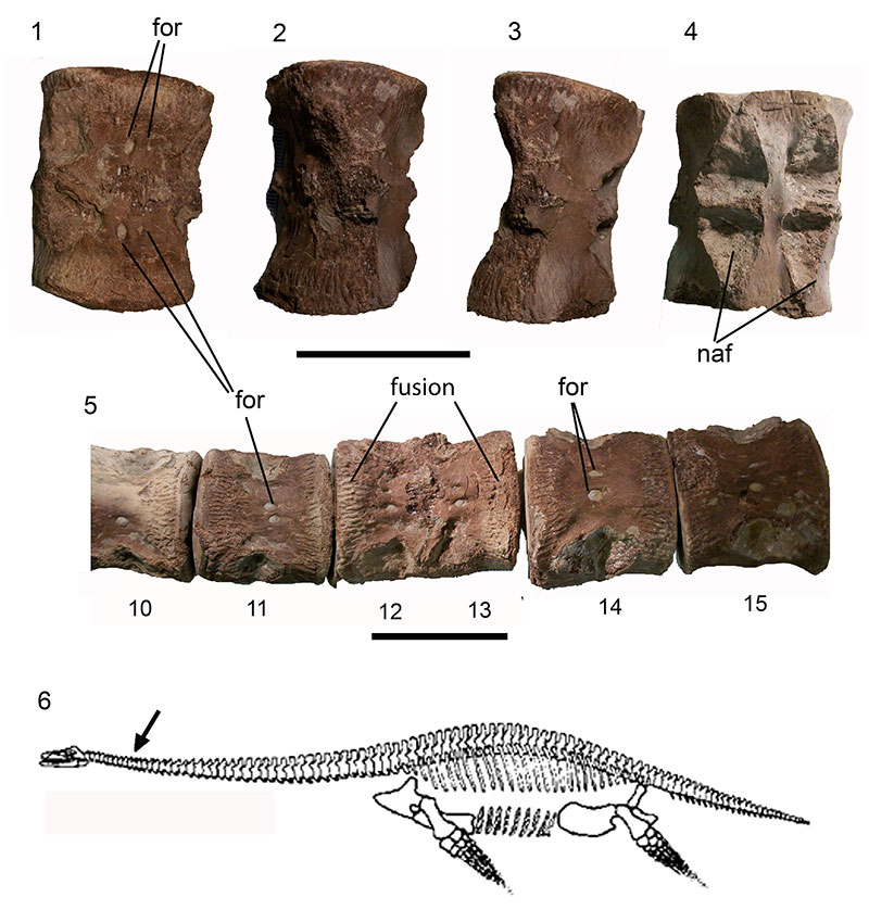
FIGURE 2. Pathological cervical vertebra from Colymbosaurus megadeirus (CAMSM J63919) in 1. anterior view 2. right lateral view 3. ventral view. 4. Left lateral view of pathological vertebrae in series, museum numbers CAMSM J63919 v, w, x, aa; estimated positions in cervical series 26 to 29. 5. Plesiosauroid (indet) (GPIT.RE.03173) terminal cervical and first pectoral in left lateral view and 6. right lateral view. Abbreviations: cv, cervical vertebra; pv, first pectoral vertebra; ve, pathological ventral expansions. Scale bar equals 3 cm.
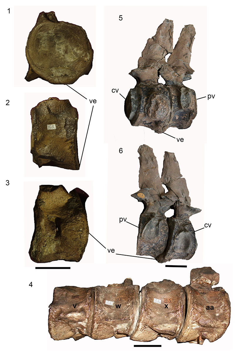
FIGURE 3. Cervical vertebral series from Callawayasaurus columbensis UCMP 38349 in ventral aspect. 1. Vertebrae 23,24,25, anterior end, to right. Scale bar, 5 cm. 2. Enlargement of osteophytic bridging between vertebrae 23-24, 24-25. Abbreviations: ns, neural spine; cr, cervical rib. Scale bar equals 5 cm.
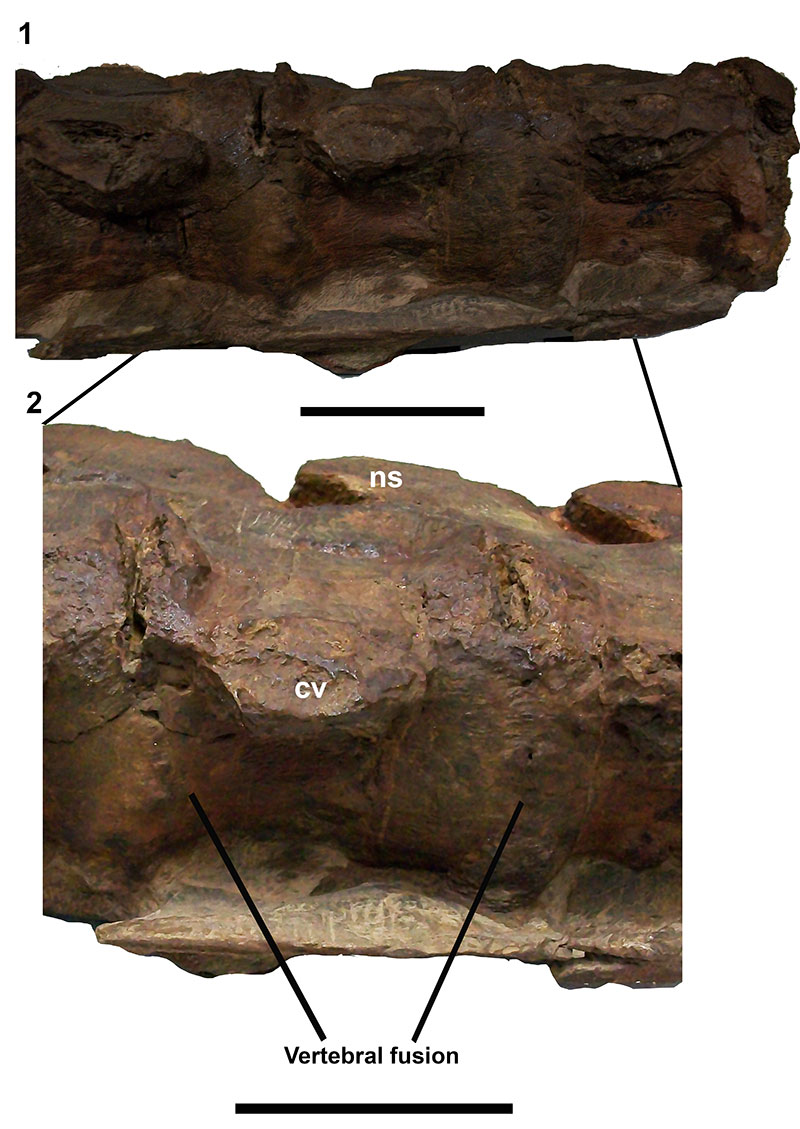
FIGURE 4. Cervical vertebral series from Callawayasaurus columbensis UCMP 38349 in ventral aspect. 1. Vertebrae 15-18, anterior end to right. Scale bar equals 5 cm. 2. Enlargement showing claw-like ventral expansion projecting antero-posteriorly between vertebrae 16-17, 17-18. 3. Ventral view of cervical series from UCMP 38349 without pathologies, vertebrae 32-35. Abbreviations: ns, neural spine; cr, cervical rib; ve, pathological ventral expansion. Scale bar equals 5 cm.
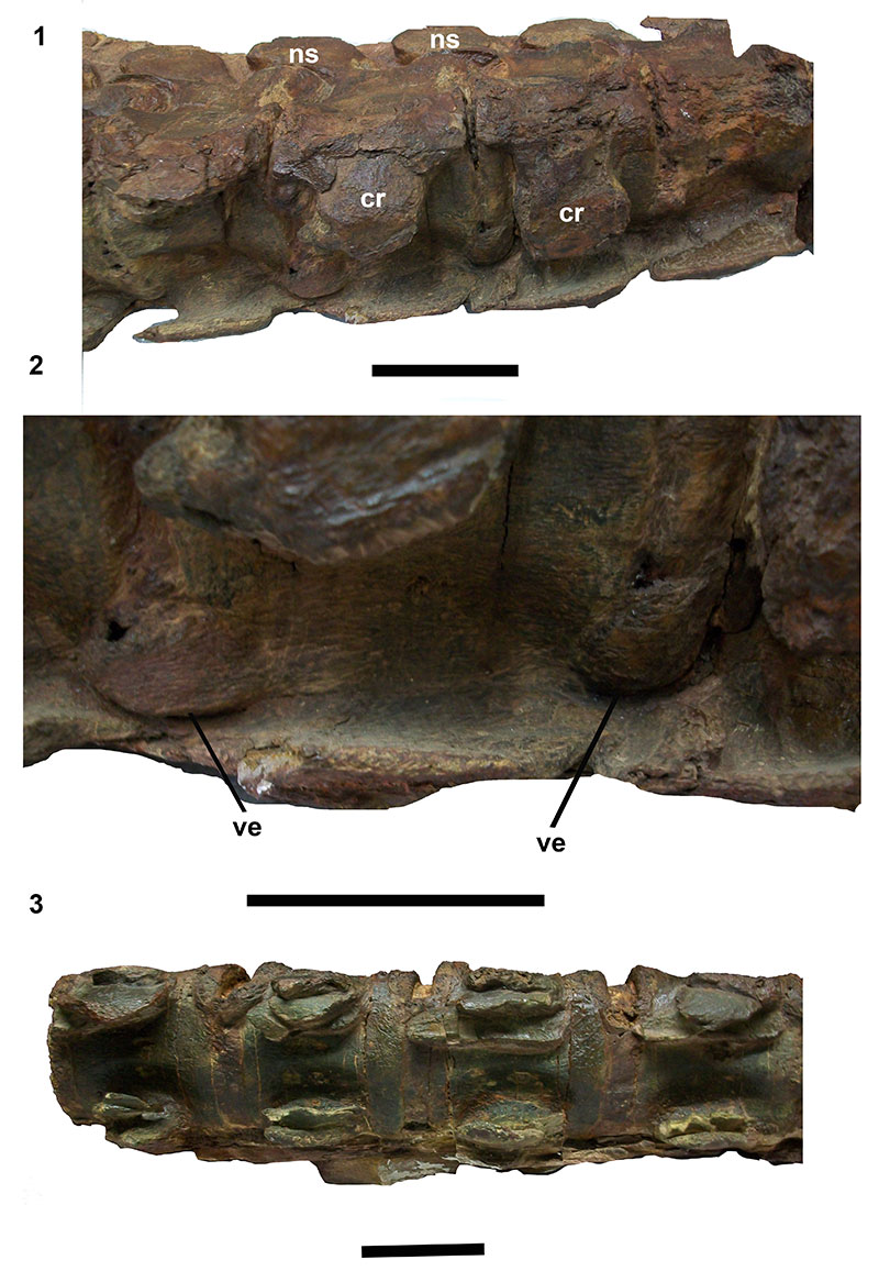
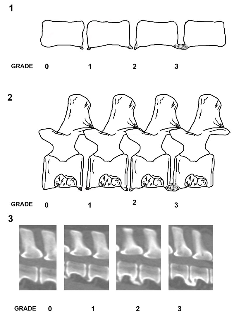
FIGURE 5. 1. General diagram of spondylosis deformans grades (based on Kranenberg et al., 2012) 2. Schematic diagram of plesiosaur cervical vertebrae showing spondylosis deformans grades. Grade 0: no enthesophytes. Grade 1: small enthesophyte at the edge of the epiphysis, not extending past the end plate. Grade 2: enthesophyte extending beyond the end plate but not connecting to enthesophyte on adjacent vertebra. Grade 3: enthesophytes on adjacent vertebrae connected forming bony bridges between vertebrae. 3. Reproduction of radiographic images of spondylosis deformans grades in a dog, for comparison. https://veteriankey.com/spondylosis-deformans/