FIGURE 1. Dunkleosteus terrelli CMNH 50322, from Late Devonian (Famennian) Cleveland Shale of northcentral Ohio, macrophotographs. 1, dorsal, external view of headshield; 2, internal view of headshield with associated ADL and anterior vertebral column (preserved synarcual and a small number of unmodified vertebrae preserved); 3, preserved synarcual oriented anteroposteriorly, and unmodified vertebrae posteriorly. Black arrows indicate anterior. Dashed line in Fig. 1.2 indicates anterior continuation of synarcual (not preserved). Abbreviations: ADL, anterodorsal plate of the trunkshield; Asg, anterior supragnathal; Ce, central; Ig, ifragnathal; L Ig, left infragnathal; Mg, marginal; Nu, nuchal; Pi, pineal; Pn, paranuchal; Pro, preorbital; Psg, posterior supragnathal; Ptn, postnasal; Pto, postorbital; R Ig, right infragnathal; Ro, rostral; So, suborbital; syn, synarcual; t.zone, transitional zone.
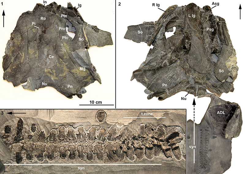
FIGURE 2. Dunkleosteus terrelli CMNH 50322, Late Devonian (Famennian) Cleveland Shale, northcentral Ohio, macrophotographs. 1, entire preserved vertebral column including synarcual. White arrowheads indicate sharp medial border of the scoop-shaped articular surface of the neural arch, black arrowhead indicates ‘waisting’ between the articular surface and the rest of the arch; 2, closeup of more anterior vertebrae within the synarcual, bone modification (via loss) results in a more 2D element and deposition of woven and perichondral bone between arch bases; 3, closeup of more posterior vertebrae, showing loss of medial wall of articular surface (compare vertebrae 14 to 13, the latter appearing crushed) and transition from more 3D to 2D vertebrae (compare vertebrae 13 and 12). Abbreviations: As in previous figures, also tub, tubercles.
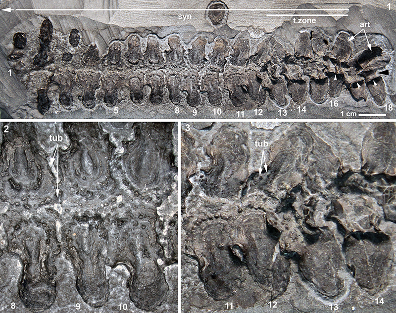
FIGURE 3. 1, Dunkleosteus terrelli CMNH 50322, Late Devonian (Famennian) Cleveland Shale, northcentral Ohio. 3D volume rendered CT-scans. 1, 2, vertebrae including unmodified outside the synarcual (vertebrae 17, 18) and through transition zone (vertebrae 12-16). Internal view, anterior to left, white asterisk indicates region of woven bone deposition; 2, vertebrae as in Figure 3.1, external view, anterior to right. 3, closeup of vertebrae in transitional zone, and just anterior to this zone (vertebrae 11-14), showing changes in vertebral morphology and deposition of new bone, internal view; 4, closeup of vertebrae 11-13, internal view. White arrows indicate anterior. Abbreviations: As in previous figures, also b.ll, second layer of deposited bone; fs, fusion of vertebrae; peri, deposition of perichondral bone; wst, waisting in vertebrae. Key to asterisks, arrows and arrowheads: Black arrowheads (Figure 3.2) indicate the dorsolateral margin of vertebrae, which is modified in vertebra 14 and those more anteriorly, but unmodified in vertebra 15 and those more posterior. White arrowheads indicate a crest along the lateral vertebral surface, progressively reduced from vertebra 16 to vertebra 13 and more anteriorly. Red arrows (vertebra 11, 12) show unfused dorsomedial margins of the arches, compared to fusion (fs) between the arches anteroposteriorly. Black arrowheads (Figure 3.3; white arrowheads in Figure 3.4) indicate tubercles developing along vertebral margin, via proposed resorption of new bone (b.ll).
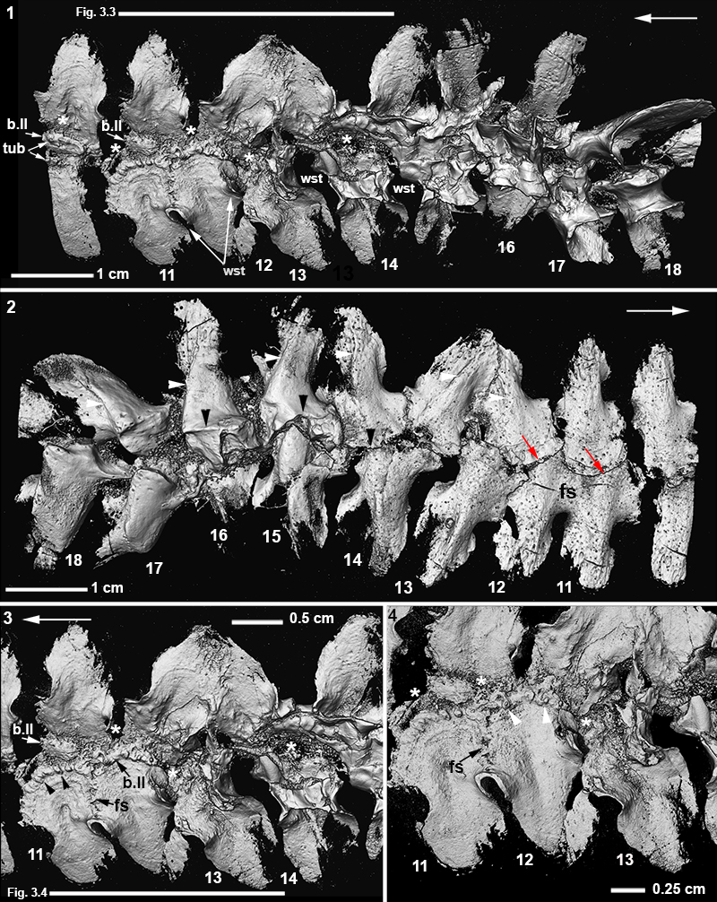
FIGURE 4. Compagopiscis croucheri NHMUK PV P52549, Late Devonian (Frasnian) Gogo Formation, Western Australia. 3D volume rendered CT-scans. 1, Headshield and anterior trunkshield in external view. Black asterisk indicates gap between head and trunkshield. 2, Internal view of median dorsal plate, showing synarcual (syn) comprising 5 modified vertebrae with minimal fusion of individual vertebrae. White arrowheads indicate position of fused vertebrae within the synarcual, paired arch bases and articular surfaces visible in this view. 3, Closeup of synarcual showing rectangular shape of articular surfaces within the synarcual, and dorsolateral processes. 4, lateral view of synarcual, showing more posterior modified and unmodified vertebra, one preserving scoop-shaped articular surface, comparable to other arthrodires such as Dunkelosteus and Eastmanosteus (Figure 5). 5, lateral view of anterior synarcual, showing more modified with weak fusion (white asterisk). Dorsal elements of the synarcual (neural arches) are modified into a keel, as in ptyctodont placoderms (Figure 6). 6, lateral view of vertebrae posterior to the synarcual, showing reduced lateral processes, but modified, rectangular articular surfaces, compared to more posterior vertebrae with scoop-shaped surface. 7, dorsal face of modified vertebrae, showing foramina and zygapophyses. White arrow indicates anterior direction. Abbreviations: as in previous figures, also k, keel on synarcual; k.MD, keel on median plate; MD, median dorsal plate; pr, process on dorsolateral surface of vertebra; vert, vertebrae posterior to synarcual; zyg, zygapophyses.
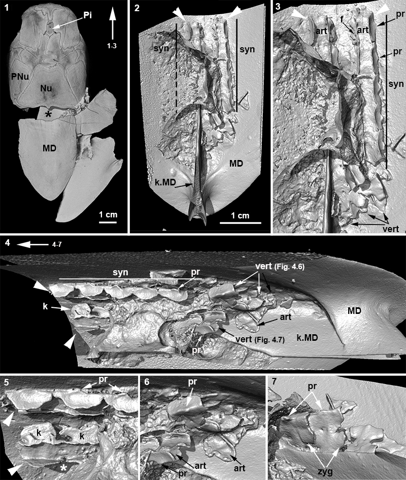
FIGURE 5. Phylogeny of the jawed vertebrates, showing distribution of the synarcual among the paraphyletic Placodermi, and among Chondricthyes, in the Holocephali and Batoidea. Comparable fusion (syncervicals) occur in a range of tetrapods (Van Buren and Evans, 2017), not shown in this cladogram. Cladogram modified from Zhu et al. (2016). Icons in cladogram from Bechard et al. (2014), Johanson and Smith (2003) and University of Washington Freshwater and Marine Image Bank. Images below cladogram (Johanson et al. 2012): 1, Rhenanida synarcual, Jagorina pandora MB.f 510.2; 2, Ptyctodontida synarcual, Austroptyctodus gardineri NHMUK PVP56665; 3, Arthrodira synarcual, Compagopiscis croucheri NHMUK PVP52549; Holocephali synarcual, Chimera monstrosi AMNH 55040; Batoidea synarcual, Dasyastis americana AMNH 30607. Red arrows indicate the position of the synarcual. Scale bars = 1cm.
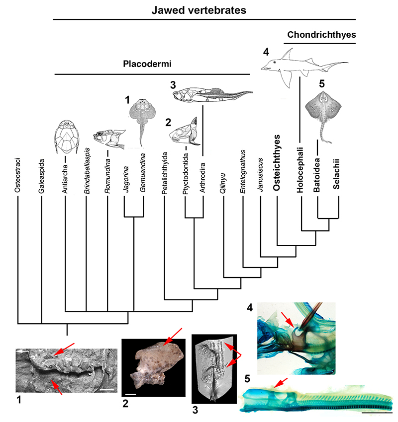
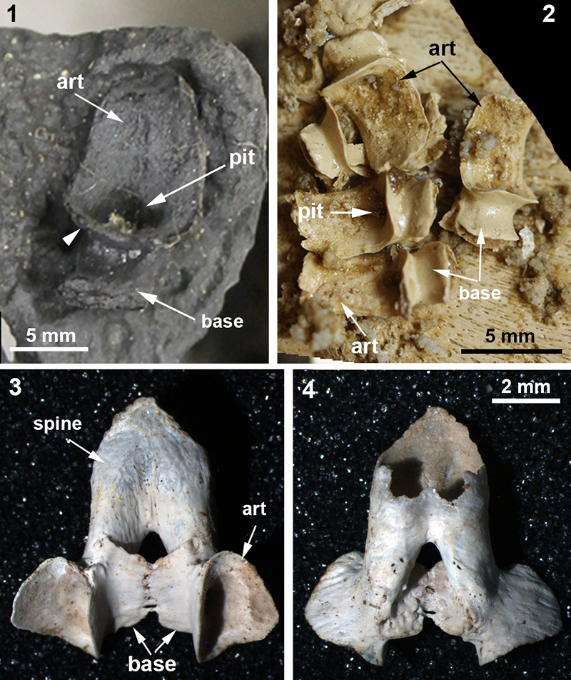
FIGURE 6. 1, Dunkleosteus terrelli CMNH 50322, Late Devonian (Famennian) Cleveland Shale, northcentral Ohio. Isolated vertebral element found near headshield plates, but believed to be from more posterior vertebral column, preserving scoop-shaped articular surface (articulating on notochord) and arch base. White arrowhead indicates sharp margin of articular surface; 2, Eastmanosteus calliaspis, NHMUK PV P54325, Late Devonian (Frasnian) Gogo Formation, Western Australia. Multiple vertebrae with scoop-shaped articular surface and reduced arch base, vertebrae located under bony trunkshield (as in D. terrelli); 3, 4, Eastmanosteus calliaspis, NHMUK PV P57642, Late Devonian (Frasnian) Gogo Formation, Western Australia. More posterior vertebra, posterior to the trunkshield plates, showing fused neural spines. 3, ventral view showing articular surface of arch bases, 4, dorsal view. Abbreviations as in previous figures, also: base, neural arch base, pit, pit within scoop-shaped articular surface.