FIGURE 1. Asterolepis orcadensis headshields; 1, 2, GSE 12828 from Monquhanny Farm quarry, Shapinsay; 1, photograph of the natural mould of the specimen; 2, line drawing of the specimen flipped horizontally to give correct orientation in life; 3, 4, NMS G.2018.8.16.1 from Taft, Westray; 3, photograph of the dorsal surface; 4, line drawing of the dorsal surface. Scale bars equal 10 mm. Abbreviations: ifc=infraorbital sensory line; L=lateral plate; mp=middle pit-line groove; Nu=nuchal plate; pp=posterior pit-line groove; PNu=paranuchal plate; Pp=postpineal plate; Pmg=postmarginal plate; Prm=premedian plate; ptc=cephalic division of the main lateral line; soc=anterior section of supra-orbital sensory line.
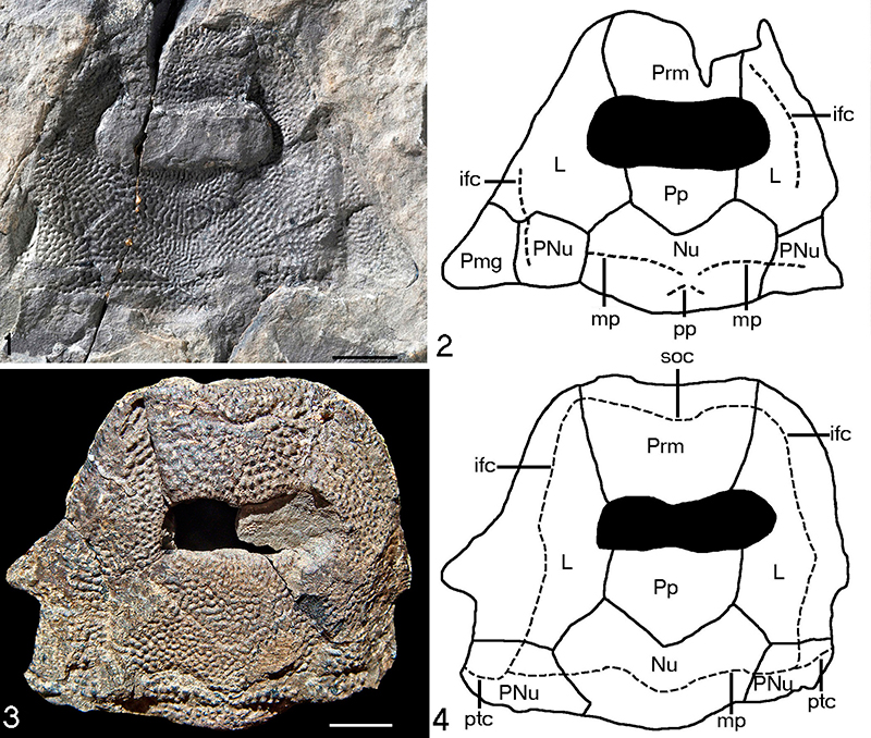
FIGURE 2. Asterolepis orcadensis NMS G.2018.8.22 a nuchal plate from Taft, Westray. Scale bar equals 10 mm.
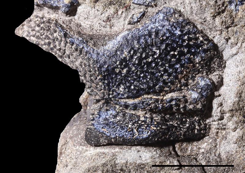
FIGURE 3. Asterolepis orcadensis, NMS G.1898.1.1 an anterior median dorsal plate from Lang Geo, Deerness; 1, viscerally preserved plate; 2, impression of the visceral surface of the same plate. Scale bar equals 10 mm. Abbreviations: ADL=anterior dorso-lateral plate; cf.ADL=area overlapping ADL; cf.MxL=area overlapping MxL; i.oa.PMD=impression of area overlapped by PMD; MxL=mixilateral plate; PMD=posterior median dorsal plate.
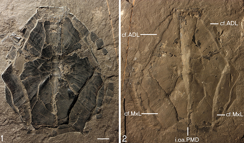
FIGURE 4. Asterolepis orcadensis, NMS G.2018.8.6 a posterior median dorsal from Stancro, Westray; 1, dorsal side; 2, natural impression of dorsal side; 3, visceral side; 4, line drawing of visceral side. Scale bar equals 10 mm. Abbreviations: cf.MxL=area overlapping MxL plate; cr.tp=posterior transversal internal crest; dmr=dorsal median ridge; l=lateral corner; MxL=mixilateral plate; pma=posterior marginal area; pt=ventral pit.
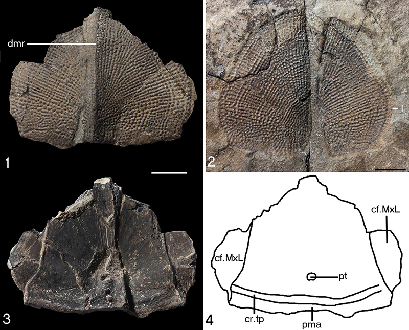
FIGURE 5. Asterolepis orcadensis, GSE12829 a right anterior dorso-lateral plate from Coopalash shore. Scale bar equals 10 mm. Abbreviations: dlm=dorsal lamina; dlr=dorso-lateral ridge; lcg=main lateral line groove; llm=lateral lamina.
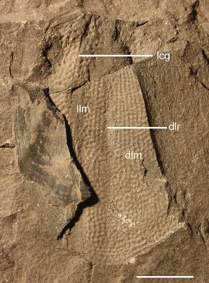
FIGURE 6. Asterolepis orcadensis. NMS G.2018.8.17.2, a natural mould of a left anterior dorso-lateral plate from Taft, Westray. Scale bar equals 10 mm. Abbreviations: dlm=dorsal lamina; llm=lateral lamina; oa.AMD=area overlapping the anterior median dorsal plate; oa.AVL=area overlapping the anterior ventro-lateral plate.
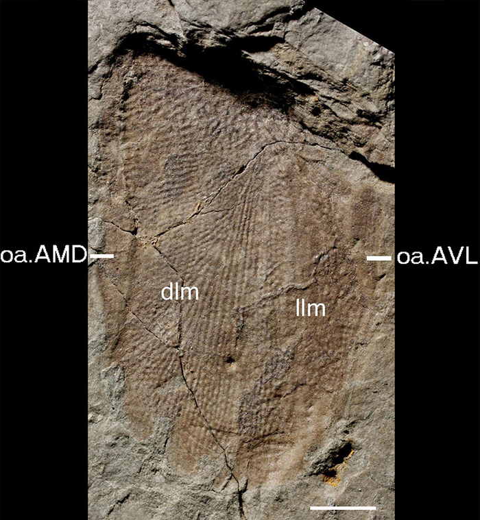
FIGURE 7. NMS G.2018.8.17.1, anterior part of a left anterior dorso-lateral plate from Taft, Westray; 1, 2, in outer view; 3, 4, in visceral view. Scale bars equal 10 mm. Abbreviations: AMD=anterior median dorsal plate; AVL=anterior ventro-lateral plate; cri=infra-articular cristae; crs=supra-articular cristae; dlm=dorsal lamina; dlr=dorso-lateral ridge; f.art=articular fossa; lcg=main lateral line groove; llm=lateral lamina; oa.AMD=area overlapping the anterior median dorsal plate; oa.AVL=area overlapping the anterior ventro-lateral plate; pnoa=postnuchal ornamented corner; pro=processus obstans.
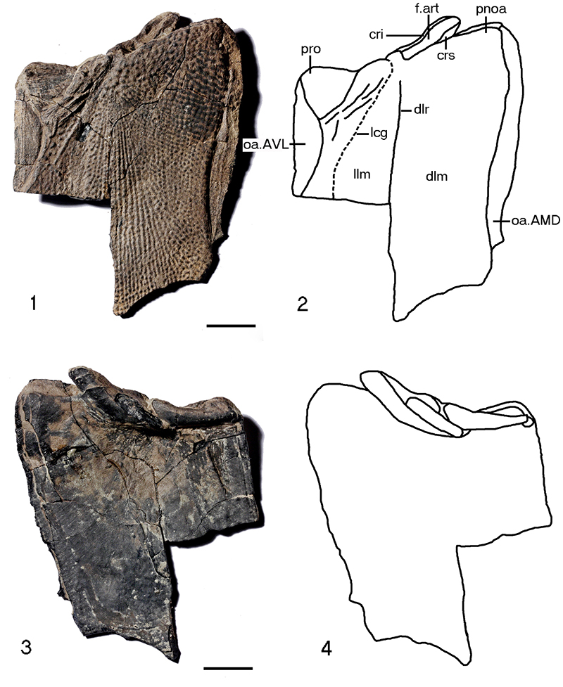
FIGURE 8. GSE 12830 from Coopalash, Shapinsay a left mixilateral plate; 1, photograph of the specimen; 2, a line drawing of the specimen. Scale bar equals 10 mm. Abbreviations: d=dorsal corner; dlm=dorsal lamina; dlr=dorso lateral ridge; lcg=main lateral line groove; llm=lateral lamina, oa.PMD=area overlapped by the posterior median dorsal plate.
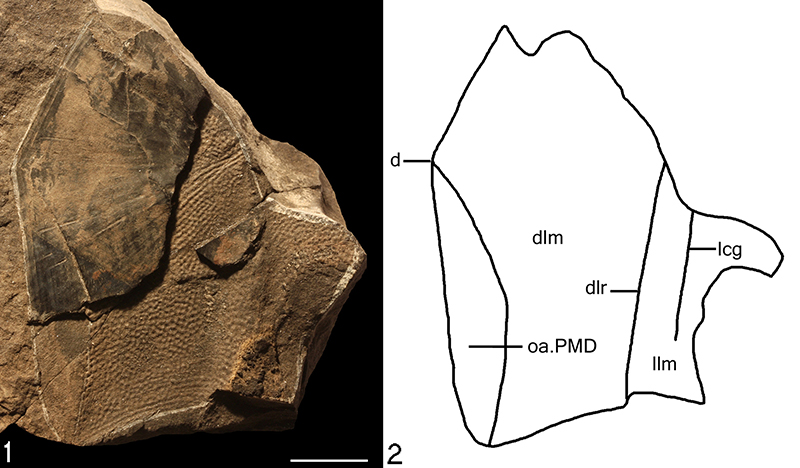
FIGURE 9. 1, 2, NMS G.2018.8.5 impression of the visceral side of a left anterior ventro-lateral plate from Loth Quarry, Sanday; 3, 4, NMS G.2019.1.1 an impression of the lateral and ventral side of a right anterior ventro-lateral plate from Coopalash, Shapinsay. Scales bars equal 10 mm. Abbreviations: c.al=antero-lateral corner; cf.MxL=area overlapping MxL; cf.MV=area overlapping MV; f.ax=axillary foramen; llm=lateral lamina; MV=median ventral plate; MxL=mixilateral plate; pbr=process brachialis; vlm=ventral lamina; vlr=ventro-lateral ridge.
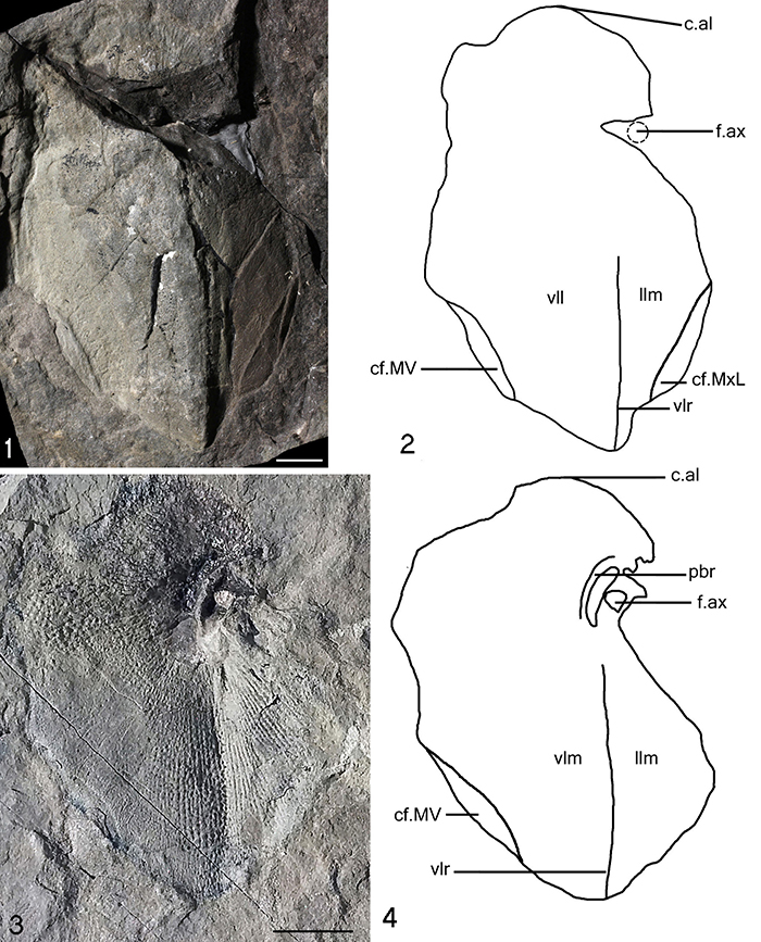
FIGURE 10. NMS G.2018.8.3, impression of the lateral and ventral side of a right anterior ventro-lateral plate from Loth Quarry, Sanday. Scale bar equals 10 mm.
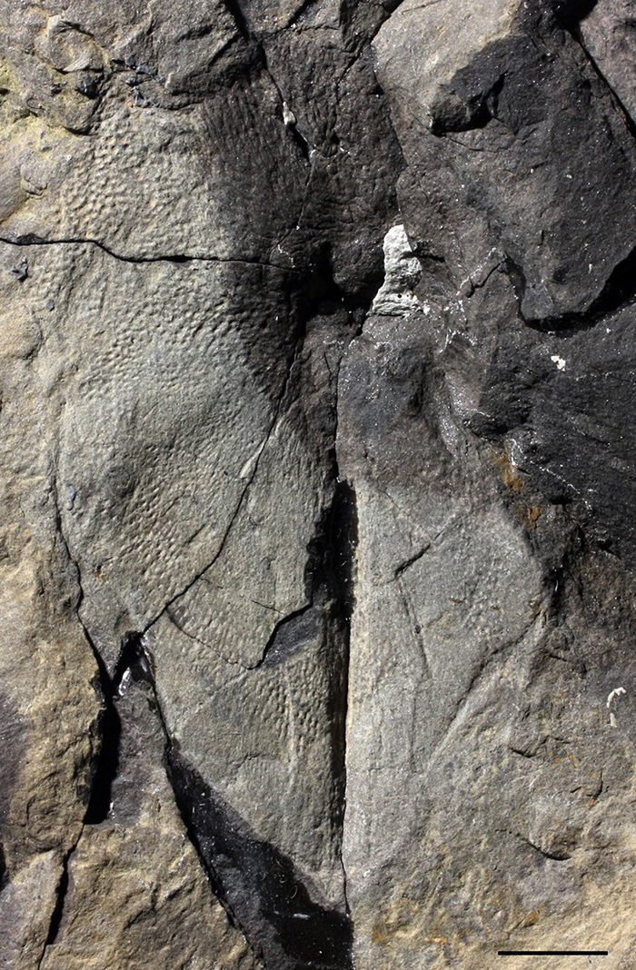
FIGURE 11. NMS G.2018.8.3 a small piece of a right anterior ventro-lateral plate from Loth Quarry, Sanday. Scale bar equals 10 mm. Abbreviations: f.ax=axillary foramen; f.art.d=dorsal half of the fossa articularis pectoralis; f.art.v=ventral half of the fossa articularis pectoralis; m.lim=margo limitans of the fossa articularis pectoralis; pbr=processus brachialis; pe=pars pedalis of processus brachialis; vlr=ventro-lateral ridge.
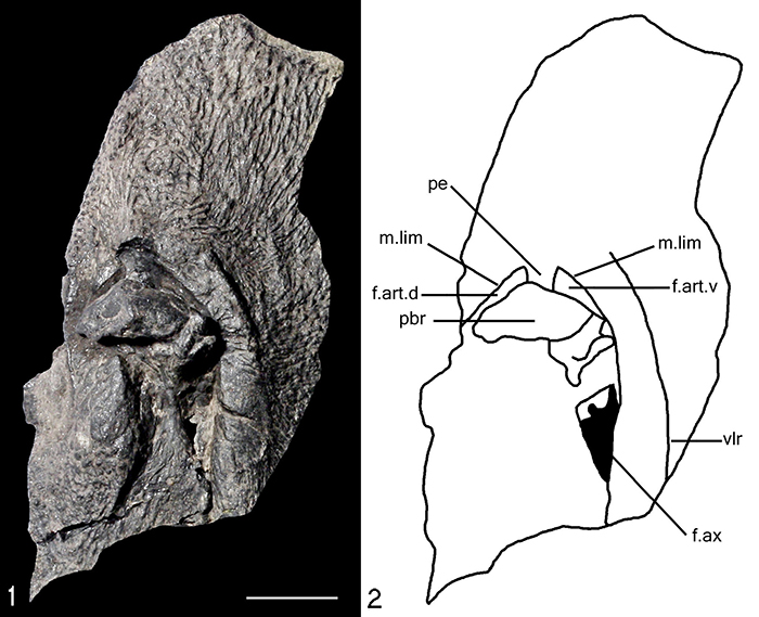
FIGURE 12. NMS G.1897.57.1 posterior part of a right posterior ventro-lateral plate from Long Geo, Orkney mainland. Scale bar equals 10 mm.
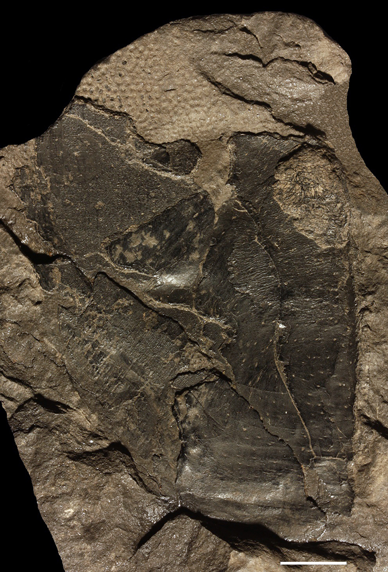
FIGURE 13. NMS G.2018.8.16.2 from Taft, Westray. Right pectoral appendage; 1, dorsal view; 2, line drawing of dorsal view; 3, in ventral view; 4, line drawing of ventral view. Scales bars equal 10 mm. Abbreviations: ar3d=internal articular area of Cd1; Cd1=dorsal central plate 1; Cd2=dorsal central plate 2; Cd3=dorsal central plate 3; Cv1=ventral central plate 1; Cv2=ventral central plate 2; Cv3=ventral central plate 3; Ml2=lateral marginal plate 2; Ml3=lateral marginal plate 3, Mm1=mesial marginal plate 1; Mm2=mesial marginal plate 2; Mm3=mesial marginal plate 3; pMm1=process on the mesial marginal plate 1.
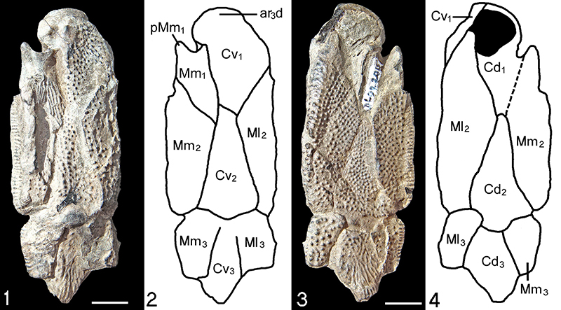
FIGURE 14. Histology of Asterolepis orcadensis NMS G.2018.8.11.4 from Stancro Shore, Westray. Scale bar equals 0.25 mm. Abbreviations: bas=basal lamellar layer; med=medial layer; rl=resting line; sup=superficial layer
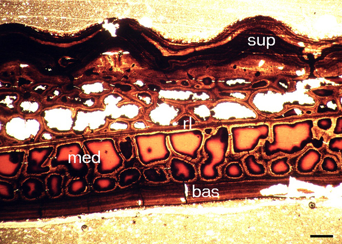
FIGURE 15. Reconstruction of Asterolepis orcadensis partly based on Watson (1932). Abbreviations: ADL=anterior dorso-lateral plate; AMD=anterior median dorsal plate; MxL=mixilateral plate; PMD=posterior median dorsal plate. For plate labelling of the headshield see Figure 1 and for the pectoral appendages Figure 13.
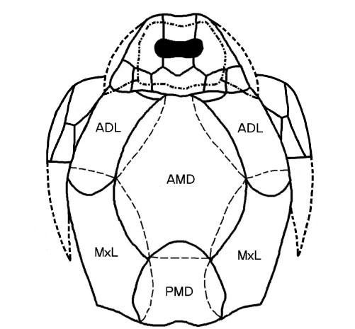
FIGURE 16. Various reconstructions of species of Asterolepis; 1, simplified drawing of Asterolepis orcadensis; 2, Asterolepis ornata, head and body trunk, redrawn and simplified from Lyarskaya 1981 (figure 74.1); 3, Asterolepis maxima, head and body trunk, Redrawn and simplified from Traquair 1894 (figure 37); 4, Asterolepis scabra, head redrawn and simplified from Nilsson 1941 (figure 3); 5, Asterolepis radiata, head redrawn and simplified from Karatajūtė-Talimaa 1963 (figure 43); 6, Asterolepis thule, head redrawn and simplified from Newman and den Blaauwen 2018 (figure 5b); 7, Asterolepis essica, head and body trunk, redrawn and simplified from Lyarskaya 1981 (figure 58); 8. Asterolepis dellei, head drawn from a photograph of a reconstruction in Karatajūtė-Talimaa 1963 (plate 4, figure 1); 9, Asterolepis sp., head redrawn and simplified from Lyarskaya 1981 (figure 63.8) which was copied from Reed 1977; 10, Asterolepis saevesoederberghi, redrawn and simplified from Stensiö 1938 (figure 7).
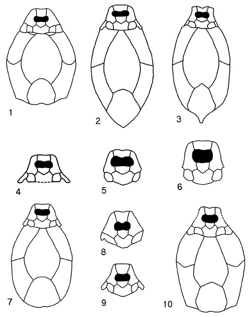
FIGURE 17. Locality map of Asterolepis orcadensis localities in the Orkney Islands; localities include, 1, Coopalash Shore; 2, Lang Geo, Deerness; 3, Long Geo; 4, Hesta Head; 5, Taft.; 6, Stancro Shore; 7, Loth Quarry.
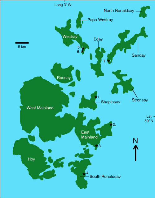
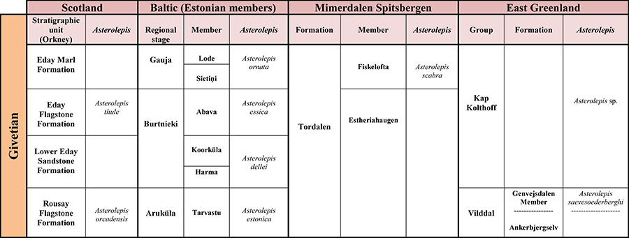
FIGURE 18. Biostratigraphical column showing Asterolepis distribution between Scotland, the Baltic area, Spitsbergen and East Greenland.