FIGURE 1. Drepanaspis reconstruction: by Gross (1963) in front (1), lateral (2), dorsal (3) and ventral (4) view; cross section (5) by Tarlo (1961); by Obruchev and Mark-Kurik (1965) in dorsal (6) and ventral (7) view; by Patten (1932) in dorsal (8) view.
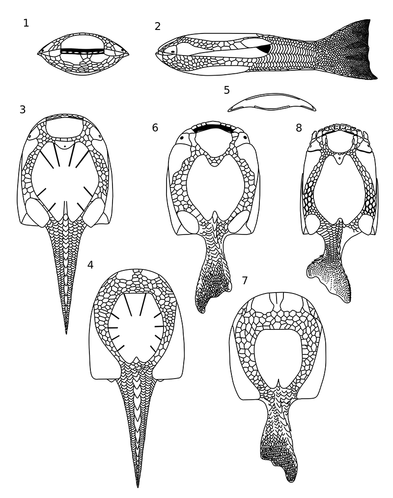
FIGURE 2. Map showing the location Podłazie Hill (1) in the Holy Cross Mountains (HCM), and map of Daleszyce (2) with the position (magnified part) of abandoned quarry at Podłazie Hill.
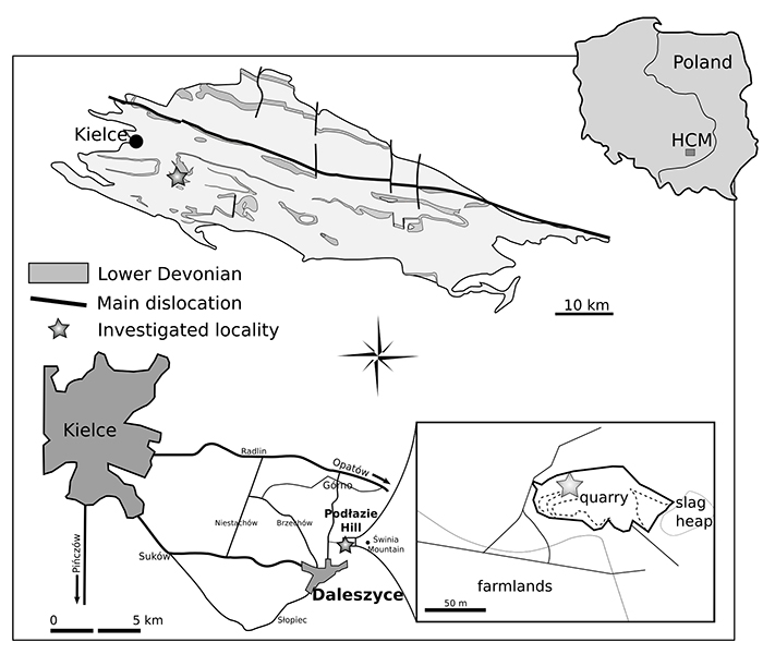
FIGURE 3. Guerichosteus kozlowskii, 1-3. dorsal plate ZPAL Ag. III/4, internal (1) and external (2) view, shape reconstruction (3); 4-7. dorsal plate ZPAL Ag. III/1, internal (4) and external (5) view, drawing of internal (6) and external (7) view with growth line marked 8-9. dorsal plate ZPAL Ag. III/2, external (8) view and drawing (9); 10-11. specimen MUZ.PIG.1733.II.13, external (10) view and drawing (12); 12-13. dorsal plate MZ-VIII/Vp-438, external (12) view and drawing (13).
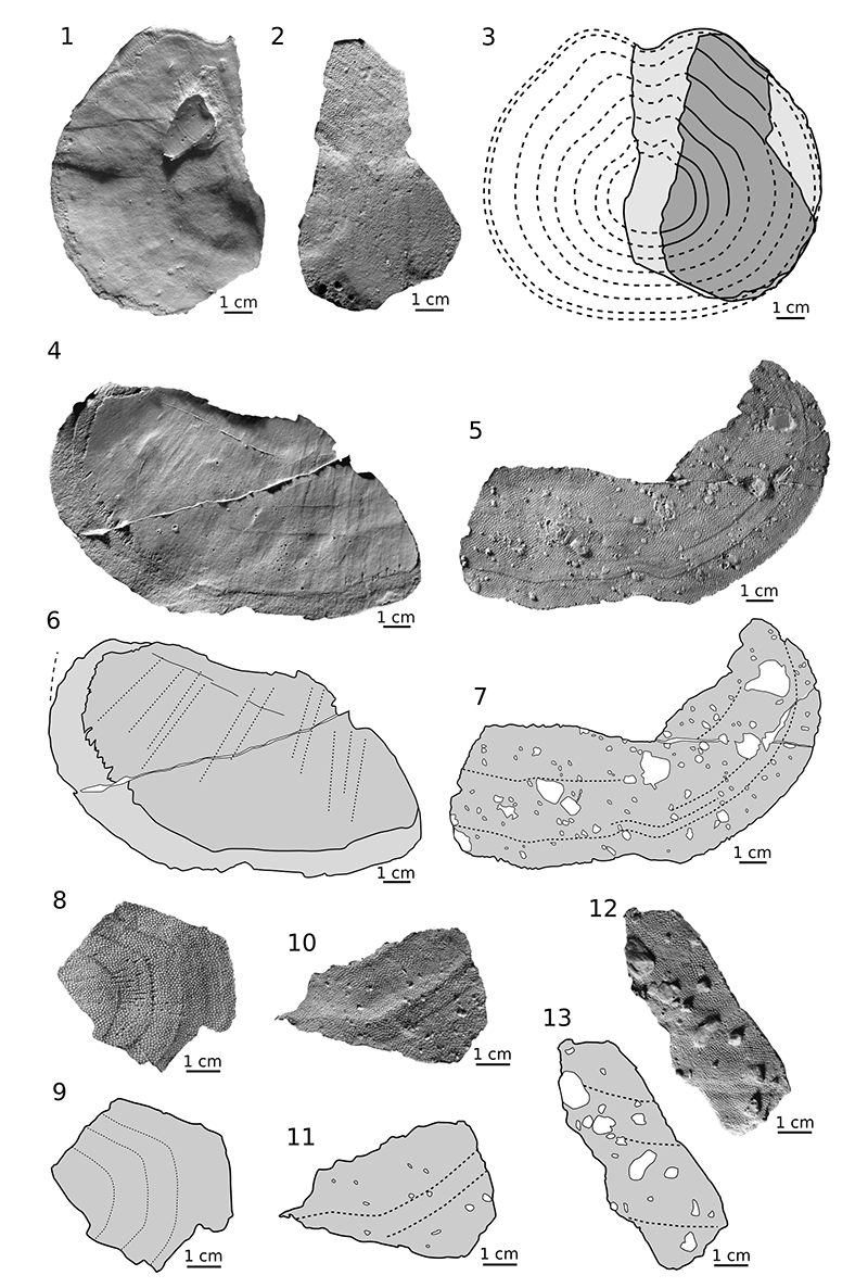
FIGURE 4. Guerichosteus kozlowskii, 1-2. ventral plate ZPAL Ag. III/45, external view, (1), shape reconstruction (2); 3-8. ventral plate MUZ.PIG.1733.II.353, external (3) and internal (5) view, drawing of external (4) view with growth line marked (hached line), drawing of internal (6) view with cross section (7), left lateral view (8).
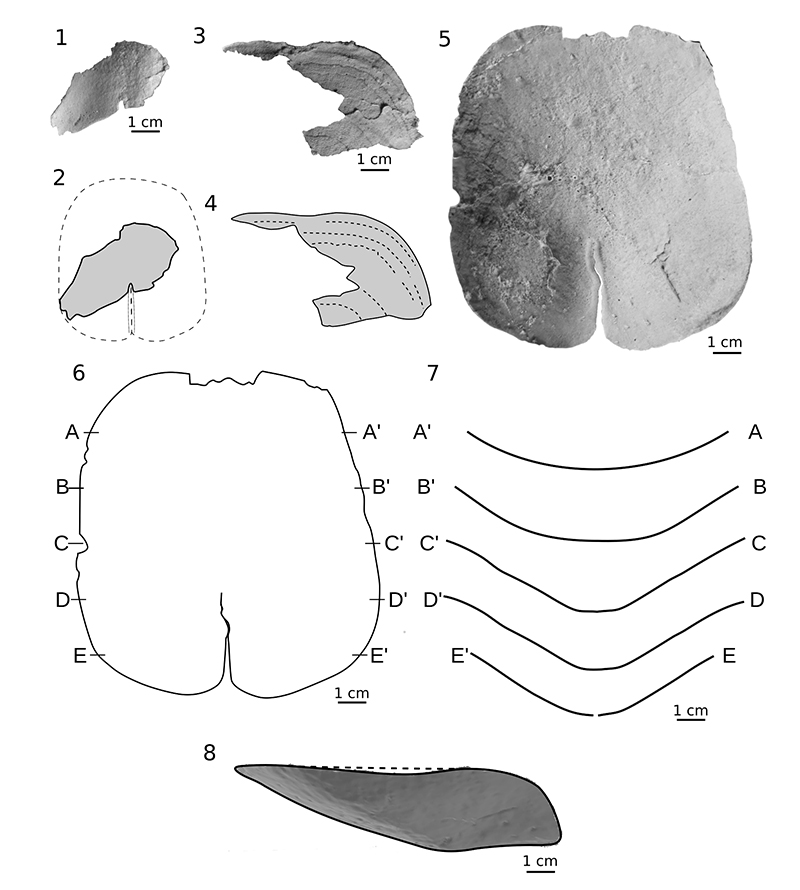
FIGURE 5. Guerichosteus kozlowskii, 1. left branchial plate ZPAL Ag. III/1089, dorsal view (1),drawing of dorsal view (2); 3. fragment of right branchial plate ZPAL Ag. III/16, dorsal view (3), drawing of dorsal view (4); 5. right branchial plate ZPAL Ag. III/15, dorsal view (5); drawing of dorsal view (6); 7. posterior tip of left branchial plate MUZ.PIG.1733.II.185, dorsal (7), ventral (9) and lateral (10) view,drawing of dorsal view (8); 11. fragment of left branchial plate ZPAL Ag. III/8, dorsal view (11), drawing of dorsal view (12); 13. posterior tip of left branchial plate MZ-VIII-Vp-533, dorsal (13) and ventral (15) view, drawing of dorsal view (14).
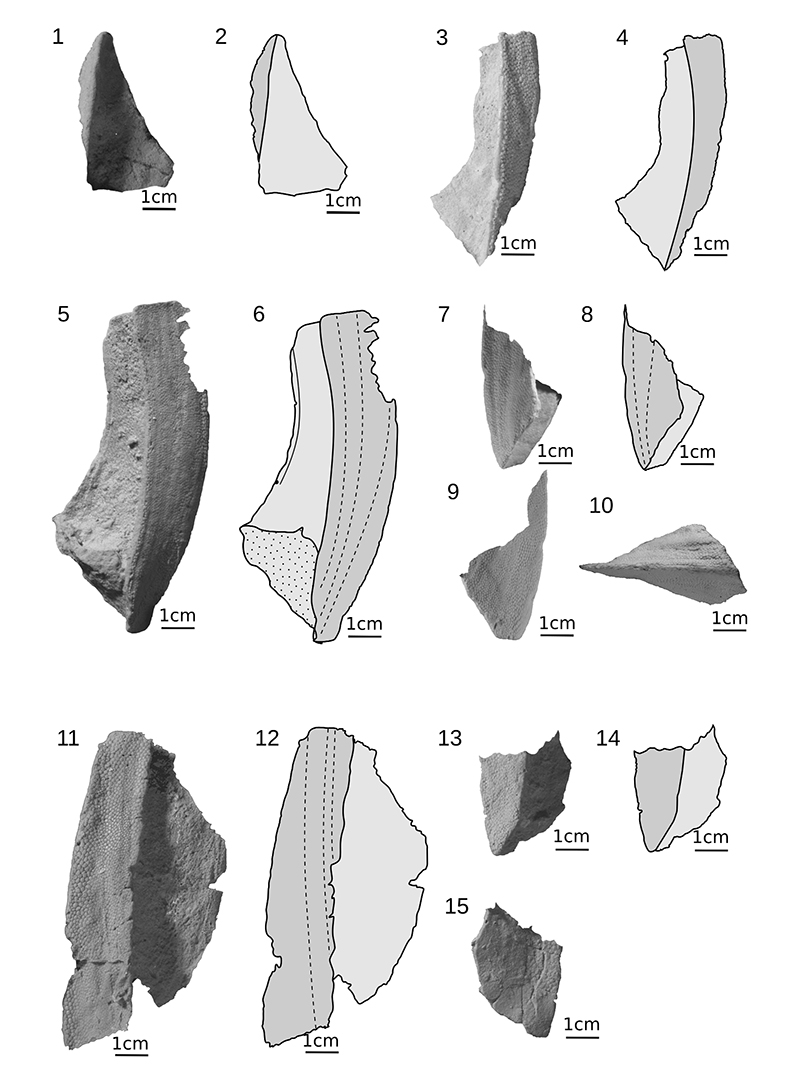
FIGURE 6. Guerichosteus kozlowskii, 1. fragment of right branchial plate ZPAL Ag. III/7, dorsal (1) ventral (2) view, drawing of dorsal (4) and ventral (3) view; 5. fragment of right branchial plate ZPAL Ag. III/9, ventral (5) and dorsal (6) view, reconstruction of ventral (7) and dorsal (8) view; 9. anterior tip of left branchial plate ZPAL Ag. III/14, ventral view (9), drawing of ventral view (10).
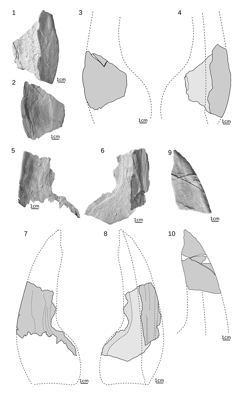
FIGURE 7. Guerichosteus kozlowskii, 1. cornual plate ZPAL Ag. III/19, dorsal view (1) with drawing (2); 3. orbital plate MUZ.PIG.1733.II.154, ventral view (3), internal view on orbital opening (4); 4. median oral plate ZPAL Ag. III/23; 6. lateral oral plate MUZ.PIG.1733.II.355, ventral view (6) and drawing of ventral view (7); 8. complex plate MUZ.PIG.1733.II.357, ventral view, cross-section (9); 10. Close-up on median oral plate and lateral oral plate of Drepanaspis gemuendenensis specimen stored in Muséum national d'Histoire naturelle in Paris.
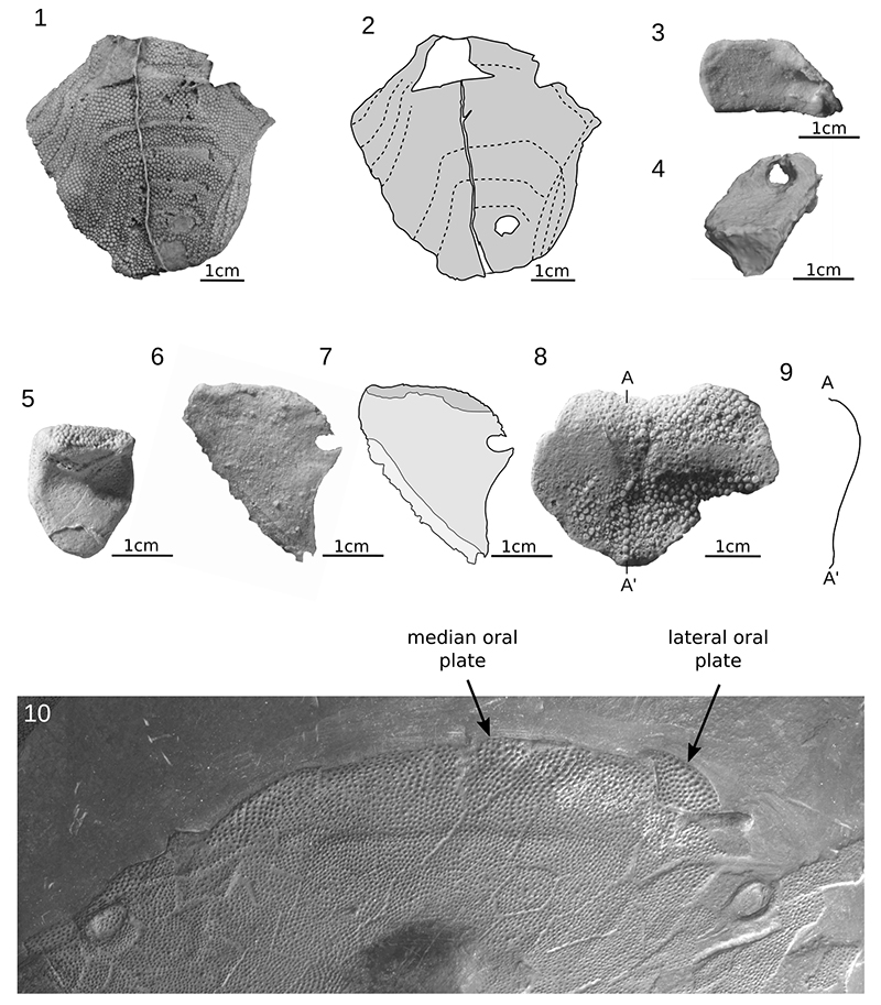
FIGURE 8. Guerichosteus kozlowskii, 1. ridge scale ZPAL Ag. III/27A, internal view (1) and drawing of internal view with photo of close-up on tubercles (2), cross-section (3); 4. ridge scale MZ-VIII/Vp-488, internal (4) and external (6) view with drawing of internal view and shape reconstruction (5); 7. ridge scale ZPAL Ag. III/26, external view (7), cross-section (8), drawing with shape reconstruction (9); 10. small ridge scale MUZ.PIG.1733.II.13, external view (10), drawing with shape reconstruction (11); 12. ridge scale MZ-VIII/Vp-335, external (12) and internal (13) view; 14. ridge scale ZPAL Ag. III/30, internal view (14), 15. drawing of internal view and shape reconstruction; 16. specimen ZPAL Ag. III/27B, external view (16), drawing of internal view with photo of close-up on tubercles (17); 18. body scale ZPAL Ag. III/33; 19. body scale ZPAL Ag. III/22 with close-up on tubercles; 20. body scale MZ-VIII/Vp-536.
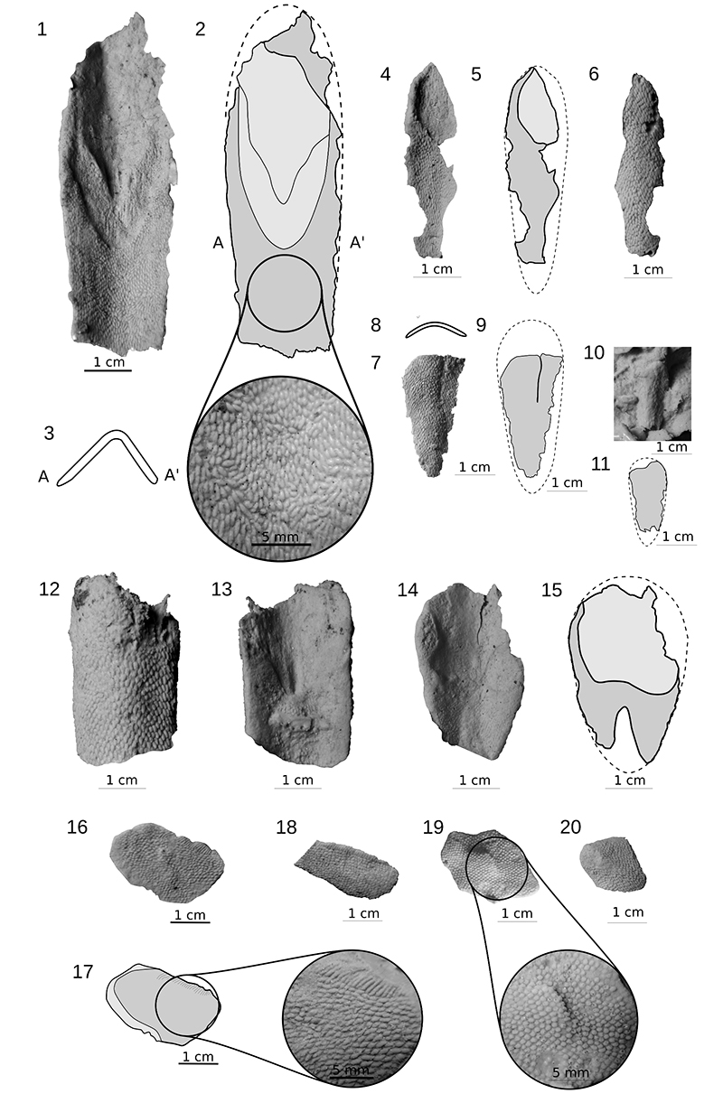
FIGURE 9. Guerichostus kozlowskii, 1. right branchial plate ZPAL Ag. III/9, ventral surface with fragments showing the detail of ornamentation (2-3); 4. left branchial plate ZPAL Ag. III/8 with fragments showing the detail of ornamentation (5).
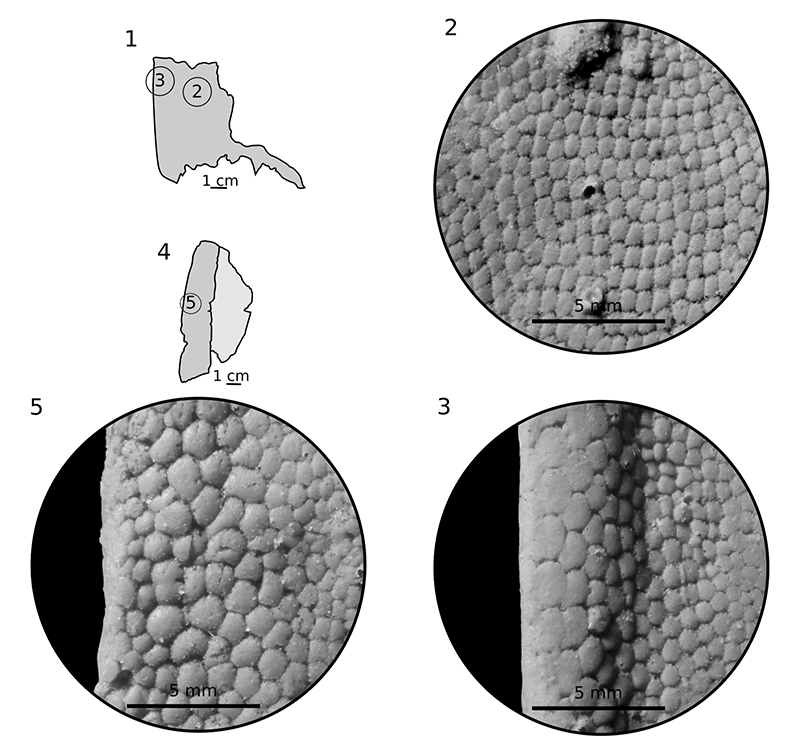
FIGURE 10. Guerichostus kozlowskii, 1. dorsal plate ZPAL Ag. III/1 with fragment showing the detail of ornamentation (2); 3. dorsal plate ZPAL Ag. III/2 with fragment showing the detail of ornamentation (4); 5. cornual plate ZPAL Ag. III/19 with fragment showing the detail of ornamentation (6).
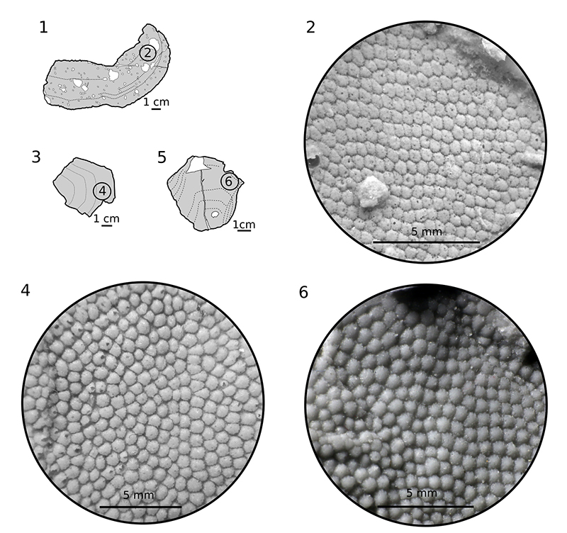
FIGURE 11. Guerichostus kozlowskii, 1. right branchial plate ZPAL Ag. III/7, dorsomedial (1) and posterior (2) view (white hatched line mark bone layer in vascular middle layer).
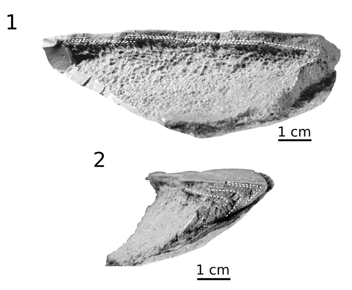
FIGURE 12. Hariosteus kielanae 1. fragment of dorsal plate ZPAL Ag. III/48, external (1) and internal (2), 3. drawing of dorsal view with growth line (hatched line); 4. external view of dorsal plate fragment MZ-VIII/Vp-549; 5. external view of dorsal plate fragment ZPAL Ag. III/50; 6. drawing of dorsal view with growth line (hatched line); 7. branchial plate ZPAL Ag. III/83, 8. drawing with growth line (hatched line).
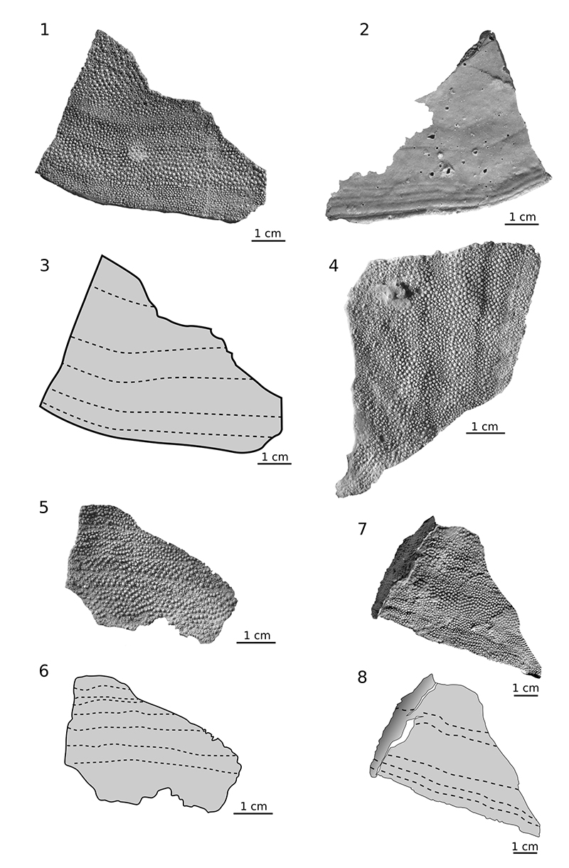
FIGURE 13. Hariosteus kielanae; 1. right branchial plate ZPAL Ag. III/17, dorsal (1) and ventral (2) view, 3 drawing of dorsal view with grow line (fine hatched line); 2. left branchial plate MUZ.PIG.1733.II.185, dorsal (4) view and drawing of dorsal view (5); 6. left branchial plate MZ-VIII/Vp-452, ventral view; 7. left branchial plate ZPAL Ag. III/24, dorsal view.
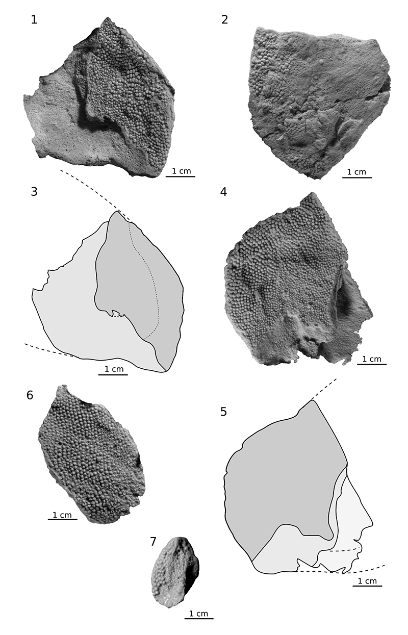
FIGURE 14. Hariosteus kielanae, 1. ridge scale ZPAL Ag. III/25, external view with close-up on tubercles, 2. drawing of the scale with shape reconstruction (hached line) and cross section (3); 4. ridge scale ZPAL Ag. III/28, visceral view with close-up on tubercles, 5. drawing of the scale with shape reconstruction (hatched line) and cross sections (6); 7. ridge scale MZ-VIII/Vp-67, internal view; 8. body scale with lateral line canal MUZ.PIG.1733.II.309, external view, 9. drawing with grow line and photo with close-up on tubercles; 10. body scale MZ-VIII/Vp-410, external view, 11. drawing with grow line and photo with close-up on tubercles; 12 trunk scale MUZ.PIG.1733.II.184, external view with drawing (13) presenting overlapping area (light grey).
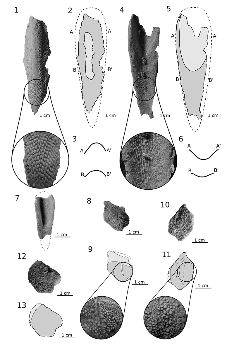
FIGURE 15. Hariosteus kielanae, 1. dorsal plate ZPAL Ag. III/2 with fragment showing the detail of ornamentation (4); 2. left branchial plate MUZ.PIG.1733.II.185, dorsal surface with fragment showing the detail of ornamentation (5, 6); 3. dorsal plate ZPAL Ag. III/50 with fragment showing the detail of ornamentation (7).
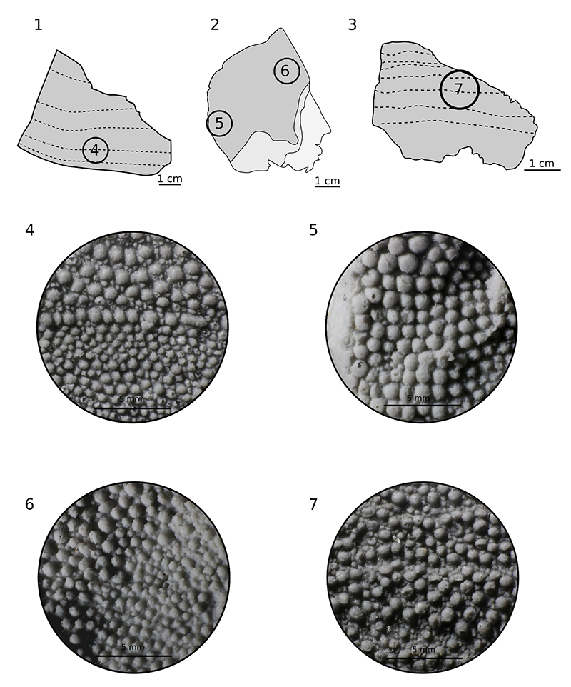
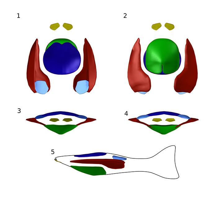
FIGURE 16. Guerichosteus kozlowskii three-dimensional reconstruction; dorsal (1), ventral (2), front (3) back (4) and left lateral (5) view.